Case Report
Volume 1 Issue 2 - 2017
Clinical Approach of Follicular Conjunctivitis in a (Cavia Porcellus)
1Centro Veterinario Villa Flaviana, Roma, Italy
2Centro Veterinario per Animali Esotici, Palermo, Italy
3School of Biosciences and Veterinary Medicine, University of Camerino , Italy
4Pet Doctors Lynfield, Auckland, New Zeland
2Centro Veterinario per Animali Esotici, Palermo, Italy
3School of Biosciences and Veterinary Medicine, University of Camerino , Italy
4Pet Doctors Lynfield, Auckland, New Zeland
*Corresponding Author: Di Giuseppe Marco, Centro Veterinario per Animali Esotici, Viale Regione Siciliana Sud-Est 422-426, 90129 Palermo, Italy.
Received: May 23, 2017; Published: July 10, 2017
Abstract
A 2-years old female guinea pig (Cavia porcellus) was presented due to mono-lateral ocular discharge unresponsive to local therapy with antibiotics and corticosteroids. Clinical examination reveled a small proliferative conjunctival mass on the right medial canthus Under general anesthesia the soft tissue proliferation was excised and sent for histopathology.
No recurrence was seen after one month from surgery and the histopathology diagnosis was follicular conjunctivitisThe conjunctiva-associated lymphoid tissue (CALT) contributes with the mucose-associated lymphoid tissue (MALT) to the first immunological response.
Follicle formation is an indication of non-specific immune stimulation. While many cases of conjunctivitis are self-limiting, some can persist and the changes within the conjunctiva can become irreversible. At this point, conjunctival biopsy is the only way to to establish the presence of any conjunctival changes and to achieve a specific diagnosis of immune-mediated conjunctivitis.
The conjunctiva slides over the cornea during the blinking and for this reason any changes in its appearance can produce an ocular damage. Moreover, failure in closing the eyelids due to protruding tissue increase the possibility of foreign body penetration and secondary infection.
Keywords: Guinea pig; Follicular conjunctivitis; CALT, MALT; Ocular mass
Abbreviations: CALT: MALT
Introduction
Conjunctivitis is a common disease in small animals and may be unilateral or bilateral, primary or secondary to another related pathology. The tissue reaction normally produce tissue inflammation with or without lacrimation and blepharospasm [1].
Primary conjunctivitis has been described in rabbits but in most cases it is secondary to other causes,: underlying dental disease, eyelid abnormalities, chronic dacryocystitis, poor housing condition[4]. Rabbits and guinea pigs have similar conjunctivitis aetiology but in the authors opinion the soft tissue proliferation is more common in guinea pigs than in other small mammals.
In literature, in dogs and cats the etiology of follicular conjunctivitis is still unknown [3]. Lymphoid hyperplasia in the conjunctiva represent a non specific immunological response in cases of drug toxicities, allergic conditions, and infections etc. The conjunctiva-associated lymphoid tissue (CALT) contributes with the mucose-associated lymphoid tissue (MALT) to the first immunological response [2]. The conjunctival epithelium undergoes to a marked hyperplasia, and this hyperplasia may by particularly relevant in guinea pig. Although the follicles can not be differentiated histologically from lymphoid follicles secondary to other causes (e.g. allergic), their presence is an indication of non-specific immune stimulation common in chronic conjunctivitis.
Clinical Case
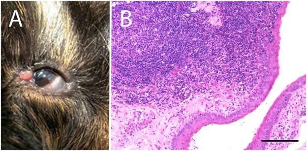
A 2-years old female guinea pig was presented for eye evaluation due to mono-lateral ocular discharge. It was bought at the age of 6 weeks old and has been the only guinea pig in the house. The guinea pig was housed in a standard 60x90 cm cage with wood pellets bedding. It was fed with proper guinea pig pellets, timothy hay ad libitum and fresh greens daily. At physical examination the animal was bright and alert and in good body condition, in the left eye a conjunctivitis with increase of tear fluid and a small mass on the right medial canthus was noticed. After the standard health check, anesthesia was induced with 4% isoflurane (Isoflo® 250 ml, Esteve, Spain) in 100% oxygen administered by face mask and maintained with 2.5% isoflurane (Isoflo® 250 ml, Esteve, Spain) and 0.5 L/minute oxygen to perform a closer investigation using a 3x magnification loupe. The mass was approximately 2 x 2 mm in size with a rounded shape originated from the conjunctival tissue (Figure 1). No obvious lesions were seen on the cornea and the fluorescein test was negative. The patient was discharged with topical tobramycin and dexamethasone eye drops (Tobradex®, ALCON ITALIA SpA) and the eye re-check after a week. One week later, the guinea pig didn’t show any improvement. At this point anesthesia was induced with the same protocol of the previous time. A drop of ossibuviprocaine eyewash (Novesina®, Novartisfarma SpA) was applied on the mass and few minutes later the soft tissue proliferation was excised with nr° # 15 scalpel blade and the bleeding was simply controlled using a small cotton tip over the surgical wound
A 2-years old female guinea pig was presented for eye evaluation due to mono-lateral ocular discharge. It was bought at the age of 6 weeks old and has been the only guinea pig in the house. The guinea pig was housed in a standard 60x90 cm cage with wood pellets bedding. It was fed with proper guinea pig pellets, timothy hay ad libitum and fresh greens daily. At physical examination the animal was bright and alert and in good body condition, in the left eye a conjunctivitis with increase of tear fluid and a small mass on the right medial canthus was noticed. After the standard health check, anesthesia was induced with 4% isoflurane (Isoflo® 250 ml, Esteve, Spain) in 100% oxygen administered by face mask and maintained with 2.5% isoflurane (Isoflo® 250 ml, Esteve, Spain) and 0.5 L/minute oxygen to perform a closer investigation using a 3x magnification loupe. The mass was approximately 2 x 2 mm in size with a rounded shape originated from the conjunctival tissue (Figure 1). No obvious lesions were seen on the cornea and the fluorescein test was negative. The patient was discharged with topical tobramycin and dexamethasone eye drops (Tobradex®, ALCON ITALIA SpA) and the eye re-check after a week. One week later, the guinea pig didn’t show any improvement. At this point anesthesia was induced with the same protocol of the previous time. A drop of ossibuviprocaine eyewash (Novesina®, Novartisfarma SpA) was applied on the mass and few minutes later the soft tissue proliferation was excised with nr° # 15 scalpel blade and the bleeding was simply controlled using a small cotton tip over the surgical wound

Figure 1A: Particular of the left eye of the guinea pig showing a 2 x 2 mm proliferative
conjunctival mass on the right medial canthus and a concurrent conjunctivitis with epiphora.
Figure 1B: Microscopic section of conjunctiva mass showing hyperplasia of the conjunctival epithelium and underlying connective tissue, and proliferation of lymph follicles. The follicles are well differentiated with some lymphoplasmacellular and heterophilic infiltrates. Stimulated (reactive) secondary follicles, shows a paler staining germinal centre with large lymphoblasts and an increased numbers of apoptotic lymphocytes and tingible body macrophages. The mantle zone surrounding the germinal centre, composed of small to medium-sized darker staining lymphocytes, is increased in size. Bar: 200 μm.
Figure 1B: Microscopic section of conjunctiva mass showing hyperplasia of the conjunctival epithelium and underlying connective tissue, and proliferation of lymph follicles. The follicles are well differentiated with some lymphoplasmacellular and heterophilic infiltrates. Stimulated (reactive) secondary follicles, shows a paler staining germinal centre with large lymphoblasts and an increased numbers of apoptotic lymphocytes and tingible body macrophages. The mantle zone surrounding the germinal centre, composed of small to medium-sized darker staining lymphocytes, is increased in size. Bar: 200 μm.
The mass was sent to the laboratory for histopathology and the animal discharged a few hours later without any medication.
The histopathology result showed: a hyperplasia in the conjunctival epithelium and in the underlying connective tissue and a proliferation of lymphoid follicles. The follicles were well differentiated with some lymphoplasmacellular and heterophilic infiltrates. Lymphoid hyperplasia involved T cell-rich follicles indicating a strong cell-mediated response. Stimulated (reactive) secondary follicles, showed a paler staining germinal center with large lymphoblasts and increased numbers of apoptotic lymphocytes and tingible body macrophages. The mantle zone surrounding the germinal center, composed of small to medium-sized darker staining lymphocytes, was increased in size. The diagnosis was lymphoid follicular conjunctivitis (Figure 2). The patient was re-checked one month after the excision but no recurrence was observed.
Discussion and Conclusion
While many cases of conjunctivitis are self-limiting, a small proportion persist, particularly when the treatment is incorrect or the inflammation is already chronic and the changes within the conjunctiva become irreversible, so that treatment is ineffective. At this point, conjunctival biopsy is the only way to to establish the presence of any conjunctival changes and to achieve a specific diagnosis of immune-mediated conjunctivitis Crispin 2002). In the authors experience guinea pigs are prone to develop bilateral conjunctivitis and usually the lymphoid tissue that is an important component of the conjunctiva may react by developing tissue proliferation.
The conjunctiva slides over the cornea during the blinking and for this reason any changes in its appearance can produce an ocular damage. Moreover, failure in closing the eyelids due to protruding tissue increase the possibility of foreign body penetration and secondary infection.
In the authors opinion, when a conjunctival proliferation is present usually it is too late for a medical treatment. In fact in many other similar cases, guinea pigs were presented at the clinic when the normal tear fluid was already increased in amount and density and in some of them the scratching caused from the discomfort determined a corneal ulcera. After many attempts, authors decided to perform surgery as the treatment of choice in conjunctivitis affecting the eyelid physiology.
References
- Crispin S. “The conjunctiva. In Manual of Small Animal Ophthalmology 2nd Edition.” BSAVA (2002): 124-127
- Liebler-Tenorio EM and Pabst R. “MALT structure and function in farm animals”. Veterinary Research 37.3 (2006): 257-280
- Swanson JS,. et al. “Nictitans abnormalities and Therapies in Small Animal Ophthalmology Secret”. Elsevier (2002): 70-71
- Wagner F. “Common Ophthalmic Problems in Pet Rabbits.” Journal of Exotic Pet Medicine 16.3 (2007): 158-167
Citation:
Di Giuseppe Marco., et al. “Clinical Approach of Follicular Conjunctivitis in a (Cavia Porcellus)”. Multidisciplinary Advances in
Veterinary Science 1.2 (2017): 77-79.
Copyright: © 2017 Di Giuseppe Marco., et al. This is an open-access article distributed under the terms of the Creative Commons Attribution License, which permits unrestricted use, distribution, and reproduction in any medium, provided the original author and source are credited.


 Scientia Ricerca is licensed and content of this site is available under a Creative Commons Attribution 4.0 International License.