Research Article
Volume 1 Issue 4 - 2017
Subclinical Mastitis: Silent Inhibitor of Milk Production in Backyard System at Puebla, Mexico.
Laboratorio de Endocrinología de la Reproducción y Malacología, Escuela de Biología, Benemérita Universidad Autónoma de Puebla, Boulevard Valsequillo y Ave, San Manuel s/n, Ciudad Universitaria, Edificio No.112-A, Puebla
*Corresponding Author: Mariana Paz–Calderón Nieto, Laboratorio de Endocrinología de la Reproducción y Malacología, Escuela de Biología, Benemérita Universidad Autónoma de Puebla, Boulevard Valsequillo y Ave, San Manuel s/n, Ciudad Universitaria, Edificio No.112-A, Puebla.
Received: October 20, 2017; Published: October 26, 2017
Abstract
The backyard livestock represents a family support system in rural and peri-urban communities in Mexico, where the whole family is involved in the care of animals at milking, but lack adequate training for optimal results mainly in the production of milk, where more commonly abounds with the presence of subclinical mastitis. The aim of this study was to determine the incidence of subclinical mastitis in dairy Basin of Santa Ana Xalmimilulco, Puebla, Mexico. 158 animals-10% of the population of milked cows in the study area (632 milk samples for each quarter of the udder) were sampled.
The samples were performed by Dark Side test, of which 62% of the animals tested were positive for subclinical mastitis, to the positive samples were subjected to culture media brand "BD": Mc Conkey, Blood Agar, Dextrose-papa and Salt-Mannitol. They differed by Gram stain of the brand "HYCEL", to which were classified using biochemical battery brand "BD" consisting of TSI, LIA, MIO, Citrate and Urea, the bacteria found were: Streptococcus agalactiae and Staphiloccocus aureus. The sub-clinical mastitis generates losses of more than $ 5.23 Mexican pesos per day per animal which decreases milk production of 23.5 to 54.6%. In conclusion subclinical mastitis thus becomes one of the anthropomorphic ecopathological abnormalities that affect the quality and safety of milk in the backyard livestock.
Keywords: Ecopathology; Milk; Dark Side; Economic loss
Introduction
Backyard cattle breeding from the time of the colony has been the livelihood of families mainly from rural and peri-urban communities, where each member of the family collaborates directly in the maintenance of the animals (Caicedo., et al. 2011). The foundation of this type of livestock system is mainly to provide all the members of the family of milk and its derivatives, and in the same way, the sale is at low cost and these products are available at any time of the year, this they obtain of being able to raise the animals in a generally reduced space.
However, lack of training and efficient supervision of trained personnel that can train family members about the care and management of their animals generates a series of diseases such as subclinical mastitis that does not show symptoms in cows, besides being the most frequent and most resistant disease (Caicedo., et al. 2011; Paz-Calderón., et al. 2013) and chronic mastitis is represented by the hardening of the mammary gland produced by the proliferation of fibrous tissue that replaced tissue healthy gland. In this type of mastitis the milk secretion is usually watery with yellowish color (pus) or coffee (blood).
This type of mastitis may adopt the modality of subclinical that appears macroscopically normal (Zurita, 1982). Both types of mastitis generate a decrease in milk production to the extent that it is impossible to continue milking the animal, which generates both health risks for the family, animals and consumers, as well as high economic losses (Paz-Calderón., et al. 2013).
Several mechanisms are known that infect the udder, but in the present work only the infection caused by bacterial contamination, coming from the environment in which the cows are developed or inhabited, this way the bacteria attack the udder, they multiply within the breast tissue and generate toxins that are the cause of injury (Loor., et al. 1999).
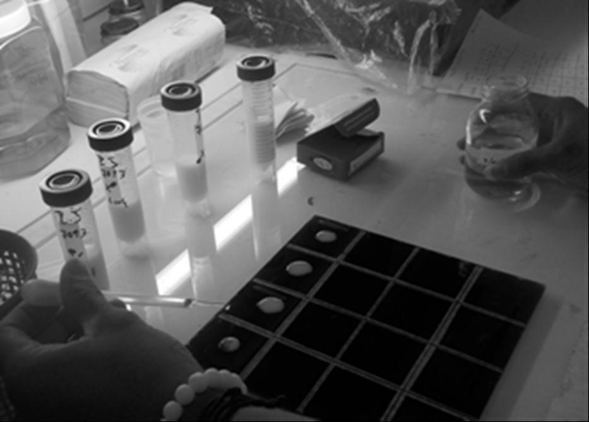
Infection of the udder or nipple has three mechanisms of invasion: 1) infection of the nipple: bacterial infection begins at the entrance of the nipple canal, which after being milked the cow is dilated for a period of approximately 15 minutes , for up to 2 hours depending on the animal, however it can remain semi-open or fully open; 2) Once the bacteria have passed through the nipple canal they invade and colonize the tissues, which make them fibrous, the bacteria are attacked by somatic cells (leucocytes and macrophages); 3) if the bacteria are not destroyed by the leukocytes enter the alveolar tissues and begins the destruction of the alveolar, therefore as the infection persists, the alveoli are kept covered and begin to diminish their size, which generates the decrease of the milk production, as shown in Figure 1; this produces irritation and causes subclinical inflammation, which by its very nature lacks symptoms (asymptomatic) and is difficult to detect, meanwhile accumulate bacterial wastes that intensify the inflammatory response, destruction of the secretory tissue, with the consequent decrease in productive output known as “agalactia”, in which a quarter or nipple of the udder or the complete udder ceases to produce in a sudden or progressive way milk.

Figure 1: The “Dark Side” test procedure is shown with 4 drops of milk and
2% of sodium hydroxide, placed on a black background.
Extensive fourth-quarter scars may render it unproductive in subsequent lactations (National Mastitis Council, 1997). Other factors that influence the proliferation of this disease are: 1) animal handling and inadequate facilities, eg excreta management is necessary, since a clean and dry bed with no accumulation of feces, urine and moisture helps to reduce the accumulation of large amounts of bacteria and their transfer to the udder (Homan and Wattiaux, 1999); 2) the coexistence of other animals in a common space generates zoonotic diseases (brucellosis and tuberculosis mainly) , since at the same time the dairy cows are in coexistence with dogs, cats, turkeys, hens, ducks, donkeys, horses, rabbits, among others.
A common problem, for example, is that the other animals sleep and defect in the food and on the floor where the cows lie, another problem is that dogs lick the udders or drink the milk that is spilled on the floor of this cows can also acquire bacteria or viruses that enter the udder and damage it (Caicedo., et al. 2011, Paz-Calderón., et al. 2013). 3). Reducing factors such as environmental stress in cows, particularly around the time of delivery, is essential for the control of mastitis.
Causes of stress in cattle are related to cortisone produced, which has immunosuppressive effects on the body, thus immunosuppressed cows become more susceptible to contracting mastitis (Wallace, 2000). Therefore, the objective of the study was to determine the presence of subclinical mastitis in dairy cows, the microorganisms involved and the incidence of subclinical mastitis in the Santa Ana Xalmimilulco dairy basin, Huejotzingo municipality, Puebla, Mexico.
Material and Methods
Characterization of the farms: Each farm has an ecological survey, where the farm is located geographically, the breed of animals, milk production, milk production mechanism, whether or not registered, vaccination to prevent diseases, number of animals in production, growth, length of lactation period, type of feed, discard of animals, frequency of mastitis, bacteriological tests, medication for mastitis, and parasitosis, abortion causes, pasture type, and sources of water on the farm.
Animals: The study was carried out in the Auxiliary Municipality of Santa Ana Xalmimilulco, Puebla, Mexico, where samples of 158 cows (Bos taurus) of the Holstein breed were taken. All farms (17 farms) under study were georeferenced with a GPS. Animals are fed with alfalfa, very few minerals are given; on the other hand, the grass used for feeding the animals is irrigated with wastewater. Little animals are only dewormed once a year. Most of the animals are under roof, where they do not give them sunlight, and they are tied with the unique freedom of lying down and standing. The animals are kept in milking for about 279 days and when they have 5 calves they are discarded.
Sampling: 158 cows from private farms were sampled in each quarter of the udder, the "White Side" test was carried out, which was modified in the Reproductive Endocrinology and Malacology Laboratory, as a "Dark Side" when using a dark background to perform the test as shown in Figure 1 (Mateus, 1983), a white background is required to place the milk, 4% sodium hydroxide is added and milk is determined to change its constitution , the modification of this technique is based on the use of a black background (instead of white) because the milk being white, it is easier to appreciate the changes on black background, because on the black background when you see it near the light is best appreciated the characteristics in the milk that presents bacteria (through clots that are made when reacted with NaOH) and is made as follows: corresponds to 4 drops of milk and 2 drops of sodium hydroxide 4% (NaOH), is removed with a toothpick If samples are positive for subclinical mastitis, coagulate or clump together, samples that are also cleared are positive for subclinical mastitis (Mateus, 1983). Positive tests were cultured on "BD" brand agar (Becton, Dickinson and Company; Madrid, Spain): Mc Conkey, Dextrose-potato, salt and mannitol and blood agar; the positive cultures were isolated and stained with the Hycel brand kit (Mexico, HYCEL de México, and S.A de C.V.), to determine if they were positive or negative cocci or bacilli. Positive samples were classified according to the literature (Bergey., et al. 1923), with the biochemical battery corresponding to the media: LIA, TSI, MIO, Citrate and Urea of the brand "BD" (Becton, Dickinson and Company, Madrid, Spain).
Results and Discussion
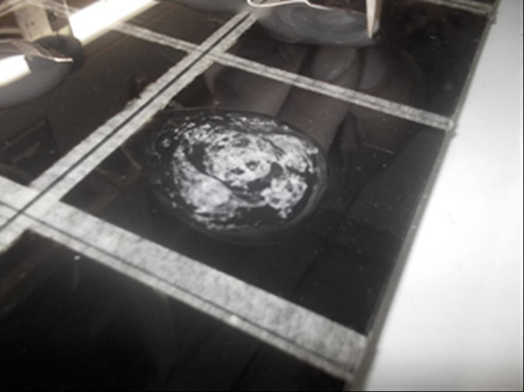
The tests of "White Side and Dark Side" determine that they are positive when the milk in contact with the sodium hydroxide changes the consistency and its characteristics, as shown in figure 2, is characterized by the formation of lumps or agglutination of the milk, in other cases it can become gelatinous and even when it becomes transparent it is determined that the test is positive (Mateus, 1983), the test of "Dark Side" becomes easier to observe, since it has to be done in a glass of black color and a light above the sample to be able to see the lumps and the consistency of the milk that changes by the greater content of somatic cells (Ávila, 1984; Pérez, 1986). After determining the positive samples with the "Dark Side" test were cultured and it was determined that the most conducive culture medium was Mc Conkey, where there was significant bacterial growth, after staining the bacteria it was determined that both cocci and Gram positive bacilli and Streptococcus agalactiae and Staphiloccocus aureus, which ferment lactose and are therefore the most frequent bacteria in subclinical mastitis, these bacteria are the most common in subclinical mastitis, because in this case, they present greater resistance with each delivery and antibiotics.

Figure 2: Changes in milk on contact with 4% sodium hydroxide
are shown if the samples show bacterial contamination.
The characteristics of these bacteria are: Staphylococcus aureus: Known as golden staphylococcus, Gram positive, facultative anaerobic, immobile, worldwide distribution, it is estimated that one in three people are colonized but not infected with this bacterium, is highly resistant, found in the skin and mucous membranes (Gil, 2000; Hurtado., et al. 2002). Streptoccocus agalactiae: Gram positive coccus, anaerobic facultative, can be found in the digestive, urinary and genital tract of adults (De la Rosa., et al. 1992).
Based on Andresen, (2001), the role of man in the spread of the disease is crucial, since the high incidence of the disease is due to poor milking, lack of maintenance in the milking machine and lack of supervision of a veterinarian. In the farms visited, the most common problems are: the dirty udder, every quarter, they stay too long in the milking machine, when it is suggested that they do not stay for more than 6 minutes in the milking machine and finish the milking manually (And the poor sealing of the nipple (Paz-Calderón., et al. 2013).
In the sampled dairy herds, it was determined that each cow produces about 15 to 23 liters of milk per day, which in 305 days of milking, generates a single cow an amount of 4 575 to 7 015 liters per year, which gives the family in your care an income of $ 60.0 to $ 92.0 pesos per day/cow, a year represents from $ 18 300 to $ 28 060 pesos/cow. Failure to properly care for animals leads to production losses, which in the end are represented in a smaller amount of income and, on the other hand, medical expenses must be made to try to restore the health of the animal, with large expenditures on medicines, medical consultations and continuous supervision.
Conclusion
The subclinical mastitis that generates economic losses for the families that are dedicated to milk production and these do not have the knowledge of how to perform asepsis of the udder, as well as the care of the corral where the cows are kept. More than 72% of the cows sampled in this study had subclinical and chronic mastitis. This is significant, since it is only a matter of time before other milking cows become infected, if there is no and appropriate and timely treatment. Both Streptococcus agalactiae and Staphylococcus aureus are resistant bacteria and are detected by the "dark background" test (modified in the Laboratory of Endocrinology of Reproduction and Malacology of the Faculty of Biology of the Autonomous University of Puebla). The udder with subclinical mastitis does not show any changes, the smell as well as the taste and color of the milk do not change, however these bacteria are lodged in the mammary gland and produce in normal glandular tissue, this is replaced progressively by fibrotic tissue and it generates that the milk production is diminished. Therefore, contamination of the udder by the transfer of bacteria from animals to animals in the backyard of the farm should be avoided; therefore it is important to have healthy animals, since economic losses and damage to public health are eluded because of the high antibiotic content that is supplied to animals with mastitis and that these antibiotics can pass into milk, this excess in the use of antibiotics produces bacterial resistance, thus affecting animal welfare and ultimately human. We have to consider that backyard breeding generates the largest quantity of milk for domestic consumption and for the production of artisanal cheese
References
- Andresen Hans S. “Mastitis: Prevención y Control”. Revista de Investigaciones Veterinarias del Perú 12.2 (2001): 55-64.
- Ávila TS. “Producción intensiva de ganado lechero. Anatomía y fisiología de la glándula mamaria”.Edit Continental México (1984): 139-157.
- Bergey DH., et al. “Bergey’s manual of determinative bacteriology. 1st edition Baltimore”. Williams and Wilkins (1923): 187-200.
- Caicedo Rivas RE., et al. “Salud animal de una cuenca lechera bajo el sistema de traspatio, Puebla, México”. AICA I (2011): 323-326
- De la Rosa M., et al. “New Granada medium for detection and identification of group B streptococci”. Journal of Clinical Microbiology 30.4 (1992): 1019-1021.
- Gil D de MM. “Staphylococcus aureus: Microbiología y aspectos moleculares de la resistencia a meticilina”. Revista Chilena de Infectología 17.2 (2000): 145-152.
- Homan Jy and Wattiaux M. “Mastitis. En su: Guías Técnicas Lecheras: Lactancia y Ordeño: Instituto Babcock para la Investigación y Desarrollo Internacional para la Industria Lechera”. Universidad de Wisconsin (1999): 61-76.
- Hurtado MP., et al. “Staphylococcus aureus: Revisión de los mecanismos de patogenicidad y la fisiopatología de la infección estafilocócica”. Revista de la Sociedad Venezolana de Microbiología Caracas 22.2 (2002): 112-118.
- Loor JJ., et al. “Aspectos básicos sobre el desarrollo de mastitis”. Instituto y Universidad Politécnica de Virginia Blacksburg (1999).
- Mateus Valles Guillermo. Mastitis en Bovinos. Volumen 1 de Boletín divulgativo PA CIAT. Boletín Informativo. Bib. Orton IICA/CATIE (1983): 1-17.
- National Mastitis Council. “A practical look at contagious mastitis”. National Mastitis Council Homepage Disponible en (1997).
- Paz–Calderón Nieto Mariana., et al. “Detección de Mastitis subclínica en el sistema de ganadería traspatio en Puebla, México. III Reunión de la Red Mexicana sobre Conservación y Utilización de los Recursos Zoogenéticos (CONBIAND)”. Memorias de Congreso (2013): 28.
- Pérez DM. “Manual sobre ganado productor de leche”. Edit Villicaña SA México (1982): 710-744.
- Wallace RL. “Production of quality milk trough environmental mastitis control”. Illini DairyNet. University of Illinois. Urbana-Champaingn (2002).
- Zurita Arevalo L. “Mastitis bovina con especial énfasis en la realidad nacional”. Monografías de Medicina Veterinaria, Chile 4.2 (1982).
Citation:
Mariana Paz–Calderón Nieto., et al. “Subclinical Mastitis: Silent Inhibitor of Milk Production in Backyard System at Puebla,
Mexico.” Multidisciplinary Advances in Veterinary Science 1.4 (2017): 169-174.
Copyright: © 2017 Mariana Paz–Calderón Nieto., et al. This is an open-access article distributed under the terms of the Creative Commons Attribution License, which permits unrestricted use, distribution, and reproduction in any medium, provided the original author and source are credited.


 Scientia Ricerca is licensed and content of this site is available under a Creative Commons Attribution 4.0 International License.