Case Report
Volume 2 Issue 3 - 2018
Low-Intensity Laser Therapy Associated with Ozone Therapy for Recurrent Atopic Dermatopathy Treatment in a Donkey. A Case Study.
1Department of Agrarian and Environmental Sciences, State University of Santa Cruz – Ilhéus, BA, Brazil
2Student of Veterinary Medicine Course, State University of Santa Cruz, Ilhéus, BA, Brazil
2Student of Veterinary Medicine Course, State University of Santa Cruz, Ilhéus, BA, Brazil
*Corresponding Author: Fernando Alzamora Filho, Department of Agrarian and Environmental Sciences, State University of Santa Cruz – Ilhéus, BA, Brazil.
Received: June 13, 2018; Published: June 20, 2018
Abstract
The present study reports the clinical presentation and the treatments applied in an Asinine with recurrent atopic dermatitis. The patient was brought to the hospital with a history of dermatitis characterized by intense pruritus and cutaneous ulcerations with chronic evolution, having been treated in several occasions without success. The history and characteristics of the lesions pointed to the diagnosis of chronic atopic dermatitis. Treatment was based on daily cleaning of lesions and complementary treatments with systemic ozone therapy and low-intensity laser therapy at the lesion site. The ozone therapy sessions were weekly and applied by intraretal gas insufflation and by minor autohemotherapy totaling five sessions. Laser therapy was applied in skin lesions with red or near infrared monochromatic light with energy and fluence varying according to the characteristics and extent of the lesion, totaling 11 sessions. At the end of the treatment, there was no more pruritus and total wound healing. It was concluded that the association of ozone therapy and low intensity laser therapy was effective in the complete resolution of this case.
Keywords: Hypersensitivity; Ozone; Photobiomodulation laser; Pruritus; Wounds
Introduction
Allergen exposition can predispose equines to dermatopathy affections which may have multifactorial origin, without breed, sex or age predilection. Insect’s hyper sensibility is caused by allergens in the Culoides spp. Saliva. Stomoxys calcitrans e Haematobia may be also involved [10]. Allergic reaction is commonly evident for hives, pruritus, alopecia, and cutaneous wounds, secondary bacterial infections and myiasis in more severe wounds [5,13]. The localization of lesions in animals are variable and can be in head, limbs, dorsal or ventral body. The diagnosis is based on seasonality, insects exposition historic, clinical signs associated with the discarding of other pruritus causes and in the treatment response [8,11]. The treatment should aim to the causal factor identification and to adopt the restriction therapy to this agent. Also, glucocorticoids therapy and insect control [10,13]. When the dermatopathy results in cutaneous wounds, others treatments shall be instituted aiming the secondary infections control and promoting healing. Complementary treatment as low intensity laser and ozone have proven promising in dermal injuries for its analgesy, inflammatory mediators reduce, angiogenesis, healing process improvement and antiseptics effects [9,2,12]. This report describes the clinical aspects of atopic dermopathy in asinine and its resolution by laser and ozone therapy.
Materials and Methods
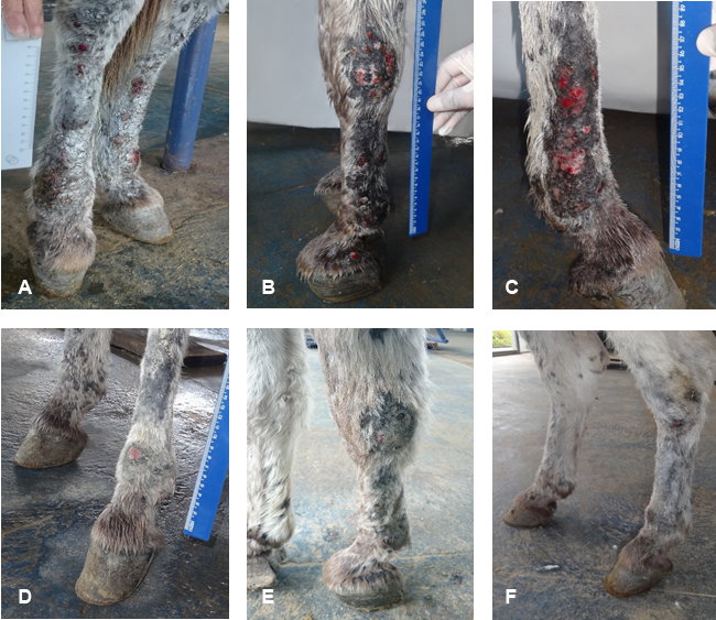
A case of recurrent atopic dermatitis is described in a 4-year-old, male, asinine, from the city of Ilhéus-Bahia and hospitalized at UESC Veterinary Hospital, due to ulcerative wounds in the thoracic and pelvic limbs. The owner reported it was a chronic condition, with two years of evolution that was treated several times unsuccessfully. The clinical aspect was characterized by crusted popular eruptions in the four limbs (Figure 1A and 1B), intense pruritus and presence of Stomoxys calcitrans flies. The patient was previously submitted to topical treatments that softened the symptoms but did not solve the problem. The diagnostic of allergic dermatitis was defined by epidemiology and clinical findings. The therapy was established by daily antisepsis with saline solution and chlorhexidine, bandaging the limbs and repellents spraying. Ozone therapy by bagging (3 times/week) and rectal insufflation (once/week) was also associated to the topical treatment.
However, due to the patient hard temperament and the intense sensitivity injured region, it was decided to change the topical ozone therapy by bagging to low intensity laser, maintaining transrectal insufflation, following these protocols: 15mg in a 30mcg/mL flow of O3 by transrectal weekly therapy totalizing 5 sessions, and minor autohemotherapy every 2 weeks (240µg of O3 in 10mL of blood intramuscular administration) totalizing 3 sessions. Laser phototherapy was applied for wound local treatment, with diode laser (0.1W and spot area of 0.028cm2). Red laser (660 nm) and infrared laser (808 nm) were applied in the thoracic and pelvic limbs wounds, in point irradiation mode. The energy (J) and fluence (J/cm2) dosages were defined according to the aspect and area of the wound (Table 1). In the first and second weeks of treatment two irradiations were applied on consecutive days (4 sessions) and from the third week on one session every 48 hours, totaling 7 sessions.
| Injury location | Wavelength λ (nm) | Energy (J) | Fluence (J/cm2) | Number of point for lesioned area (1 cm of space between the points) |
| Right front limb | 660 | 0.1 | 17.9 | 5 |
| 808 | 0.5 | 125 | 7 | |
| Left front limb | 660 | 0.2 | 42.9 | 6 |
| 808 | 0.5 | 107.1 | 6 | |
| Right posterior limb | 660 | 0.2 | 121.4 | 17 |
| 808 | 0.5 | 142.9 | 8 | |
| Left posterior limb | 660 | 0.2 | 100 | 14 |
| 808 | 0.5 | 178.6 | 10 |
Table 1: Laser dosage parameters per injured limb in asinine with recurrent atopic dermatopathy.
Discussion
Comparing the blood cells count pre and post treatment is possible to attest modulation of the inflammatory process, with reduction of global leucocyte count, reactive lymphocytes and eosinophilia. In red cells, ozone therapy improves ductility properties and consequent promotion of blood perfusion and greater oxygen and nutrients supply, benefiting healing. The positive effect of ozone in tissue perfusion is related to its antioxidant action on the erythrocyte membrane that improves the red cell ductility [12].
The O3 therapy protocol applied fits in the mid dosage, which according to Madrid Declaration on Ozone therapy, produce immune modulatory and antioxidants effects [6]. The atopic wounds, according to this publication, fits in the 3rd diseases category of ozone therapy indication. The response in healing process was already reported in the 2nd laser therapy session, with edema reduction, structural tissue rearrangement and inicial epithelialization (Figure 1C). These results are assigned to laser effect in promoting microcirculation stimuli and inflammation reduction [2,7]. Colombo., et al. [1] reported improvement collagen deposition in surgical wounds treated with 660 nm laser, immediately applied 24/24 hours after surgery. Pereira., et al. [7] observed vessel expansion in the first 12 hours after 670 nm laser irradiation and significant reduce of edema 3 days after laser therapy. After the third session it was visible the analgesic effect on the wounds, as the patient improved its behavior, allowing the treatment of the wounds standing calm. Petersen., et al. [9] also subjectively observed that surgical wounds treated with laser therapy presented less edema, pain and exudate than control wounds, particularly in bay horses.
As the number of laser therapy sessions advanced, a reduction in healing time (Figure 1D) was observed and at the end of treatment all wounds were epithelialized, with growing hair and absence of pain and pruritus (Figure 1E and 1F). In the present case, 11 therapy session were performed for tissue repair, corroborating to Ferreira [3], who reported a range of 1 to 20 session, mainly 1 to 7 session for surgery wound healing. Ozone therapy contributes for the best tissue perfusion due to its half-life effects and blood cells resistance [6]. Hadad [4] found an increase in hematocrit values after ozone therapy sessions. The improvement in tissue perfusion possibly influenced positively the cutaneous lesions healing. Therefore, the combination of laser therapy with ozone therapy prevented wounds infection, reduced hospitalization time and promoted welfare to the patient by controlling pruritus and pain.

Figure 1: Asinine. A: Multiple wounds in posterior limbs before the treatment begin. B: Wounds localized on right front limb, day 3. C: Wounds on right posterior limb; in healing process during treatment, day 9. D: Wound in regression stage during the treatment period, with hair growing, day 17. E and F: Epithelialized wounds and presence of new hair on previously injured areas, day 25.
Conclusion
It was concluded that the association of ozone and laser therapy was effective in the presented case, with the complete lesions healing, being a protocol of easy application, fast action and excellent cost-benefit relation. However, but preventive hematophagous flies control are necessary to avoid a possible recurrences.
References
- Colombo F., et al. “Effect of low-level laser therapy (660 nm) on angiogenesis in wound healing: an immunohistochemical study in a rodent model”. Brazilian Dental Journal 24.4 (2013): 308-312.
- Meneguzzo DT. Fototerapia com laser em baixa intensidade em processo inflamatório agudo induzido por carragenina em pata de camundongos – estudos de dosimetria. (Doctoral dissertation) – Instituto de Pesquisas Energéticas e Nucleares - IPEN-CNEN/SP, University of São Paulo, Brazil (2010).
- Ferreira AGA. Aplicação do laser de baixa intensidade no processo de cicatrização de ferida cirúrgica: padronização dos parâmetros dosimétricos. MSc Thesis, Federal University of Minas Gerais, Minas Gerais, Brazil (2016).
- Hadad MA. “Efeitos da ozonioterapia sobre parâmetros clínicos, hematológicos e da bioquímica sanguínea em equinos”. 138f. MSc Thesis, Federal University of Viçosa, Faculty of Veterinary Medicine, Viçosa, Brazil (2006).
- Lomas HR and Robinson PA. “A Pilot Qualitative Investigation of Stakeholders’ Experiences and Opinions of Equine Insect Bite Hypersensitivity in England”. Veterinary Sciences 5.1 (2018.): 3-17.
- Madrid Declaration on Ozone Therapy. https://www.drsozone.com/wp-content/uploads/2014/02/
- Madrid-Declaration-updated-July-30 (2010).
- Pereira MCMC., et al. “Influence of 670 nm low-level laser therapy on mast cells and vascular response of cutaneous injuries”. Journal of Photochemistry and Photobiology B: Biology 98.3 (2010): 188-192.
- Pessoa AFA., et al. “Doenças de pele em equídeos no semiárido brasileiro”. Pesquisa Veterinária Brasileira 34.8 (2014): 743-748.
- Petersen SL., et al. “The effect of low level laser therapy (LLLT) on wound healing in horses”. Equine Veterinary Journal 31.3 (1999): 228-231.
- Reed SM and Bayly WM. Equine Internal Medicine. 2nd edition. Guanabara Koogan (2000): 442-480.
- Smith BP. Tratado de Medicina Interna de Grandes Animais. 3rd edition. Manole (2006): 1728.
- Smith, NL., et al. “Ozone therapy: an overview of pharmacodynamics, current research, and clinical utility”. Medical Gas Research 7.3 (2017): 212-219.
- White SD and Yu AA. Equine Dermatology. In-Depth: Selected topics in Dermatology 52 (2006): 457-500.
Citation:
Fernando Alzamora Filho., et al. “Low-Intensity Laser Therapy Associated with Ozone Therapy for Recurrent Atopic Dermatopathy
Treatment in a Donkey. A Case Study.” Multidisciplinary Advances in Veterinary Science 2.3 (2018): 373-376.
Copyright: © 2018 Fernando Alzamora Filho., et al. This is an open-access article distributed under the terms of the Creative Commons Attribution License, which permits unrestricted use, distribution, and reproduction in any medium, provided the original author and source are credited.


 Scientia Ricerca is licensed and content of this site is available under a Creative Commons Attribution 4.0 International License.