Case Report
Volume 3 Issue 3 - 2018
Reinforced Single Complete Maxillary Denture.
1Intern - Rural Dental College, Loni, PIMS
2Professor - Dept. of Prosthodontics
3Professor and Head - Dept. of Prosthodontics
4Intern - Rural Dental College, Loni, PIMS
2Professor - Dept. of Prosthodontics
3Professor and Head - Dept. of Prosthodontics
4Intern - Rural Dental College, Loni, PIMS
*Corresponding Author: Pranit Bora, Intern - Rural Dental College, Loni, PIMS, India.
Received: May 17, 2018; Published: June 02, 2018
Abstract
The edentulous state of the oral cavity is equivalent to the absence of any other body part with specific morphological and psychological sequelae. The dentist has a difficult mission in evaluating the biomechanical differences in the supporting tissues for the two arches and applying the appropriate procedures to produce and maintain the conditions necessary for long-term treatment success. The dental literature evidences suggest that the maxillary arch exhibits earlier tooth loss due to various factors and that the mandibular anterior teeth are preserved the longest, so this case focuses on the oral condition in which the maxillary arch is edentulous and opposed by a natural mandibular dentition [2].
The main problem is that there are important qualitative and quantitative differences between natural tooth and complete denture support. The natural dentition is capable of specialized responses to occlusal demands that preserve its function, whereas the residual ridge is not and it will respond in a variable way depending on age, sex and racial category. Therefore, the replacement of the missing maxillary dentition must provide an optimum distribution of the occlusal forces in order to minimize the negative effects in the compromised edentulous arch. There has been so much debate regarding the Masticatory loads and forces transferring in the case of a single complete denture opposing a natural dentition. In most of the times flexural fatigue and stress concentration is found to be major cause of denture fractures [3]. So a single complete denture opposing natural dentition should be reinforced to that extent that it should withstand the huge occlusal forces acting on it. Metal can be added in form of wires, bars, mesh or plates. Metal strengthener has a beneficial effect on the fracture resistance of the poly-methyl-methacrylate.
Main objective of any prosthetic treatment should be based on DE VANS statement that, “Perpetual preservation of what remains of the human masticatory apparatus is important rather than meticulous replacement of what is lost.” This is especially true in terms of completely edentulous jaw opposing natural dentition. A single complete denture is a complete denture that occludes against some or all of the natural teeth, a fixed restoration, or a previously constructed Removable Partial Denture or Complete Denture. – GPT. [1]
Case Report
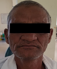
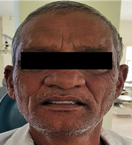
A 58 years old male patient was reported to Loni, Department of Prosthetic Dentistry Rural Dental College, (figure 1). His chief complaint was to replace his missing upper teeth .The main reason for missing teeth was generalized periodontitis. He had beedi smoking habit and no significant medical and systemic illness. History of using maxillary denture which had been fabricated 3 months ago and was fractured.
Intraoral and radiological examination
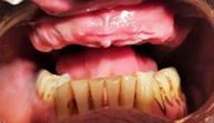
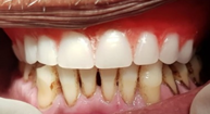
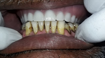
The intraoral findings revealed completely edentulous upper arch (figure 2). No teeth were missing in lower arch. Radiographic findings showed good prognosis with lower teeth. [4]
The intraoral findings revealed completely edentulous upper arch (figure 2). No teeth were missing in lower arch. Radiographic findings showed good prognosis with lower teeth. [4]
Treatment Planning
As there were no irregularities in the occlusion of mandibular teeth no occlusal corrections were required. Along with this, patient had a history of midline fracture of maxillary denture which was fabricated 3 month ago. Then decision was made to fabricate a new upper complete maxillary denture with reinforced metal-meshwork in order to avoid further fracture of the prosthesis. [5]
As there were no irregularities in the occlusion of mandibular teeth no occlusal corrections were required. Along with this, patient had a history of midline fracture of maxillary denture which was fabricated 3 month ago. Then decision was made to fabricate a new upper complete maxillary denture with reinforced metal-meshwork in order to avoid further fracture of the prosthesis. [5]
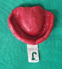
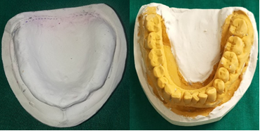
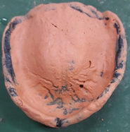
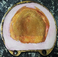
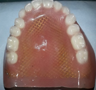
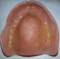
After proper intraoral examination, preliminary impression of upper arch was made in impression compound (figure 3). Diagnostic impression of lower arch was made in irreversible hydrocolloid alginate impression material and both cast were obtained (figure 4). Upper special tray was fabricated, border molding was recorded & final impression of maxillary arch was made in Zinc oxide eugenol impression paste (figure 5). Final cast was retrieved (figure 6). Record base and occlusal wax rim was fabricated on the maxillary cast. Jaw relation was recorded & casts were mounted on the articulator. Maxillary teeth were arranged in relation with lower dentulous arch. No occlusal corrections were required for lower arch. Try-in was done. Dewaxing was completed followed by adaptation of maxillary metal meshwork on to the maxillary cast before denture packing (figure 7). Further packing of the maxillary denture was completed in High Impact heat cure acrylic denture material (LUCITONE). Curing was done and maxillary denture was obtained (figure 8). Finishing and polishing was done, followed by denture insertion. Occlusion was evaluated by clinical remount procedure using articulating paper. Patient was satisfied with his denture.
Conclusion
Due to biomechanical differences in the supporting tissues for opposing arches the patient requiring single denture opposing a natural or restored dentition faces a challenging job for the dentist. Thus the treatment planning and the prosthesis to be given should be evaluated, fibricated and corrected to provide a stable prosthesis having stable functional relationships for controlling the resorption, discomfort and re-establishing esthetics, phonetics, masticatory functions and other lost functions of the patient (figure 9).
References
- Glossary of Prosthodontics Terms.
- Ellsworth Kelly. “Changes caused by a mandibular removable partial denture opposing a maxillary complete denture.” The Journal of Prosthetic Dentistry 90.3 (2003): 213-219.
- Kenneth D., et al. “Occlusion and single denture”. The Journal of Prosthetic Dentistry 30.1 (1973): 4-11.
- Robert G Vig. “A modified chewing and functional impression technique”. The Journal of Prosthetic Dentistry 14.2 (1964): 214-220.
- Sandhya Shroff., et al. “Single complete denture-A corrective prosthodontics: a case report.” International Journal of Healthcare and Biomedical Research 4.4 (2016): 104-107.
Citation:
Pranit Bora., et al. “Reinforced Single Complete Maxillary Denture.” Oral Health and Dentistry 3.3 (2018): 648-652.
Copyright: © 2018 Pranit Bora., et al. This is an open-access article distributed under the terms of the Creative Commons Attribution License, which permits unrestricted use, distribution, and reproduction in any medium, provided the original author and source are credited.


 Scientia Ricerca is licensed and content of this site is available under a Creative Commons Attribution 4.0 International License.