Case Report
Volume 1 Issue 2 - 2017
Nontuberculous Mycobacteria: A Case Report and Overview
Attending physician, Pulmonary Physicians of Norwich, Hartford Healthcare. Norwich, CT. 06360 USA
*Corresponding Author: Ankit Gupta MD MS, Attending physician, Pulmonary Physicians of Norwich, Hartford Healthcare. Norwich,
CT. 06360 USA.
Received: December 17, 2017; Published: December 28, 2017
Abstract
Nuntuberculous Mycobacteria (NTM) were first identified in the late nineteenth century but it is only recently that they are getting recognized as human pathogens. They are present in our environment in the water, soil etc and are usually benign. They are not contagious and the disease is acquired from the environment. Patients with underlying lung disease and immune compromised status are more prone to develop pulmonary disease. Symptoms are nonspecific, actual disease is often confused with contamination and treatment can be more debilitating than the disease itself.
In our experience, NTM should be included as a differential of any idiopathic chronic cough in the right clinical context. A multi-disciplinary approach with inputs from pulmonary, infectious disease and primary care can help smoothen the management and allay patient anxiety. We present the case of a NTM infected patient that does not fit the usual demographics.
Keywords: Nontuberculous mycobacteria; Chronic cough; Mycobacterium avium complex
Case Report
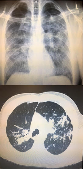
We present the case of a 39 y/o otherwise healthy male with no previous comorbidities who presented to our office with six months of progressive cough and an abnormal CXR. He had a previous short incarceration a few years prior to the presentation. He had no fevers, chills, phlegm production or night sweats. He complained of an unintentional ten-pound weight loss and progressive dyspnea on exertion. He had a twenty-pack year smoking history and occasional marijuana use. He was hemodynamic ally stable and his examination was unremarkable. His HIV panel, LFTs, CBC with diff, BMP were within normal limits. His CXR (Figure 1) showed Coarse diffuse interstitial changes with ill-defined opacities. A follow up CT chest (Figure 1) showed extensive bilateral streaky opacities with no enlarged lymphadenopathy. His cardiac workup was benign. His office spirometry showed mild restriction with no bronchodilator response, low lung volumes and diffusion. Sputum cultures were non-diagnostic. Quantiferon was indeterminate.
Bronchoscopy showed airway erythema/inflammation but no endobronchial lesions. Endobronchial biopsies and bronchoalveolar lavage failed to demonstrate any infectious etiology or malignancy initially. Ten days after the initial procedure, mycobacterium avium complex were isolated from the bronchial lavage and biopsies. Sensitivities were obtained and a multidrug regimen was prescribed. At 3 months follow up, the patient is doing marginally better with improved office spirometry and negative sputum AFB cultures.
Nontuberculous Mycobacterium in the immunocompetent patient
Nontuberculous Mycobacterium (NTM) are mycobacteria other than Mycobacterium tuberculosisand Mycobacterium leprae. NTM are found everywhere in our surroundings with an increasing frequency in the non -HIV population [1]. NTM is increasingly recognized and identified and its management is complex and controversial. There is no evidence of human to human transmission and the disease is not reportable [2] which makes the actual incidence difficult to estimate. Human disease is largely due to environmental exposures.
Nontuberculous Mycobacterium (NTM) are mycobacteria other than Mycobacterium tuberculosisand Mycobacterium leprae. NTM are found everywhere in our surroundings with an increasing frequency in the non -HIV population [1]. NTM is increasingly recognized and identified and its management is complex and controversial. There is no evidence of human to human transmission and the disease is not reportable [2] which makes the actual incidence difficult to estimate. Human disease is largely due to environmental exposures.
Although over 140 species of NTM have been identified, M. avium complex (MAC) and M. kansasii are the commonest species isolated in the United States and are slow growing organisms. The incidence of NTM has been increasing [3] and is reported to be as high as 47 cases per 100,000 individuals [4]. Immune status is especially important in disease pathogenesis and clinical presentation.
MAC is free living and has been isolated from soil, water sources and animals [5]. Common presentations of NTM include progressive pulmonary disease, skin/soft tissue/lymph node disease and disseminated disease in the immunosuppressed patients with multi organ involvement [6]. Chronic lung infections are present in a clear majority of patients with NTM/MAC. The most common presentation is an elderly Caucasian male with underlying lung disease presenting with symptoms similar to tuberculosis (cough, purulent sputum, hemoptysis, malaise and weight loss) [7]. Another commonly described presentation is an elderly nonsmoking woman with interstitial involvement [8]. Bronchiectasis, chest cavitation, nodularity and infiltrates have all been described radiologically. Radiological data and microbiological evidence from sputum/bronchial wash/ transbronchial biopsies are included in the diagnostic criteria [6]. Most NTM will grow within 2-3 weeks on subculture but it can take much longer. Gene sequencing is the most accurate method for identifying various NTM sub-species. Drug sensitives should be obtained especially before initiating treatment but is often controversial because of differences between in vivo and in vitro outcomes. Macrolide resistance is not uncommon. Treatment is long and involves multiple antibiotics seldom with limited clinical benefit. Some patients can be safely observed without treatment unlike mycobacterium tuberculosis. The treatment should be under taken after discussion with the patient as compliance is paramount to minimize drug resistance. Based on drug tolerance, up to four drugs can be used for treatment concurrently. Drug toxicity monitoring and sputum cultures should be routinely performed and treatment is often continued a year beyond negative sputum cultures [9,10].
Infectious disease consultation should be obtained early on. Sputum cultures need to be frequently monitored on therapy to document clearance. Therapy is often required for over one year based on response. Treatment for disseminated disease is even longer.
An often-used initial regimen for MAC lung disease is a three-times a week regimen including clarithromycin or azithromycin, ethambutol, and rifampin. The goal of therapy is to achieve negative sputum cultures for a 12 month period. Surgical options including de-bulking are often reserved for the more complicated patients with poor antibiotic response.
Conclusions
Prevalence of NTM lung disease is on the rise. Symptoms are often nonspecific and can often be confused with tuberculosis. Any patient with chronic unexplained cough should be investigated for NTM. Clinical, microbiological and radiological criterion need to be met before treatment. NTM are often secondary to contamination than because of the disease. However, even these species can, under some circumstances cause clinical disease. The physician should therefore consider the clinical picture and likelihood of disease before embarking on a long and often toxic treatment.
References
- Gruft H., et al. “Recent experience in the epidemiology of disease caused by atypical mycobacteria”. Reviews of Infectious Diseases 3.5 (1981): 990-996.
- Tanaka E., et al. “Familial pulmonary Mycobacterium avium complex disease”. American Journal of Respiratory and Critical Care Medicine 161.5 (2000): 1643-1647.
- Brode SK., et al. “The epidemiologic relationship between tuberculosis and non-tuberculous mycobacterial disease: a systematic review”. International Journal of Tuberculosis and Lung Disease 18.11 (2014): 1370-1377.
- Adjemian J., et al. “Prevalence of nontuberculous mycobacterial lung disease in U.S. Medicare beneficiaries”. American Journal of Respiratory and Critical Care Medicine 185.8 (2012): 881-886.
- Wolinsky E. “Nontuberculous mycobacteria and associated diseases”. The American Review of Respiratory Disease 119.1 (1979): 107-159.
- Griffith DE., et al. “An official ATS/IDSA statement: diagnosis, treatment, and prevention of nontuberculous mycobacterial diseases”. American Journal of Respiratory and Critical Care Medicine 175.4 (2007): 367.
- Teirstein AS., et al. “Pulmonary infection with Mycobacterium avium-intracellulare: diagnosis, clinical patterns, treatment”. Mount Sinai Journal of Medicine 57.4 (1990): 209-215.
- Prince DS., et al. “Infection with Mycobacterium avium complex in patients without predisposing conditions”. The New England Journal of Medicine 321 (1989): 863-868.
- Griffith DE., et al. “Azithromycin activity against Mycobacterium avium complex lung disease in patients who were not infected with human immunodeficiency virus”. Clinical Infectious Diseases 23.5 (1996): 983-989.
- Griffith DE., et al. “Azithromycin-containing regimens for treatment of Mycobacterium avium complex lung disease”. Clin Infect Dis 32.11 (2001): 1547-1553.
Citation:
Ankit Gupta. “Nontuberculous Mycobacteria: A Case Report and Overview”. Pulmonary Research and Respiratory Care 1.2 (2017):
121-124.
Copyright: © 2017 Ankit Gupta. This is an open-access article distributed under the terms of the Creative Commons Attribution License, which permits unrestricted use, distribution, and reproduction in any medium, provided the original author and source are credited.


 Scientia Ricerca is licensed and content of this site is available under a Creative Commons Attribution 4.0 International License.