Research Article
Volume 1 Issue 1 - 2016
Role of Red Blood Cell Distribution Width for Assessment of Severity of Critically Ill Patients in Intensive Care Unit and To Correlate with Acute Physiology and Chronic Health Evaluation-Ii (Apache-Ii) Score
1Medical Officer, Department of Medicine, Sir Salimullah Medical College Hospital, Dhaka
2,3Assistant Professor, Department of Clinical Pathology, Bangabandhu Sheikh Mujib Medical University, Dhaka
4Associate Professor, Department of ENT and Head-Neck Surgery, Bangabandhu Sheikh Mujib Medical University, Dhaka
5,6Associate Professor, Department of Clinical Pathology, Bangabandhu Sheikh Mujib Medical University, Dhaka
7MS in Microbiology, Bangladesh University of Health Sciences, Dhaka
8Chairman, Department of Haematology, Bangabandhu Sheikh Mujib Medical University, Dhaka
*Corresponding Author: Mesbah Uddin Ahmed, MS in Microbiology, Bangladesh University of Health Sciences, Dhaka.
Received: June 18, 2016; Published: July 06, 2016.
Abstract
Morbidity and mortality rate is high in ICU patients. Various scoring system is used like APACHE-II, SAPS-II, SOFA score to predict outcome of ICU patients and helps physicians for patient admission and management but they contain multiple variable, which are expensive, time consuming. This cross sectional study was carried out in the Department of Clinical Pathology, in collaboration with Department of Anaesthesia, Analgesia and Intensive Care Medicine, BSMMU, DMC and BIRDEM, Dhaka. Study population was 60 patients. After taking informed written consent from patient’s attendant blood sample were obtained with all aseptic precaution from all patients and processed in hematology auto analyzer. Red cell distribution width, mean cell volume, total leucocytes count, heamatocrit were performed. The study was conducted to assess the RDW as a marker for severity of disease among critically ill patients in ICU. Among the more severely ill patients half were > 60 years age group (50.0 %). On the other hand among the less severely ill patients majority were 31 to 60 years age group (65.6 %). The higher tertile of RDW were observed in older critical ill patients in ICU. This study showed that the both RDW and APACHE-II are increased in above four groups. The primary finding of this study showed that increasing RDW levels can serve as a marker to assess the severity of ICU patients. This association remained significant even after adjustment for APACHE-II score. RDW is less expensive and common measurement found on the complete blood count. The RDW fared better than APACHE II for morbidity or mortality prediction of ICU patients. RDW is widely available test, no additional cost and found in routinely performed with complete blood cell count and is highly reproducible, RDW has the potentially clinical utility to predict outcome for ICU patients. Our study revealed that RDW was significantly associated with severity of diseases of ICU patients. So RDW may be a reliable marker for assessment the severity of critically ill patients at ICU and to correlate with APACHE-II score.
Keywords: Red Blood Cell Distribution Width; Critically Ill Patients; Chronic Health Evaluation-II (Apache-II) Score.
Introduction
Red cell distribution width (RDW) is a quantitative measure of red cell size variation co-efficient. Higher RDW indicates greater variation of red blood cell size (RBC). It is expressed as percentage and reported as RDW. It is routinely done by physician in clinical practice as a part of the complete blood count (CBC) [1-3]. Recently several studies showed that high RDW value predicts the severity of diseases like morbidity and mortality in critically ill patients in intensive care unit (ICU). In recent study, RDW has a potential prognostic power in critically ill patients (Jackson C E., et al. 2009). RDW is a strong and independent predictor of morbidity and mortality in ICU patients [4-6]. In Bangabandhu Sheikh Mujib Medical University (BSMMU) 2012, the mortality rate of ICU patients was 46% (From ICU data 2012) and 32.5% in BIRDEM (From statistical Year Book 2011-12 of DAB). Critically ill patients are provided with the highest level of monitoring, care, and treatment which are very expensive. It also consumes many hospital resources. So various scoring system have been used for assessing the severity of critically ill patients in different ICU in the world like APACHE (acute physiology and chronic health evaluation) score. But an accurate scoring system is required for assessing the severity of patients and proper effective therapies. It contains variables those can independently predict the outcome or severity of diseases [7]. RDW includes only one variable, widely available to clinician [3]. It is computed directly from the red blood cell histogram [3,8]. APACHE-II or SOFA or other score contain multiple variables and those are more expensive, very costly and time consuming. On the other hand, RDW is the only one variable, simple and cheaper test. It is easily available and can be done rapidly along with complete blood count (CBC). Bangladesh is a developing country and most of the patients are unable to bear the costly investigation. So it will be beneficial for the patients and helpful for the physicians. With this aim, this study was undertaken to evaluate the role of red cell distribution width (RDW) for the assessment of severity of critically ill patients in ICU and to correlate with APACHE-II score.
Methods
Study design: Cross sectional study.
Place of study: This study was done in the Department of Clinical Pathology BSMMU in collaboration with ICU of Anaesthesiology Department, BSMMU and Dhaka Medical College Hospital (DMCH) and ICU of Critical care medicine unit of Bangladesh Institute of Research and Rehabilitation for Diabetes, Endocrine and Metabolic Disorder (BIRDEM), Dhaka.
Study duration: One year (1) from September, 2012 to August, 2013.
Study population: All adult ICU admitted patients in BSMMU, DMCH and Critical care medicine unit of BIRDEM.
Sample size: Sample size was determined by power analysis for a single proportion. In initial stage of study we planned to enrol 60 patients.
Sampling Technique: Purposive sampling was used. As per inclusion criteria the patients were enrolled in this study. The particulars of the patients and clinical data were recorded in a pre-designed data-sheet and were kept until the end of the study. The whole procedure was explained to the patient and informed written consent was taken.
Inclusion criteria: Eligible patients were those needed to be hospitalized to ICU who were transferred from the emergency department or the other hospital including medical and trauma patient like-
- Adult ICU patients (Age > 18 years)
- Patient with more than cut - off value of RDW & APACHE-11(cut-off value RDW is ≥ 14.8% and APACHE-11 is ≥ 15 points).
- Normal mean corpuscular volume (MCV) (76-96 fl).
Exclusion criteria
- Age < 18 years
- Patients with less than cut-off value of RDW & APACHE-11 score (cut-off value RDW is ≥ 14.8% and APACHE-11 is ≥ 15 points).
- Pregnancy
- Known hematological disease (Leukaemia, Myelodysplastic disease, Metastatic cancer in bone marrow, some anaemia like iron deficiency anemia, thalassaemia).
- History of recent blood transfusion (less than 2 weeks).
Grouping of Patients: Enrolled patients were divided into four (4) groups according to ICU admission. Initially we thought each group will contain 15 patients, but finally we got
- Group 1: 10 cases of sepsis (septic shock).
- Group 2: 21 cases of neurological (stroke, sub-aracnoid hemorrhage, meniningitis, encephalitis, brain tumor).
- Group 3: 18 cases of cardiovascular (myocardial infarction, heart failure, hypertension).
- Group 4: 11 cases of Trauma (head injury, road traffic accidents).
- After measuring RDW value, ICU patients were divided into two tertiles. Patients who had ≤ 17.5 considered as less severe group and who had > 17.5 considered as more severe.
Laboratory assay
RDW (Red cell distribution width) and others.
APACHE-II score includes the followings clinical and laboratory monitoring parameters.
Laboratory monitoring parameters were Complete blood count (CBC), Serum creatinine, Serum electrolyte, Alanine Aminotransferase (ALT/SGPT), Arterial blood gas (ABG) analysis.
Data collection: Data were collected by a predesigned Proforma. Blood samples were collected from all ICU patients during admission within 24 hours. Patient’s information were obtained through using patient’s information sheet which involves, questionnaire, clinical findings and laboratories reports.
Data presentation: Data and result were presented in the form of tables and graph where applicable.
Laboratory procedure: 7.0 ml blood was collected by veni-puncture and 1.0 ml arterial blood was collected with all aseptic precaution. All the tests were done in the concern department of ICU for APACHE-II score except CBC, RDW and ALT which were done in Clinical Pathology, BSMMU.
Specimen collection and Processing: Among 7.0 ml venous blood, 2.0 ml venous sample with EDTA for RDW and CBC and another 3.0 ml for electrolyte and 2.0 ml for biochemical test (1.0 ml for alanine aminotransferase and 1.0 ml for serum creatinine examination) were collected.
Time of sample collection- Blood was collected after admission within 24 hours.
For RDW and CBC measurement:Specimen collection and Processing: 2.0 ml venous sample with EDTA for RDW and CBC.
Test procedure: Measurement of RDW and CBC were done after collection. RDW and CBC was done by automated hematology analyzer, calibrated according to manufacturer instruction. Sample were presented to the haematology analyzer Sysmex-XT 4000i, where 20 microlitre (μl) blood was aspirated directly. CBC includes Hb, red cell count, white blood cell count, platelete count, hematocrit, MCV, MCH, MCHC and RDW was done. RDW was derived from pulse height analysis and can be expressed as the coefficient of variation (CV) (%) of the measurement of the red cell volume. RDW plotted as a histogram, deviation from a unimodal gaussion curve was inspected.
Method: Fully automated hematology analyzer.
Principle: Based on haematology autoanlyzer (Flow cytometry).
For serum electrolyte measurement: Specimen collection and Processing 3.0 ml venous blood was released into a test tube containing 2.0 ml liquid paraffin touching with nozzle at the side of test tube in 45 degree angle. Allow to clot and serum was separated by centrifugation.
Test procedure: Test was done immediately by spectrophometric principle. Calibrated according to manufacturer instruction sample was presented to the instrument, where blood was aspirated directly.
Method: electrolyte auto-analyzer (Biolyte machine).
Principle: Based on electrolyte analyzer (Atomic absorption spectrophotometry).
For estimation of serum Alanine aminotransferase (ALT/SGPT) and creatinine:
Specimen collection and Processing: 1.0 ml venous blood was collected for ALT allow to clot and separate serum by centrifugation at room temperature.
Specimen collection and Processing: 1.0 ml venous blood was collected for ALT allow to clot and separate serum by centrifugation at room temperature.
Storage-This serum was stored at -20°c until analysis. The test was done on several occasions.
Test procedure for ALT: Serum test was done on several occasions by semi-automated biochemical analyzer. Calibrated according to manufacturer instruction sample was presented to the instrument, where blood was aspirated directly.
Method: Enzymatic method by semi-automated biochemical analyzer
Principle: Enzymatic method.
For estimation of serum creatinine: Specimen collection and Processing
1.0 ml venous blood was collected for creatinine allow to clot and separate serum by centrifugation at room temperature.
Storage-This serum was stored at -20°c until analysis. The test was done on several occasions.
Test procedure for serum creatinine: Serum test was done on several occasions by semi-automated biochemical analyzer. Calibrated according to manufacturer instruction sample was presented to the instrument, where blood was aspirated directly.
Method: Enzymatic method by semi-automated biochemical analyzer (Picrate).
Principle: Picrate method.
For Arterial blood gas analysis
Specimen collection and Processing: 1.0 ml arterial blood was collected in a heparinized wash syringe and immediately was capped and examination was done within 15 minutes. If any delayed sample was kept on ice with sealed nozzle and test as early as possible.
Specimen collection and Processing: 1.0 ml arterial blood was collected in a heparinized wash syringe and immediately was capped and examination was done within 15 minutes. If any delayed sample was kept on ice with sealed nozzle and test as early as possible.
Test procedure: Arterial blood was done by arterial blood gas analyzer. Calibrated according to manufacturer instruction sample was presented to the instrument, where blood was aspirated directly.
Method: Arterial blood gas autoanalyzer.
Principle: Autoanalyzer based principle.
Data management: Data editing, clearing and analysis was done by Statistical Package for Social Science (SPSS)-17.
Statistical analysis: All data were recorded systematically in a preformed data collection sheet and quantitative data was expressed as mean ± Standard deviation and qualitative data was expressed as frequency and percentage. Spearman correlation coefficient test was used for correlation analysis. We divided RDW into two groups on basis of severity below 17.5 RDW and above 17.5 RDW values and compared demographics, clinical characteristics, laboratory test result and APACHE-II score with analysis of variance and chi-square test, ANOVA and t-test for categorical variables. All statistical computations were performed by using SPSS 17.
Safety precautions: All samples received for analysis were considered potentially positive for infectious agents including HIV and the hepatitis B virus. Universal precaution was observed. Gloves, lab coat, and safety glasses were worn when handling all human blood products and infectious organism. Disposable plastic, glass, paper and gloves that contact blood was placed in a biohazard bag or discard pan to be autoclaved. Working surfaces were disinfected with 10% bleach solution. Disposed other potentially contaminated materials in a biohazard bag. Disinfected other non-disposable material at the end of the working day. Pipette by mouth was avoided. Eating, drinking or smoking was avoided in designated working areas. Wash hands thoroughly were practiced after removal of personal protective devices.
Result
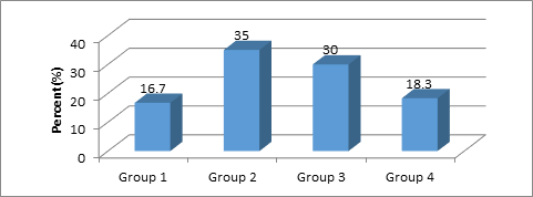
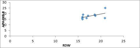
Table-I shows age distribution of the study patients and among the more severely ill patients half were > 60 years age group (50.0%). On the other hand among the less severely ill patients majority were 31 to 60 years age group (65.6%). But these differences were not statistically significant in chi-square test. Mean age was found 53.81 ± 15.15 years in less severe group and 53.56 ± 19.44 years in more severe group. The mean age difference was statistically significant (P < 0.05) between the two groups in unpaired t- test. Figure 1 shows distribution of patients in different groups. Among the total patients (n = 60) more than one third 21 (35.0%) was in group-2, more than one fourth 18(30%) was in Group-3, more than eighteen percent 11(18.3) were in Group-4 and 10(16.7%) were in Group-1. Table 2 shows mean RDW was found 16.04 ± 0.70 in less severe group and 19.75 ± 1.90 in more severe group. The mean RDW difference was statistically significant (p < 0.001) between the two groups in unpaired t test which indicates raised RDW was associated with severity of disease. Table 3 reveals mean RDW in group-1 was found 16.30 ± 0.60 in less severe group and 19.75 ± 1.90 in more severe group patients, 15.90 ± 0.696 was in less severe, 21.23 ± 1.87 was in more severe in group-2, 15.97 ± 0.89 was in less severe, 19.08 ± 1.05 was in more severe in group-3 and 16.28 ± 0.53 was in less severe, 19.85 ± 0.65 was in more severe group patient in group-4. The mean RDW difference in less and more severe group patients among the individual group were statistically significant (p < 0.05) in unpaired t-test. Table 4 shows that in group-1 RDW mean ± SD were 18.71±2.47, group -2 were 17.93 ± 2.93, group -3 were 17.53 ± 2.01, group-4 were 17.58 ± 1.97. The relation among the groups and RDW were not statistically significant (p > 0.05). On the other hand tables 4 shows that in group 1 APACHE-II were 20.40 ± 5.27, group 2 were 18.85 ± 3.16, group 3 were 19.27 ± 3.52, and group 4 were 17.72 ± 2.76. The relation of APACHE-II score among the groups were not also statistically significant (p > 0.05). Figure 2 shows positive correlation between RDW and APACHE-II. Pearson correlation test showed that it is statistically highly significant (p < 0.01, r = 0.733). This result indicates when APACHE-II score is increased than RDW value also increased stepwise. Figure 3 shows positive correlation between RDW and APACHE-II in Group 1. Pearson correlation test showed that it is statistically highly significant (p < 0.01, r = 0.923). Figure 4 shows positive correlation between RDW and APACHE-II in Group 2. Pearson correlation test showed that it is statistically highly significant (p < 0.01, r = 0.823). Figure 5 indicates positive correlation between RDW and APACHE-II in Group 3. Pearson correlation test showed that it is statistically significant (p < 0.05, r = 0.538). Figure 6 shows positive correlation between RDW and APACHE-II in Group-4. But Pearson correlation test showed that it is statistically significant (p < 0.05, r = 0.576).
| Age (in years) | Less severe* n (%) | More severe** n (%) | χ2 | df | p value |
| 18-30 | 4 (12.5) | 2 ((7.1) | 5.211 | 2 | 0.071 |
| 31-60 | 21 (65.6) | 12 (42.9) | |||
| > 60 | 7 (21.9) | 14 (50.0) | |||
| Total | 32 (100.0) | 28 (100.0) | |||
| Mean ± SD | 53.81 ± 15.15 | 53.56 ± 19.44 | 0.019s | ||
Table 1: Relationship of age with severity of disease.
*Less severe group = Patient with lower RDW value ≤ 17.5, here 32 patients were included.
**More severe group = patient with higher RDW value > 17.5, here 28 patients were included.
*Less severe group = Patient with lower RDW value ≤ 17.5, here 32 patients were included.
**More severe group = patient with higher RDW value > 17.5, here 28 patients were included.

Group 1: sepsis (e.g, septic shock), 10 patients.
Group 2: Neurological diseases (e.g, stroke, subdural haemorrhage, meningitis, and encephalitis) 21 patients.
Group 3: Cardiovascular diseases (e.g, myocardial infarction, heart failure, and hypertension) 18 patients.
Group 4: Trauma patients (e.g, head injury, road traffic accidents) 11 patients.
Figure 1: Distribution of patients according to group (group-1 = 10, group-2 = 21, group-3 = 18, group-4 = 11).
| RDW | Less severe | More Severe | t | df | p value |
| Mean ± SD | 16.04 ± 0.70 | 19.75 ± 1.90 | -10.85 | 58 | 0.001 |
Table 2: Relation of mean RDW between in less and more severe group in total patients.
p value reached from unpaired t-test.
p value reached from unpaired t-test.
| Group | Less severe (RDW ≤ 17.5) Mean ± SD |
More severe (RDW >17.5) Mean ± SD |
p value |
| Group-1 (n = 10) | 16.30 ± 0.60 | 19.74 ± 2,20 | 0.003 |
| Group-2 (n = 21) | 15.90 ± 0.696 | 21.23 ± 1.87 | 0.001 |
| Group-3 (n = 18) | 15.97 ± 0.89 | 19.08 ± 1.05 | 0.001 |
| Group-4 (n = 11) | 16.28 ± 0.53 | 19.85 ± 0.65 | 0.008 |
Table 3: Relation of RDW of individual group with severity of the disease.
p value reached from independent t-test.
p value reached from independent t-test.
| Value of test score | Group-1 Mean ± SD |
Group-2 Mean ± SD |
Group-3 Mean ± SD |
Group-4 Mean ± SD |
F | df | p value |
| RDW (%) | 18.71 ± 2.47 | 17.93 ± 2.93 | 17.53 ± 2.01 | 17.58 ± 1.97 | .563 | 3.58 | 0.641 |
| APACHE-II | 20.40 ± 5.27 | 18.85 ± 3.16 | 19.27 ± 3.52 | 17.72 ± 2.76 | .99 | 3 | 0.40 |
Table 4: Average value of RDW and APACHE-II in different groups.

Figure 2: The scatter diagram showing relationship (r = 0.733, p < 0.01) between RDW and
APACHE-II of the total study patients (n = 60).

Figure 3: The scatter diagram showing relationship (r = 0.923, p < 0.01) between RDW and APACHE-II of the group-1 study patients (n = 10).

Figure 4: The scatter diagram showing relationship (r = 0.823, p < 0.01) between RDW and APACHE-II of the study patients in group 2 (n = 21).

Figure 5: The scatter diagram showing relationship (r = 0.538, p < 0.05) between RDW and APACHE-II of the study patients group 3 (n = 18).

Figure 6: The scatter diagram showing relationship (r = 0.576, p < 0.05) between RDW and APACHE-II of the study patients in group 4 (n = 11).
Discussion
This cross sectional study was conducted to assess the RDW as a marker for severity of disease among critically ill patients in ICU. Our study was enrolled in 60 patients from intensive care unit, Dhaka. Among 60 patients who participated in our study were divided into four (4) groups on the basis of ICU admission primarily based on clinical evaluation and lab diagnosis. Among them 10 (16%) patients were in group-1(sepsis), 21 (35%) were in group-2 (neurological disease), 18 (30%) were in group-3(cardiovascular diseases) and 11 (18.3%) were in group-4 (trauma patients). This cross sectional study evaluated RDW for assessing the severity of diseases of ICU patients that correlated with APACHE-II score. After measuring RDW value, ICU patients were divided into two tertiles. Patients who had ≤ 17.5 (RDW), considered as less severe patients and who had > 17.5 (RDW) considered as more severe patients [8]. In our study, mean age was 53.81± 15.15 in less severe patients and 53.56± 19.44 in more severe patients. The mean age difference was statistically significant (p<0.05) between less and more severe patients in unpaired t-test which indicate increase RDW was associated with critically ill patients with age. Similar findings were found in other studies [3,8-10. According to their study mean age was 70.39 ± 16.73years, 67.4 years, 63 years, and 61.7 ± 18.3 years respectively. These finding were nearly consistent with our study. This study revealed that mean RDW value in less severe patients were 16.04 ± 0.70 and mean RDW value in more severe patients were 19.75 ± 1.90. The mean RDW difference in both groups was statistically significant (p < 0.001) in unpaired t-test which indicates raised RDW was associated with severity of disease. Also significant finding was observed the other studies [3,6,8-11]. Previous studies showed that high mortality rate were associated with higher RDW tertile compared with low RDW tertile. This finding was consistent with our study. In our study we have found the mean RDW 16.30 ± 0.60 in less severe group and 19.75 ± 1.90 in more severe group patients, 15.90 ± 0.696 in less severe, 21.23 ± 1.87 in more severe in group-2, 15.97 ± 0.89 in less severe, 19.08 ± 1.05 in more severe in group-3 and 16.28 ± 0.53 in less severe, 19.85 ± 0.65 in more severe group patient in group-4. The mean RDW difference in less and more severe group patients among the individual group were statistically significant (p < 0.05) in unpaired t-test which indicate raised RDW was associated with severity of disease. p value < 0.05 were observed in the other studies [6,11,9,10]. Their studies showed that higher mortality rate were associated with higher RDW tertile compared with low RDW tertile. This finding was similar to our study. In septicaemia, RDW is high in relation with mortality is more due to pro-inflammatory cytokine [6]. Septicaemia occur mainly due to mechanical ventilation that induces infection and causes increased RDW and also increased mortality in ICU patients [12]. Our study revealed that the mean RDW difference in less and more severe group patients in septicaemia was statistically significant (p value < 0.05) in unpaired t-test which indicates raised RDW was associated with severity of septicaemia. p value < 0.05 was observed in the other studies [6,10]. These studies showed that higher mortality rate were associated with higher RDW tertile compared with low RDW tertile. This finding was consistent with our study. Among the patients with neurological disorders like stroke, RDW is high due to inflammatory states, infection of atherosclerotic plaque that causes cytokines production and those cytokine impede RBC maturation. Elevated RDW predict mortality in person with known stroke [10,11]. Our study showed that mean RDW difference in both less and more severe group in neurological diseases was statistically significant (p < 0.05). This result indicates raised RDW was associated with severity of neurological diseases were demonstrated that higher mortality rate was associated with higher RDW tertile compared with low RDW tertile. p value was < 0.05 in their studies. This finding was similar to our study. In cardiovascular diseases increased RDW is not clear but RDW may represent inflammation. In some previous study, RDW is found to be a new prognostic marker in heart failure, coronary cardiac diseases [6,7]. Higher value of RDW were associated with increased risk of death [13]. The present study revealed that mean RDW difference in both less and more severe group in cardiovascular diseases was statistically significant (p value < 0.05), which indicates raised RDW was associated with severity of cardiovascular diseases. p value was < 0.05 in their studies. Previous studies showed that higher mortality rate were associated with higher RDW tertile compared with low RDW tertile. This finding was similar with our study. Our study revealed that mean RDW difference in both less and more severe group in trauma patients were statistically significant (p < 0.05). This finding indicates raised RDW was associated with severity of trauma patients. The APACHE-II score system has shown positive correlation with ICU mortality and is one of the most common model to evaluate patient’s disease severity. In accordance with previous studies APACHE-II score had demonstrated a strong power to predict ICU mortality. Patients with high APACHE-II score and high RDW levels carry the higher risk of mortality. For predicting ICU mortality, predicting power of RDW was relatively low than APACHE-II. But previous studies had shown that there was stepwise increased APACHE-II score with increased RDW tertiles (all p < 0.05) and when adding RDW to APACHE-II score strongly predict ICU mortality [3,8]. In our study, RDW ranged from 14.70 to 23.00 (median 17.00; mean 17.87 ± 2.41), and the mean APACHE-II score was ranged from 15.0 to 32.0 (median 18.00; mean 19.03 ± 1.60). This was nearly consistent with the finding (mean RDW = 14.5 ± 20.1 and mean APACHE-II = 13.2 ± 7.0) of other studies done by Wang F., et al. (2011), Sadaka F., et al. (2012). In our study, we found positive correlation among the total patients between RDW and APACHE-II in pearson correlation test that is statistically highly significant (p < 0.01, r = 0.77). This result was consistent with other studies done by Wang F., et al. (2011), Sadaka F., et al. (2012). In our study, we also found positive correlation among the individual group between RDW and APACHE-II in pearsons correlation test that is statistically highly significant (p < 0.01, r = 0.923) in group-1, (p < 0.01, r = 0.823) in group-2, (p < 0.01, r = 0.538) in group-3 and (p < 0.01, r = 0.576) respectively. These results were consistent with other studies done by Wang F., et al. (2011), Sadaka F., et al. (2012).
Limitations
This study was done in limited time of span. The sample size was small. Sample were collected from tertiary hospital, hence may not represent the whole population of country. The cause of increased RDW was not investigated, such as iron or vitamin B12 deficiency, which could confound the relationship between RDW and adverse outcome.
Conclusion
This study revealed that RDW is an effective parameter for prediction the adverse outcome in ICU patients. Increased RDW likely reflects the presence of pro-inflammatory cytokines and oxidative stress. There was a positive correlation between RDW and APACHE-II score. RDW is better than APACHE-II for morbidity or mortality prediction in ICU patients, moreover RDW is less expensive, available, routinely done with CBC and no additional cost is needed and highly reproducible. It is also helpful for daily follow up of the patients by only evaluating CBC. From this study, we concluded that higher RDW levels can serve as a marker for assessing the severity of diseases and can be used for prediction of adverse outcomes of ICU patients like APACHE-II score.
References
- Patel KV., et al. “Red blood cell distribution width and the risk of death in middle-aged and older adults”. Archives of Internal Medicine 169.5 (2009): 515–523.
- Forhecz Z., et al. “Red cell distribution width in heart failure: predictionof clinical events and relationship with markers of ineffective erythropoiesis, inflammation, renal function, and nutritional state”. American Heart Journal 158.4 (2009): 659-666.
- Wang F., et al. “Red cell distribution width as a novel predictor of mortality in ICU patients”. Annals of Medicine 43.1 (2010): 40-46.
- Perlstein TS., et al. “Red blood cell distribution width and mortality risk in a community-based prospective cohort”. Archives of Internal Medicine 169.6 (2009): 588-594.
- Braun E., et al. “Elevated red cell distribution width predicts poor outcome in young patients with community acquired pneumonia”. Critical care 15.4 (2011): R194.
- Jackson CE., et al. “The novel biomarker red cell distribution width (RDW) has incremental prognostic value, in addition to B-type natriuretic peptide (BNP), in patients with acute decompensated heart failure”. European Heart Journal 30 (2009): 14.
- Al-Najjar Y., et al. “Red cell distribution width an inexpensive and powerful prognostic marker in heart failure”. European Journal of Heart Failure 11.12 (2009): 1155–1162.
- Sadaka F., et al. “Red Cell Distribution Width and Outcome in Patients With Septic Shock”. Journal of Intensive Care Medicine 28.5 (2013): 1-7.
- Hunziker S., et al. “Red cell distribution width improves the simplied acute physiology score for risk prediction in unselected critically ill patients”. Critical care16.3 (2012).
- Bazick HS., et al. “Red cell distribution width and all-cause mortality in critically ill patients”. Critical Care Medicine 39.8 (2011): 1913-1921.
- Ani C., et al. “Elevated red blood cell distribution width predicts mortality in persons with known stroke”. Journal of the Neurological Sciences277 (2009): 103–108.
- Silva E., et al. “Prevalence and outcomes of infections in Brazilian ICUs: a subanalysis of EPIC II study”. Revista Brasileira de Terapia Intensiva 24.2 (2012): 143-150.
- Pascual-Figal DA., et al. “Red blood cell distribution width predictslong-term outcome regardless of anaemia status in acute heart failure patients”. European Journal of Heart Failure 11.9 (2009): 840–846.
Citation:
Mesbah Uddin Ahmed., et al. “Role of Red Blood Cell Distribution Width for Assessment of Severity of Critically Ill Patients in Intensive Care Unit and To Correlate with Acute Physiology and Chronic Health Evaluation-Ii (Apache-Ii) Score”. Anaesthesia, Critical Care and Pain Management 1.1 (2016): 7-16.
Copyright: © 2016 Mesbah Uddin Ahmed., et al. This is an open-access article distributed under the terms of the Creative Commons Attribution License, which permits unrestricted use, distribution, and reproduction in any medium, provided the original author and source are credited.


 Scientia Ricerca is licensed and content of this site is available under a Creative Commons Attribution 4.0 International License.