Case Report
Volume 2 Issue 1 - 2018
MR Imaging in Multiple System Atrophy-Cerebellar Type
1Assistant Proffesor, Department of Radio-diagnosis, Dr RPGMC, Kangra at Tanda. Himachal Pradesh. India
2Professor, Department of Radio-diagnosis, Dr RPGMC, Kangra at Tanda. Himachal Pradesh. India
3Resident, Department of Radio-diagnosis, Dr RPGMC, Kangra at Tanda. Himachal Pradesh. India
4Senior resident, Department of Anaesthesia, DRPGMC, Kangra at Tanda, Himachal Pradesh, India
2Professor, Department of Radio-diagnosis, Dr RPGMC, Kangra at Tanda. Himachal Pradesh. India
3Resident, Department of Radio-diagnosis, Dr RPGMC, Kangra at Tanda. Himachal Pradesh. India
4Senior resident, Department of Anaesthesia, DRPGMC, Kangra at Tanda, Himachal Pradesh, India
*Corresponding Author: Pooja Gurnal, Senior resident, Department of Anaesthesia, DRPGMC, Kangraat Tanda, Himachal Pradesh, India.
Received: June 08, 2018; Published: June 22, 2018
Abstract
We present a case of 54-year-old man with resented with two years’ history of gait unsteadiness associated with dysarthria and bladder incontinence of one-year duration. MRI of brain of the patient was done which shows “Hot cross bun” sign in the form of cruciform hyper intensity in the pons on T2W images characteristic of MSA-C (Multiple systemic atrophy-Cerellar type). Cerebellum and middle cerebellar peduncles are is atrophied which again favoured MSA-C.
Keywords: Multiple system atrophy; Cerebellar
Introduction
Multiple system atrophy is a sporadic neurodegenerative disease characterised by varying degrees of cerebellar ataxia, autonomic dysfunction, parkinsonism and corticospinal dysfunction and MSA-C predominance of cerebellar symptoms olivopontocerebellar atrophy [1,2,6]. Key Diagnostic Features of MSA-C shows Cruciform shape of hyperintense signal in pons on T2WI and atrophy of pons, inferior olives, and cerebellum is also seen [3].
Case Report
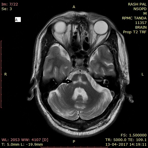
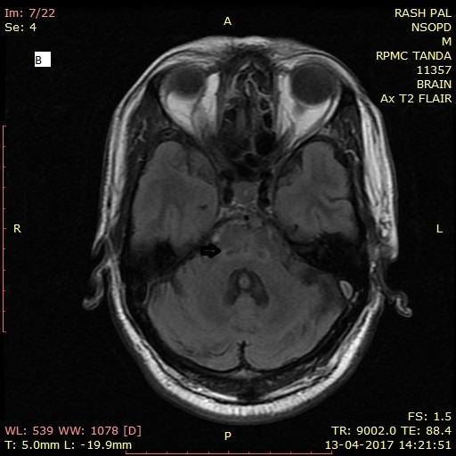
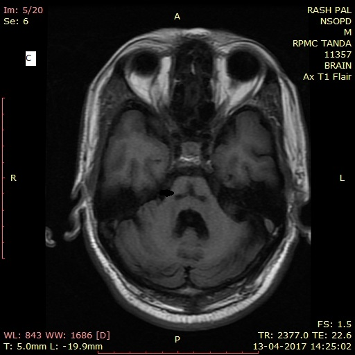
A 54-year-old man presented with two years’ history of gait unsteadiness associated with dysarthria and bladder incontinence of one-year duration. After clinical assessment the patient was referred to our department for MRI brain and find out cause of the above mentioned clinical symptoms. We did routine MRI scan in GE 1.5 Tesla machine which included axial T1W, FLAIR, DWI, axial and coronal T2W andT1W post contrast sequences. MRI of brain of the patient showed as in Figures (1) T2WI(A) shows “Hot cross bun” sign in the form of cruciform hyperintensity in the pons which is seen as hypointensity on T1W(C) and FLAIR(B) axial sequences (Thin arrow). Cerebellum(Thick arrow) as seen on T2W saggital images and midle cerebellar peduncles are is atrophied. On the basis characteristic findings of Cruciform shape of hyperintense signal in pons on T2WI and atrophy of pons, inferior olives, and cerebellum diagnosis of MSA-C was made.
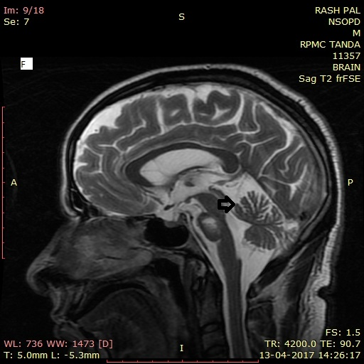
MRI of brain of the patient : T2WI(A) shows “Hot cross bun” sign in the form of cruciform
hyperintensity in the pon which is seen as hypointensity on T1W(C) and FLAIR(B) axial
sequences(Thin arrow).Cerebellum(Thick arrow) as seen on T2W saggital images and
midle cerebellar peduncles are is atrophied.
Discussion
Multiple system atrophy (MSA) is a sporadic neurodegenerative disease characterised by varying degrees of cerebellar ataxia, autonomic dysfunction, parkinsonism and corticospinal dysfunctionand MSA-C predominance of cerebellar symptoms olivopontocerebellar atrophy [1,2,7,8]. Multiple systemic atrophy is a sporadic disease, with a prevalence of 4 per 100,000. Typically symptoms begin in 4th or 5th decade. Clinical presentation is variable, but typically presents in one of three patterns MSA-C demonstrates primarily cerebellar dysfunction Shy-Drager syndromeis used when autonomic symptoms predominate and in striatonigral degeneration shows predominant parkinsonian features. Multiple systemic atrophy results from abnormalities of alpha-synuclein metabolism, resulting in intracellular deposition which are found not only in neurons but also in oligodendroglia [3]. An MRI of the brain in T2/ PD/FLAIR sequences reveals the classical ‘hot-cross bun’ sign in the pons characterized by cruciate hyperintensity secondary to atrophy of the transverse pontine fibers [4,5].
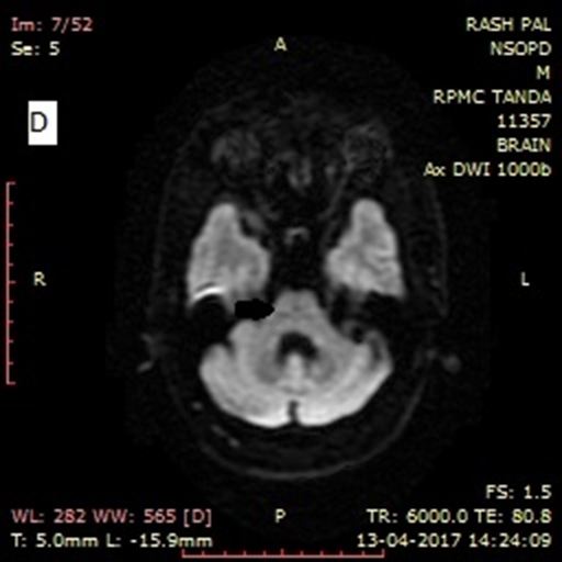
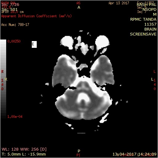
There was no restricted diffusion of the signal changes on apparent diffusion coefficient (ADC) mapping. These features were seen in our case with atrophy of pons, inferior olives, and cerebellum which is also key diagnostic feature.Differentials are firstly Cerebello-olivary Atrophy which has cortical cerebellar degeneration with Selective atrophy of lateral cerebellum ("fish-mouth deformity" on parasagittal sections) and superior vermis [4,5]. Other differential can be Friedreich Ataxia which shows severe atrophy of spinal cord and medulla oblongata and mild atrophy of vermian and paravermian structures is seen.As far as treatment is concernedunfortunately no effective treatment is currently available and the disease progresses relentlessly culminating in death usually within decade of diagnosis [6-8].
Conclusion
Multiple system atrophy (MSA) is a sporadic neurodegenerative disease with MSA-C predominance of cerebellar symptoms olivopontocerebellar atrophy. Key Diagnostic Features on MRI of MSA-C are Cruciform shape of hyperintense signal in pons giving a characteristic “Hot cross bun” sign along with atrophy of pons, inferior olives, and cerebellum.
Acknowledgments
We are grateful to almighty for his blessings.
We are grateful to almighty for his blessings.
References
- Matsusue E., et al. “Cerebellar lesions in multiple system atrophy: postmortem MR imaging-pathologic correlations”. American Journal of Neuroradiology 30.9 (2009): 1725-1730.
- Ozawa T., et al. “The spectrum of pathological involvement of the striatonigral and olivopontocerebellar systems in multiple system atrophy: clinicopathological correlations”. Brain 127. Pt12 (2004): 2657-2671.
- Schulz JB., et al. “Multiple System Atrophy: Natural history, MRI morphology, and dopamine receptor imaging with 123IBZM-SPECT”. Journal of Neurology, Neurosurgery, and Psychiatry 57.9 (1994): 1047-1056.
- Savoiardo M., et al. “Magnetic resonance imaging in progressive supranuclear palsy and other parkinsonian disorders”. Journal of Neural Transmission. Supplementa 42 (1994): 93-110.
- Schrag A., et al. “Clinical usefulness of magnetic resonance imaging in multiple system atrophy”. Journal of Neurology, Neurosurgery, and Psychiatry 65.1 (1998): 65-71.
- Bhattacharya K., et al. “Brain magnetic resonance imaging in multiple system atrophy and Parkinson disease: A diagnostic algorithm”. Archives of Neurology 59.5 (2002): 835-842.
- Nicoletti G., et al. “Apparent diffusion coefficient measurements of the middle cerebellar peduncle differentiate the Parkinson variant MSA from Parkinson's disease and progressive supranuclear palsy”. Brain 129. Pt10 (2006): 2679-2687.
- Paviour DC., et al. “Longitudinal MRI in progressive supranuclear palsy and multiple system atrophy: rates and regions of atrophy”. Brain 129.Pt4 (2006): 1040-1049.
Citation:
Pooja Gurnal., et al. “MR Imaging in Multiple System Atrophy-Cerebellar Type”. Medical Research and Clinical Case Reports 2.1
(2018): 149-154.
Copyright: © 2018 Pooja Gurnal., et al. This is an open-access article distributed under the terms of the Creative Commons Attribution License, which permits unrestricted use, distribution, and reproduction in any medium, provided the original author and source are credited.


 Scientia Ricerca is licensed and content of this site is available under a Creative Commons Attribution 4.0 International License.