Case Report
Volume 2 Issue 3 - 2018
Cryptococcoma’s on Mr Imaging-A Case Report
1Assistant Proffesor, Department of Radio-diagnosis, Dr RPGMC, Kangra at Tanda, Himachal Pradesh, India
2Professor, Department of Radio-diagnosis, Dr RPGMC, Kangra at Tanda, Himachal Pradesh, India
3Senior Resident Department of Radio-diagnosis, Dr RPGMC, Kangra at Tanda, Himachal Pradesh, India
4Resident Department of Radio-diagnosis, Dr RPGMC, Kangra at Tanda, Himachal Pradesh, India
5Associate Professor, Department of Radio-diagnosis, Dr RPGMC, Kangra at Tanda. Himachal Pradesh, India
*Corresponding Author: Lokesh Rana, Assistant Proffesor, Department of Radio-diagnosis, Dr RPGMC, Kangra at Tanda, Himachal
Pradesh, India.
Received: July 15, 2018; Published: August 13, 2018
Abstract
CNS cryptococcosis results from infection of the central nervous system with the yeast-like fungus Cryptococcus neoformans. It is the
most common fungal infection and second most common opportunistic infection of the central nervous system. The disease tends to
be predominant in immunocompromised individuals such as those with AIDS. In immunocompetent patients, there is usually history
of close contact with birds. We present a case of 18-year-old immunocompromised males suffering from AIDS and having CD4 count
70 cells/μL with protracted cryptococcal infection of the central nervous system, MR imaging showed presence of crytococcomas in
the bilateral basal ganglia region. Purpose of this study is to present relatively rare opportunistic infection of brain in HIV positive
patient with typical MR imaging characterisctics [1,3].
Introduction
Intracranial cryptococcosis primarily manifests itself as meningitis, CNS cryptococcosis is a particularly important neurologic problem
in patients with the acquired immunodeficiency syndrome (AIDS) since Cryptococcus neoformans ranks as the third most frequent
CNS pathogen in these patients following only the human immunodeficiency virus (HIV) and Toxoplasma gondi [1-4]. AIDS patients are
not only at increased risk to develop cryptococcal infection, but they also tend to present with more disseminated forms of the infection5.
MR is the imaging modality of choice in diagnosing the CNS manifestation of cryptococcosis and reliably differentiate between cryptococcomas
and gelatinous pseudocysts [4,5].
Case Report
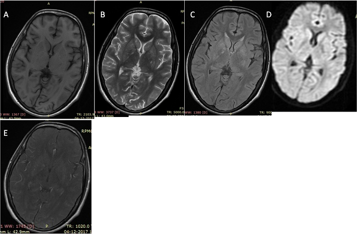
We present a case of an 18-year-old male who is HIV positve with a 2-month history of headache and 1 episode of seizures. Neurologic
examination showed bilateral papilledema. CD4 count was 70cells/μL and CSF study findings showed marginally elevated protein
levels. Brain MR imaging showed multiple small variable sized liesons in basal ganglia region which were hyointense on T1W images
and hyperintence on T2W images, not suppressed on FLAIR, showed no restriction on DWI and on post contrast images showed subtle
faint contrast enhancement. The patient underwent stereotactic biopsy at more higher institute which revealed on histopathology a
granulomatous lesion with the typical appearance of cryptococcoma. The patient was treated with amphotericin-B, intravenous steroids,
and anticonvulsants.

A 18-year-old male who is HIV positve with a 2-month history of headache and 1 episode
of seizures. Showed multiple small variable sized liesons in basal ganglia region which are
hyointense on T1W (A) images and hyperintence on FLAIR (C) showed no restriction on
DWI(D) and on post contrast images (E) showed subtle faint contrast enhancement
Discussion
Cryptococcosis, caused by an encapsulate yeast like fungus and is the most common CNS mycotic infection. Most patients have an underlying chronic illness, such as AIDS, diabetes mellitus, collagen vascular disease, chronic renal disease, alcoholism, or malignant neoplasms, particularly lymph reticular disorders, or they are on immunosuppressive medication, although the infection may occur in immunocompetent persons. It typically results from haematogenous spread from the lungs [1-4]. In HIV/AIDS patient’s cryptococcal infection of the CNS usually occurs when the CD4+ count drops below 100 cells/µL. The disease can have either meningeal or parenchymal involvement with the former being the primary manifestation [6]. With meningeal involvement, a grayish, mucinous exudate accumulates over the involved brain surface. There are three dominant CNS forms to the disease which are: meningitis, cryptococcoma sand gelatinous pseudo cysts [5].
One of the most common finding is hydrocephalus and there is a tendency for the disease to spread along the perivascular spaces [7,9]. With parenchymal involvement, there can be often formation of parenchymal cryptococcomas that commonly involve the midbrain and basal ganglia as in our case. Gelatinous pseudocysts and choroidal ependymal granulomas may also develop [6,9]. CT findings can be often non-specific and with normal scans seen in a significant proportion of patients Hydrocephalus and mass lesions may also each be present in approximately 10% of cases. MRI is better at assessing dilated perivascular spaces, one of the most frequently described feature on MRI, and basal ganglia pseudo cysts. These findings are more common in immunocompromised patients. Cryptococcomas are of low signal T1W images and of high signal on T2W and FLAIR images and on post contrast images show variable, ranging from no enhancement to peripheral nodular enhancement. This typical characteristic was seen in our case. Gelatinous pseudo cysts give a "soap bubble" appearance which are low to intermediate signal on T1W images and high in T2W images and suppressed on FLAIR images and this feature on FLAIR differentiate it from crptococcomas which are of high signal. Treated with intravenous amphotericin B or fluconazole. It is fatal if left untreated [6,7,8,10].
Conclusion
Cryptococcomas is more frequently encountered infection in AIDS patients especially when the CD4 count fall below 100 cells/µL. MR is the imaging modality of choice in diagnosing the CNS manifestation of cryptococcosis and reliably differentiate between cryptococcomas and gelatinous pseudo cysts particularly differentiated on FLAIR images in which cryptococcomas are not suppressed while pseudo cyst are suppressed i.e. they show hypo intense signals.
References
- Everett BA., et al. “Cryptococcal infection of the central nervous system”. Surgical Neurology 9 (1978): 157-163.
- Harper CG., et al. “Cryptococcal granuloma presenting as an intracranial mass”. Surgical Neurology 11.6 (1979): 425-429.
- Long JA., et al. “Cerebral mass lesions in Torulosis demonstrated by computed tomography”. Journal of Computer Assisted Tomography 4.6 (1980): 766-769.
- Fujita NK., et al. “Cryptococcal intracerebral mass lesions: the role of computed tomography and nonsurgical management.” Annals of Internal Medicine 94.3 (1981): 382-388.
- Cornell SH and Jacoby CG. “The varied computed tomographic appearance of intracranial cryptococcosis”. Radiology 143 (1982): 703-707.
- Garcia CA., et al. “Cryptococcal intracerebral mass lesions: CT-pathologic considerations”. Neurology 35.5 (1985): 731-734.
- Waterson JA and Gill igan BS. “Cryptococcal infections of the central nervous system: a ten year experience”. Clinical and experimental neurology 23 (1987): 127-137.
- Tan CT and Kuan BB. “Cryptococcus meningitis, clinicai-CT scan considerations”. Neuroradiology 29.1 (1987): 43-46.
- Popovich MJ., et al. “CT of intracra nial cryptococcosis”. American Journal of Roentgenology 154.3 (1990):139-142.
- Tien RD., et al. “Intracranial cryptococcosis in immunocompromised patients: CT and MR findings in 29 cases”. American Journal of Neuroradiology 12.2 (1991): 283-289.
Citation:
Lokesh Rana., et al. “Cryptococcoma’s on Mr Imaging-A Case Report”. Medical Research and Clinical Case Reports 2.3 (2018):
218-220.
Copyright: © 2018 Lokesh Rana., et al. This is an open-access article distributed under the terms of the Creative Commons Attribution License, which permits unrestricted use, distribution, and reproduction in any medium, provided the original author and source are credited.


 Scientia Ricerca is licensed and content of this site is available under a Creative Commons Attribution 4.0 International License.