Case Report
Volume 2 Issue 4 - 2018
Granular Cell Tumor of the Breast: A Rare Tumor Mimicking Breast Cancer
1Department of Surgery, Local Health Unit of the Alentejo Coast, Portugal
2Department of Surgery, Local Health Unit of the Alentejo Coast, Portugal
3Department of Surgery, Local Health Unit of the Alentejo Coast, Portugal
4Department of Surgery, Local Health Unit of the Alentejo Coast, Portugal
5Department of Pathology, Local Health Unit of the Alentejo Coast, Portugal
6Department of Surgery, Local Health Unit of the Alentejo Coast, Portugal
*Corresponding Author: Andreia Ferreira, Department of Surgery – Local Health Unit of the Alentejo Coast, Monte do Gilbardinho EN261, 7540-230 Santiago do Cacém, Portugal.
Received: December 13, 2018; Published: December 31, 2018
Abstract
Granular cell tumor is a rare tumor, first described by Abrikossoff in 1926, being localized in the breast in about 5% of the cases. The purpose of this article is to present a case of a granular cell tumor of the breast. As this tumor clinically and radiologically mimics breast carcinoma, it should be part of the differential diagnosis. In the authors opinion this case is clinically important because it emphasizes the importance of diagnosing this rare neoplasm in the breast as it simulates carcinoma, which can lead to a radical surgery. The authors made a review of the patient clinical process and a brief review of the literature.
Key words: Granular cell tumor; Breast carcinoma; Abrikossoff tumor; Benign tumor; Breast tumor
Introduction
Granular cell tumor was first described by Abrikossoff in 1926 [1], being a rare tumor that can arise throughout the body, most frequently in the head and neck and occurring in breast in 5% of the cases [2,3]. It was thought it had a muscular origin, being called “granular cell myoblastoma”, but it is now known that it originates in Schwann cells due to their similarities in ultrastructural features and the positivity of the tumor for S-100 protein [3-7]. In the breast, it is more common in the inner upper quadrant, corresponding to the supraclavicular nerve territory. It is usually benign, showing malign transformation in about 1% of cases [4]. It can mimic carcinoma on physical examination, mammography, ultrasonography and macroscopic evaluation of specimen [8]. The authors present a case of granular cell tumor of the breast which clinically and radiologically mimicked carcinoma.
Clinical Case
We present a case of a fifty-nine-year old woman, with personal history of oligophrenia, G0P0, which had menopause at fifty-four-year old and with no family history of breast diseases. She was referenced to Senology consultation with complaint of a nodule in the right breast. Physical examination showed a 2cm mass in the inner upper quadrant, which was irregular and firm.
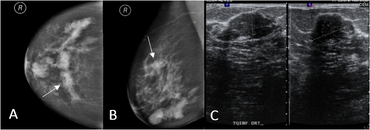
Mammography revealed an asymmetric and dense mass with structural distortion, spiculated margins and microcalcifications in the upper inner quadrant of the right breast (Figures1 A and B). Ultrasonography with color doppler showed a nodular hypoechoic lesion with acoustic shadowing and peripheral vascularization, in the same localization as the density described in the mammography, measuring 20,9x16,6x16,3mm (Figure 1C). It was classified as Breast Imaging Reporting and Data System (BI-RADS) 4c.

Figure 1A and B: Mammography showing an asymmetric mass with spiculated margins and micro calcifications in the inner upper quadrant of the right breast (arrow).
C. Ultrasonography showing a nodular hypoechoic lesion with acoustic shadowing.
C. Ultrasonography showing a nodular hypoechoic lesion with acoustic shadowing.
A core biopsy was performed for histologic diagnosis, which revealed cells with a small nucleus and an abundant, eosinophilic and granular cytoplasm. Mitosis and necrosis were not observed. They were positive for S-100 protein, vimentin and smooth muscle actin in the immunohistochemical analysis, compatible with a granular cell tumor of the breast (Figure 2).

Figure 2A and B: H&E. Tumor cells with small nucleus and abundant granular cytoplasm.
C. Immunohistochemistry showing positivity for S-100 protein.
C. Immunohistochemistry showing positivity for S-100 protein.
The patient underwent a tumorectomy with extemporaneous examination showing normal breast tissue around the lesion. Histopathological examination confirmed the diagnosis of a granular cell tumor of the breast (Abrikossoff tumor). There were no intercurrences in the postoperative period. Follow-up showed no sign of recurrence.
Discussion
Granular cell tumors of the breast are more common in women, but there are cases reporting this lesion in men [2,7,8]. Literature shows a slight preponderance in premenopausal women, although it can occur at any age [7]. Clinically, they present as a hard, painless nodule, mimicking breast carcinoma [2,4]. Retraction of the overlying skin can occur when it is superficial [7]. Granular cell tumors appear mainly in the inner upper quadrant, as in the presented case, corresponding to the cutaneous sensory territory of the supraclavicular nerve, which differs from carcinoma that arise more commonly in the upper outer quadrant [2-5]. On mammography, these tumors can appear as well defined or spiculated margin nodules, such as breast carcinoma [2,6,7]. Microcalcifications are not common [2], although they were present in our case. On ultrasound they also can appear as a solid, poorly marginated lesion or as a more benign-appearing well circumscribed mass [4]. An acoustic shadowing distal to the solid and hypoechoic mass can mimic a malignant lesion [7,9], as we found in the presented case.
Granular cell tumors of the breast are rarely malignant (1% of the cases), but there are some clinical and imaging features that should make us suspect of malignancy, such as the presence of an adenomegaly, the size of the tumor (more than 40mm), and infiltration of adjacent tissues [2,10].
The diagnosis of this tumor is histopathological and immunohistochemical. Usually, it presents as abundant granular eosinophylic cytoplasm cells with round nuclei [2,6,11], showing S-100 protein positivity and cytokeratin negativity [4], as seen in this case. A high proportion of granular cell tumors are reactive for vimentine, which is detectable in few carcinomas, and granular cell tumor is usually negative for estrogen and progesterone receptors [3,5]. In the presented case, we were able to do the diagnosis in the preoperative period with core needle biopsy, allowing better surgical planning.
Malignant granular cell tumors can be histopathologicaly differentiated from benign lesions with evaluation of the following six features: necrosis, spindling, vesicular nuclei with large nucleoli, increased mitotic activity (>2mitosis per 10HPF), high nuclei/cytoplasm ratio and nuclear pleomorphism. Atypical granular cell tumors must have two of these six features, and malignant tumors must have at least three of the six criteria [2]. In the presented case none of these features were present, which is compatible with the benign granular cell tumor diagnosis.
Treatment of benign granular cell tumor of the breast consists on wide excision, as subtotal excision can lead to local recurrences [3,4,8]. The prognosis is favorable, with recurrence occurring in 2-8% of individuals after excision with wide margins [10].
Conclusion
Granular cell tumor of the breast is a rare condition, usually benign, that mimics breast cancer, so it should be considered in the differential diagnosis to prevent overtreatment [2,4,10]. Preoperative diagnosis with core needle biopsy is important because it can prevent a radical surgery.
Conflict of interest
The authors declare that they have no conflicts of interests.
The authors declare that they have no conflicts of interests.
References
- Abrikossoff A. “Uber myome ausgehend von der quergestreiften wilkurlicken muskulatur”. Virchows Arch. (Pathol. Anat.) 260 (1926): 215-233.
- Scaranelo A., et al. “Granular cell tumor of the breast: MRI Findings and review of the literature”. The British Journal of Radiology 80.960 (2007): 970-974.
- Filipovski V., et al. “Granular cell tumor of the breast: a case report and review of literature”. Cases Journal 2 (2009).
- Pergel A., et al. “A Therapeutic and Diagnostic Dilemma: Granular Cell Tumor of the Breast”. Case Reports in Medicine (2011).
- Simone N., et al. “Granular Cell Tumor of the Breast: Clinical and Pathological Characteristics of a Rare Case in a 14-Year-Old Girl”. Journal of Clinical Oncology 29 (2011): 656-657.
- Ilvan S., et al. “Benign granular-cell tumor of the breast”. Canadian Journal of Surgery 48 (2005): 155-156.
- Baidya R., et al. “Granulosa Cell tumor of the male breast”. JPN 1 (2011): 161-163.
- Mariscal A., et al. Granular Cell Tumor of the Breast in a male patient. AJR 165 (1995): 63-64.
- Siegel J., et al. “Unusual Sonographic Features of Granular Cell Tumor of the Breast”. Journal of Ultrasound in Medicine 18 (1999): 857-859.
- Fujiwara K., et al. “Granular cell tumor of the breast mimicking malignancy: a case report with literature review”. Acta Radiologica Open 7.12 (2018): 1-5.
- Pathania M and Bhargava C. “Granular Cell Tumor of Breast”. A Mimic of Carcinoma. MJAFI 66 (2010): 292-294.
Citation:
Andreia Ferreira., et al. “Granular Cell Tumor of the Breast: A Rare Tumor Mimicking Breast Cancer”. Medical Research and Clinical Case Reports 2.4 (2018): 294-297.
Copyright: © 2018 Andreia Ferreira., et al. This is an open-access article distributed under the terms of the Creative Commons Attribution License, which permits unrestricted use, distribution, and reproduction in any medium, provided the original author and source are credited.


 Scientia Ricerca is licensed and content of this site is available under a Creative Commons Attribution 4.0 International License.