Case Report
Volume 2 Issue 5 - 2019
Integrative Approach to Assessing the Results of PET/CT of the Human Body.
Clinic of urology. Tyumen Medical University. Russia
*Corresponding Author: VB. Berdichevskу, Clinic of urology. Tyumen Medical University. Russia.
Received: December 06, 2018; Published: January 05, 2019
Abstract
Introduction: The reason for this study was our interest in publications in which authors draw our attention to the functional nature of PET/CT studies, which allows visualizing and evaluating a number of biological processes occurring at various levels of a person in real time [1, 2, 5-7].
Aim: The purpose of the study is to analyze the possibilities of combined positron emission (PET) and computer (PET-CT) tomography with 11C-choline in a visual and mathematical evaluation of the lipid metabolism of morpho-functional kidneys in patients with suspected prostate cancer [3-4].
Materials and Methods: The results of 100 PET-CT studies with 11C-choline isotope all over the body of patients with suspected prostate cancer were analyzed, depending on the activity of isotope inclusion in the parenchyma of intact kidneys.
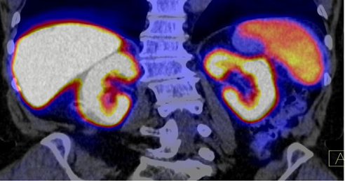
Results: According to the results of visual comparative CT and PET-CT studies of the patient’s entire body, it was established that intact parenchymal organs included the lipid metabolite in the process of realizing their physiological needs. The parenchyma of the liver, kidneys and spleen showed the greatest tropism for 11C-choline. Figure 1 and 2.
Key words: PET/CT with C11-choline; Kidney metabolism

Figure 2: Fragment of PET/CT of the whole human body with 11C-choline (visual color ranking of parenchymatous organs by the level of capture of C11-choline).
In the kidneys, hypermetabolism of 11C-choline was observed in the cortical and medulla of the parenchyma, less in the tissues of the calyx and the pelvis relative to the projection of their cavities (10 cu - 8 cu - 2 cu), which excluded the excretory character of 11C-choline imaging Figure 3.
Results
An integrative approach to evaluating the results of PET/CT studies with 11C-choline of the whole body of patients with suspected prostate cancer allowed simultaneously visualization of the metabolism of 11C-choline labeled biomolecules in the intact kidney parenchyma, which can significantly expand the range of diagnostic capabilities of this technology in clinical and experimental nephrology and urology.
References
- Blery M. “Functional cellular imaging: revolution for oncology/M. Blery”. Presse Med 35.9 (2006): 1339-1346.
- Mecca, C. PET/CT in diagnostic oncology/C. Mecca et al.//Q. J. Nucl. Med. Mol. Imaging. 48.2 (2004): 66-75.
- Pozitronno-ehmissionnaya tomografiya: rukovodstvo dlya vrachej/pod red. A. M. Granova i L. A. Tyutina. Moskva: Foliant 368 (2008).
- Aslanidi IP., Pursanova D.M., Muhortova O.V. i dr. Rol 'PEHT / KT s 11S-holinom v rannej diagnostike progressirovaniya raka predstatel'noj zhelezy//Medicinskaya radiologiya i radiacionnaya bezopasnost' 59.5 (2014): 37–54.
- Blackshaw, G. Prospective comparison of endosonography, computed tomography, and histopathological stage of junctional oesophagogastric cancer/G. Blackshaw, W.G. Lewis, A.N. Hopper et al.//Clin. Radiol. –63.10 (2008): 1092-1098.
- Nikitin YU.P., Kurilovich S.A., Davidik G.S. Pechen 'i lipidnyj obmen. Izdatel'stvo nauka. Moskva (1985) 192 s. (Russian)
- Luppa H. Osnovy gistohimii. Izdatel'stvo "Mir" Moskva (1980): 343 s. (Russian)
Citation:
VB. Berdichevskу. “Integrative Approach to Assessing the Results of PET/CT of the Human Body.” Medical Research and Clinical Case Reports 2.5 (2019): 298-300.
Copyright: © 2019 VB. Berdichevskу. This is an open-access article distributed under the terms of the Creative Commons Attribution License, which permits unrestricted use, distribution, and reproduction in any medium, provided the original author and source are credited.


 Scientia Ricerca is licensed and content of this site is available under a Creative Commons Attribution 4.0 International License.