Oral Health and Dentistry
ISSN: 2573-4989
Case Report
Volume 6 Issue 1
A Rare Combination of Tooth Agenesis in Association with Anomalous Supernumerary Tooth: Report of a Rare Case
-
Professor and Consultant, “Garike” Dental Care
*Corresponding Author: Dr. Nagaveni NB, Professor and Consultant, “Garike” Dental Care, Davangere, Karnataka, India.
Abstract
Isolated existence of dental anomalies is very frequent finding during clinical practice. However concomitant occurrence of two anomalies in a single patient is a rare entity and clinician should be aware of such anomalies to diagnose and render proper treatment for the patient. The purpose of this paper is to present a rare combination of both tooth agenesis (of mandibular central incisors) and a supernumerary tooth (rudimentary, anomalous mandibular premolar) in the same arch of Indian patient.
Keywords: Tooth agenesis; Supernumerary; Short root anomaly; Central incisors; Premolars
Introduction
Mandibular premolars exhibit numerous variations in their structure, size and shape including both crown and root portion.[1] These are the teeth most commonly reported as congenitally missing, next to the third molars. However, these are the teeth which show variations in root structures too like two or three rooted or two / three canaled.[2] The one more clinical condition with a greater prevalence of more than two premolars is termed as a supernumerary tooth. Mandibular supernumerary premolars are well reported and many prevalence studies have been published in the literature.[3] They occur more frequently in mandible, with supplemental type being the most common. In maxillary arch, they usually appear conical or smaller than normal tooth. Supernumerary tooth occurring in the premolar region may be single or in multiple form and can be erupted or impacted (75%). [3]
Congenital tooth agenesis is also an uncommon clinical entity found during clinical practice. The most commonly involved teeth are third molars, mandibular premolars and maxillary lateral incisors. Agenesis of mandibular central incisors is an infrequent finding and most commonly involves unilateral absence of incisor. The first published data on congenitally missing two mandibular incisors is given by Newman in 1967 [4], and it is also reported that congenital agenesis of lower central incisors is common in certain populations like Korean, Chinese and Japanese groups.[5]
Literature shows isolated data on any one of the above conditions mentioned in the literature. [1-4] The author of this paper reported many case reports on isolated cases of congenitally missing of mandibular central incisors [6] and this condition in association with other dental anomalies.[7] But literature on concomitant occurrence of congenital agenesis both central incisors along with rudimentary, anomalous mandibular supernumerary premolar is not reported so far. Therefore, the aim of this article is to present such a rare combination of both tooth agenesis along with extra supernumerary tooth in the same dental arch of an Indian patient.
Case Report
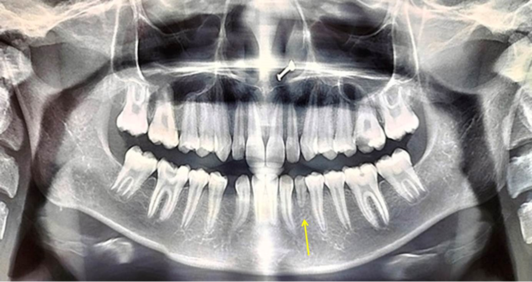
A 17-year-old female patient reported to a private dental clinic complaining of pain in the upper left back tooth region since a week. Patient was well nourished, moderately built with no signs and symptoms of medical, systemic or syndromic features. On intraoral examination, deep dental caries in relation to maxillary left first molar and mandibular right first molar was evident. Spacing was seen in the mandibular arch between right first premolar and right canine. On further examination mandibular both central incisors (right and left) appeared to be missing. History of previous extraction of the same teeth or early exfoliation was absent. Both right and left lateral incisors were erupted in place of central incisors. To rule out whether these are central and lateral incisors, clinically the both teeth were examined thoroughly. The clinical features were similar to lateral incisors. On contra lateral side, one small rudimentary extra premolar was noticed. Both first and second premolars were present beside to this small tooth structure. The tooth was very small in size having two small cusps as one buccal and one lingual cusp. Patient was subjected to radiographic examination pertaining to those teeth. On orthopantomograph, congenital agenesis both right and left central incisors was evident (Figure 1).
The small tooth present in the left quadrant had single root which was also short in length. When the small tooth was measured on the radiograph the length of the root appeared almost equal to the length of the crown. This extra tooth did not resembled to any other tooth. Root dilacerations was noticed in the mandibular both right and left permanent canines. Therefore based on literature search this tooth was diagnosed as rudimentary, anomalous supernumerary premolar. Pertaining to absence of central incisors the condition was diagnosed as idiopathic, non-syndromic, congenital agenesis of both central incisors was made for this patient. As dental caries was involving the pulp, with widening of periodontal ligament space, root canal treatment was advised for both carious molars. Since the rudimentary premolar was normal with no associated symptoms no treatment was considered. With respect to agenesis of both central incisors, as there was no gap evident in the midline and was almost closed by lateral incisors no treatment was considered.
Discussion
Supernumerary teeth appearing in the premolar region have peculiar characteristics when compared to supernumeraries of other teeth pertaining to various factors such as epidemiology, characteristics, etiology, complications associated with it, clinical presentation, radiographic presentation and treatment. A review article shows approximately 8-9% of all supernumerary teeth occur in the premolar region of non-syndromic patients. It is also shown that it is more in African patients and 1% of Southern Nigerians have one or more supernumerary premolar teeth. [3,8]
The supernumerary premolar appeared very small in size in our case. The root also appeared short and was almost equal to the length of the crown. The differential diagnosis considered for this condition is the short root anomaly (SRA) or Rhyzomicroly (RM). RM or SRA is a rare radicular anomaly seen only in permanent teeth and characterized by teeth having short and blunt root along with closed and round apices and reduced crown to root ratio (C:R). The crown to root ratio usually ranges from 1:1 or less and completely formed roots with closed apices are the unique feature found in this rare anomaly. [9] It occurs most commonly unilateral than bilateral and found frequently in the maxillary central incisors followed by maxillary premolars and maxillary lateral incisors. Mandibular premolars, canines and molars are the least affected teeth with this condition. SRA affected tooth show normal size and shape in morphology on clinical examination. Even the surround soft tissues appear normal and asymptomatic.[9,10] Therefore, when these features are considered, the supernumerary premolar found in our report was small in size and shape. The occlusal anatomy did not resemble the normal premolar. Hence, keeping all these facts the SRA was ruled out and a diagnosis of rudimentary, anomalous supernumerary premolar was made in the present case. Literature shows association of tooth mobility with SRA. But this finding was not found in our case.
Plenty of isolated reports on either congenital agenesis of mandibular central incisors or supernumerary premolars can be found in the literature.[6,7] But association of two or more anomalies occurring within the same patient is a rare entity. Previously, the current author published concomitant occurrence of agenesis of both lower central incisors and type I left mandibular canine transmigration in a 14 year old Indian male patient. [7] Later the same author published an unusual occurrence of a combination of dental anomalies including congenital missing of 14 numbers of permanent teeth excluding third molars, canine transmigration and many other anomalies in a non-syndromic 13 year old female Indian patient.[11] Previously there was a publication on congenital agenesis of only both central incisors in four Indian patients.[6] This clearly indicates the appearance of dental anomalies is common in people of Indian races and hence more and more prevalence studies are required in the context of Indian population.
The exact etiology behind occurrence of missing lower incisors is not known. Literature review suggests the following factors as etiological factors: 1. Anomalies present in the development of the mandibular symphysis may disturb the dental tissues during tooth formation of lower incisors, 2. A decrease in the size of dentition as Nature’s attempt to fit the shortened dental arch. 3. Disturbance of the endocrine system resulting in a localized ectodermal dysplasia, 4. Localized infection or inflammation in the jaw destroying the tooth buds, 5. Heredity or familial distribution of congenital absence of lower incisors.[4-6]
Clinically, this condition does not lead to any pathognomonic features. The place of central incisors is usually occupied by lateral incisors there by resulting in no space or little space in the dental midline. If the space is little once all the permanent teeth erupt into the oral cavity is closed on its own. If the space is more, a removable partial denture or implant or a fixed prosthesis can be considered once the growth of the patient completes.[4-6] These are the possible treatment modalities considered or suggested for the congenital agenesis of lower central incisors. In the present case, the gap in the midline was not present except for the little gap between the canine and first premolar on the right side. This was not aesthetically visible and hence no treatment was suggested. Regarding the supernumerary premolar the tooth was kept for observation as there was no mobility or any other symptoms were present. In addition to these two anomalies, root dilacerations were noticed in the permanent mandibular both canines which is also a rare phenomenon.
Conclusion
An awareness and knowledge regarding occurrence of congenital missing of lower incisors and supernumerary premolars characterized by anomalous structure is essential among all clinicians and general practitioners. Rendering meticulous clinical diagnosis and proper treatment for the patient having dental anomalies is very important.
References
- Nagaveni NB., et al. “Molarization of the mandibular second premolar in an Indian patient: Report of a rare case”. Annals of Bioanthropology 3.1 (2015): 33-35.
- Nagaveni NB and Ashwini KS. “Bifurcated Mandibular Second Premolars: Report of Unusual Root Anomaly (Supernumerary Root) – A Case Series”. Journal of Dentistry and Oral Health 10 (2023): 1-7.
- Khalaf k., et al. “A Review of Supernumerary Teeth in the Premolar Region”. International Journal of Dentistry (2018): 6289047.
- Newman GV. “Congenitally missing mandibular incisors: treatment procedures”. American Journal of Orthodontics 53.7 (1967): 489-491.
- Niswander JD and Sujaku C. “Congenital Anomalies of Teeth in Japanese Children”. American Journal of Physical Anthropology 21.4 (1963): 569-574.
- NB Nagaveni and KV Umashankara. “Congenital bilateral agenesis of permanent mandibular incisors: case reports and literature review”. Archives of Orofacial Sciences 4.2 (2009): 41-46.
- Nagaveni NB., et al. “Concomitant occurrence of canine transmigration and symmetrical agenesis of incisors – A case report”. Bangladesh Journal of Medical Science 10.2 (2011): 133-136.
- Kawashita Y and Saito T. “Nonsyndromic multiple mandibular supernumerary premolars: a case report”. Journal of Dentistry for Children 77.2 (2010): 99-101.
- Lamani E., et al. “Short Root Anomaly - A Potential "Landmine" for Orthodontic and Orthognathic Surgery Treatment of Patients”. Annals of Maxillofacial Surgery 7.2 (2017): 296-299.
- Puranik CP., et al. “Characterization of short root anomaly in a Mexican cohort--hereditary idiopathic root malformation”. Orthodontics & Craniofacial Research 18 (2015): 62-70.
- Nagaveni NB. “An unusual occurrence of multiple dental anomalies in a single nonsyndromic patient: a case report”. Case Reports in Dentistry (2012): 426091.
Citation:
Dr. Nagaveni NB. “A Rare Combination of Tooth Agenesis in Association with Anomalous Supernumerary Tooth: Report of a Rare Case”. Oral Health and Dentistry 6.1 (2023): 18-21.
Copyright: © 2023 Dr. Nagaveni NB. This is an open-access article distributed under the terms of the Creative Commons Attribution License, which permits unrestricted use, distribution, and reproduction in any medium, provided the original author and source are credited.
 Scientia Ricerca is licensed and content of this site is available under a Creative Commons Attribution 4.0 International License.

