Oral Health and Dentistry
ISSN: 2573-4989
Case Report
Volume 6 Issue 1
Intraosseous Transmigration of Permanent Maxillary Right Canine - Report of a Rare Case
-
Professor and Consultant, “Garike” Dental Care
*Corresponding Author: Dr. Nagaveni NB, Professor and Consultant, “Garike” Dental Care, Davangere, Karnataka, India.
Abstract
Unusual dental anomalies are quite interesting from different aspects including genetic, anthropological and from clinical perspective. Transmigration of maxillary canine is an uncommon dental phenomenon involving eruption scenario. Information pertaining to this clinical entity is sparse in the dental literature. Therefore, the aim of present article is to report an infrequent development of impacted maxillary canine transmigrating towards opposite side of the maxillary dental arch in an Indian female patient.
Keywords: Canine impaction; Dental anomalies; Maxillary canine; Tooth eruption phenomenon; Transmigration
Introduction
Transmigration of canine is an unusual dental anomaly characterized by intraosseous migration of impacted teeth across the dental midline to the opposite side of the dental arch [1]. The teeth most commonly affected by this condition are the permanent teeth and mostly involving mandibular canines [2]. Transmigration occurring in primary teeth is not reported so far. However, there are some reports showing transmigration of mandibular lateral incisor and second premolar too apart from cases of mandibular canines [3-5]. Mandibular lateral incisor is usually transmigrated in a distal direction and found transposed with the canine. Whereas second premolar most of the time migrates distally and found in the gonial angle, ramus of mandible and at the coronoid process. With respect to mandibular canine, the transmigration is usually seen in mesial direction crossing the mandibular symphysis and transmigrating to the opposite side of the dental arch [6,7]. Maxillary canine transmigrating intra-osseously across the mid-palatal suture to the opposite side of the maxilla is not found with enough evidence in the literature. Literature review revealed countable number of maxillary canines’ transmigration in the existing database [8-10]. Canines when get impacted inside the alveolar bone fail to erupt and usually seen with retained primary canines in the oral cavity beyond their normal exfoliation period due to lack of resorption process within the roots. However, other dental anomalies have been reported along with transmigration of maxillary canines [8-10].
Although these maxillary canines’ transmigration is not classified, as there is no classification system given so far because of low frequency of canines transmigrated which are not sufficient to draw out proper classification and also including its etiology, demographic features and associated anomalies or syndromes. This shows highly requirement of large number of studies to dissipate more information about this uncommon dental phenomenon. Therefore, with the intention of reporting a rare occurrence of transmigration of maxillary canine in a 27-year-old Indian female patient, the present article is prepared. This will encourage other authors to publish such rare anomalies which they come across during their routine clinical or academic practice.
Case Report
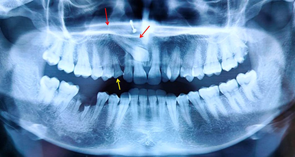
A 27-year-old female patient reported to a private dental clinic complaining of decayed tooth in the right lower back tooth region. On physical examination patient appeared absolutely normal, well-built and nourished with no history of any systemic or metabolic disorders. Features of any syndromes were also not found. On intra oral examination permanent mandibular right first molar was grossly destructed due to long standing dental decay. On left side also maxillary third molar was carious. Patient had complete permanent dentition with all third molars erupted. On further examination a retained primary canine was observed in the maxillary right quadrant and permanent canine was appeared to be missing or impacted. On opposite side of the arch, permanent canine was present. To rule out missing canine and to see the status of decayed first and third molars an orthopantomograph radiograph was advised. Examination of orthopantomograph radiograph showed impaction of permanent maxillary right canine (Figure 1). Along with being impacted the one-third crown portion of canine was transmigrated crossing the mid-palatal suture to the opposite side.

Figure 1: Orthopantomograph radiograph showing intraosseous transmigration of permanent maxillary right canine (one-third portion of the crown) crossing the mid-palatal suture to the opposite side of the arch (red arrows). Retained primary right canine can also be seen (yellow arrow).
The root was comparatively long compared to left side. The root of the retained primary canine did not show any resorption. Root stumps in relation to mandibular first molar was noticed with associated any pathology. The case was diagnosed as congenital, idiopathic transmigration of maxillary canine in association with radiculomegaly (Figure 1). Patient was informed about the impaction of maxillary canine. As patients’ main complaint was about her decayed tooth, extraction of root stumps of permanent mandibular right first molar was scheduled under local anesthesia. As the impacted and transmigrated maxillary canine was associated with any cyst or pathology periodic observation was advised. As the retained primary canine was in good condition with no mobility, no treatment was planned till its exfoliation.
Discussion
Current literature on occurrence of maxillary canine transmigrating to the opposite side of the maxillary arch by crossing the mid-palatal suture is very sparse. However, there are studies showing impaction of maxillary canines in different population [11,12]. Recently one or two prevalence studies or case presentations are peeping into the literature. However complete information pertaining to its etiology, pathogenesis, genetic influence, classification systems, clinical manifestations, associated anomalies or pathologies, demographic factors about transmigrated maxillary canines is not obtained. Aras et al in 2007 [13] evaluated 6000 patients for the presence of transmigrated maxillary canines in order to assess its right/left localization, gender or age prevalence, retained primary canines and presence of any other associated pathology. From this investigation, including large number of patients (6000 patients), transmigration of maxillary canines was detected only in 12 patients (0.2%). Among these 12 patients, there were 6 males and 6 females with age ranging from 15 to 57 years.
Only unilateral appearance was observed with equal predilection for both right and left side of the dental arch. None of the patients showed presence of retained primary canines or any associated pathology with impacted canines. In the case described here, although no associated pathology was observed but retained primary canine was observed in the same arch where permanent canine was transmigrated. In this case, maxillary right canine was transmigrated from right to left side of the maxillary arch. Similar to the above study, Celikoglu et al in 2010 [14], examined panoramic radiographs of 2,215 Turkish orthodontic patients consisting of 940 males and 1,275 females. The maxillary transmigrated canine was associated with impacted mesiodens. The percentage of maxillary canine impaction was more prevalent in females compared to males.
Transmigration of maxillary canines is always detected on routine radiographic examination when patient is subjected to radiographic survey advised for some other purpose. Most of the time, retained primary canines are usually seen beyond the normal expected period of primary teeth duration in the oral cavity. Such retained primary canines exhibit absence of root resorption on radiographic examination and also do not show any mobility on clinical examination of the particular tooth [1-5]. Even in the present case, primary canine was retained even at the 27th age of patient which was firm and did not show root resorption or clinical tooth mobility. This finding strictly warns every clinician that whenever they come across such retained primary teeth anywhere in the oral cavity and if any permanent tooth is clinically missing, first thing it should strike the mind of a clinician for the presence of unusual dental findings or phenomenon within the alveolar bone. In the present case too, radiograph was taken to study the status of decayed tooth, following which an impacted and transmigrated permanent maxillary canine was diagnosed.
To detect transmigration of maxillary canines, always orthopantomography radiograph should be considered for its correct evaluation. On periapical radiographs such transmigrated canines particularly in the maxillary arch, usually escape from identification because impacted canine most of the time, are horizontally positioned very high in the palatal vault, and close to the floor of the nasal cavity because of less surface area compared to mandibular arch [12,13]. Therefore, they will not be detected on a routine periapical radiograph. Although maxillary canine impaction is having been found to occur 20 times more often in the maxilla compared to mandible, but this impacted canine transmigrating by crossing the mid-palatal suture and found in the opposite side of the dental arch of the maxilla is not seen. Few authors have reasoned for this finding as the maxillary mid-palatal suture acts as a strong barrier, which prevents the palatally impacted canine from crossing to the opposite side of the arch. However, it is still a big controversy regarding how the migration of maxillary canine occurs whether it is labial or palatally impacted [12,13].
Impacted and transmigrated canines always remain asymptomatic with no evidence of pain or obvious pathology and hence most of the time they missed from diagnosis there by indicating reason for the low reports in the dental literature. The transmigration of maxillary canine displays varied type of migration from its original position. It ranges from its cusp tip being located adjacent to the lateral incisor root apex, or may be found near the central incisor root apex. It is sometimes even found at or close to the mid-palatal suture. Most of the case reports have shown similar findings and have stated that the maxillary canine after crossing the central incisor root tip usually stops at the mid-palatal suture as it acts as a barrier. However, sometimes the impacted and migrating canines due to the moving force make a tunnel in the mid-palatal suture and cross the midline and found in the opposite side of the maxillary arch. [12,13] This has been proved by a computed tomography scan and can be evaluated during surgical exposure of the impacted canine if patient agrees for suggested treatment.
The whole tooth crossing this suture is not reported so far. Only small portion of the crown part crossing this suture have been published. Therefore, the proper classification system is lacking as compared to mandibular transmigrated canines which easily cross the dental midline. In the case presented here, one third portion of maxillary canine crown was found crossing the mid-palatal suture at the level of root tip of maxillary central incisor and transmigrated to the opposite side of the maxillary arch which was seen in close to the root of maxillary left central incisor.
Impaction of maxillary canines has been studied recently by various investigators, as a result various classification systems have been given for impacted canines. Yamamoto et al [11] in 2003, proposed a classification system for impacted canines and second premolars. For maxillary canines he gave seven types from Type I to Type VII, based on the orientation of the long axis of the impacted canine and the occlusal plane. Based on this classification the present case was categorized as Type IV, as the impacted canine was horizontally placed with crown facing mesially and root projected distally above the roots of permanent maxillary central and lateral incisors. Recently Alassiry et al in 2020 [12] studied the prevalence and pattern of impaction of maxillary canines using 5000 orthopantomograms in Saudi population according to Yamamoto’s classification. They found a prevalence of 46% of type I followed by 37% of type II. Similar such studies are highly essential pertaining to transmigration of maxillary canines in order to research about various factors such as pathogenesis, prevalence in different ethnic groups, demographic factors, clinical features, classification system and associated pathology.
The present author previously published 3 cases of mandibular canine transmigration in Indian patients. In one patient, the transmigrated canine found was type I according to Mupparapu’s classification [15] which was found in female patient aged 56 years old. In this case, the transmigrated canine was remained asymptomatic till this age and was diagnosed accidentally on routine radiographic examination when taken for assessment of root stumps in partially edentulous dental arch [16]. Another case was published in 2012 in a 13-year-old girl patient with various unusual occurrence of dental anomalies like occurrence of taurodontism in permanent mandibular molars, congenital agenesis of 14 numbers of permanent teeth excluding third molars, canine impaction, type I mandibular canine transmigration, presence of pyramidal roots in primary molars, generalized microdontia and diastema [17].
The third case was reported in 2011 which was diagnosed in 12-year-old male patient characterised by symmetrical bilateral agenesis of mandibular central incisors and mandibular canine transmigration [18]. From the present case report, from above two published reports and from the results of previous studies it is clear that female predilection of canine impaction and transmigration was observed. The exact etiology for this clinical presentation is not mentioned in the available literature. Apart from the above cases, the present author also published a case of transmigration of maxillary central incisor in Indian patient [19]. Recently author reported a case of transmigration of maxillary canine in association with bilateral agenesis of permanent mandibular canines in a 35-year-old female patient. Occurrence of these number of cases in Indian patients signifies the urgency for need of prevalence studies to be undertaken among Indian ethnic population to evaluate in detail to dig more literature about this rare dental anomaly [20].
There is no particular well reported treatment modality for these transmigrated teeth. Clinician should not make an attempt to orthodontically move such impacted and transmigrated teeth as the prognosis is not good. If these teeth found symptomatic or associated with some pathology like cyst or tumours, surgical removal of the transmigrated tooth along with pathology is indicated.
Conclusion
Detailed awareness and knowledge about occurrence of transmigrated canines in individuals is highly required among clinicians in order to diagnose and provide appropriate treatment. Clinicians or academicians should show interest in documenting and reporting such unusual anomalies which further help in adding up more evidence to the existing literature.
References
- Becker A and Chaushu S. “Etiology of maxillary canine impaction: a review”. American Journal of Orthodontics and Dentofacial Orthopedics 148.4 (2015): 557-567.
- Kazor-Urbanowicz KE., et al. “Transmigration of mandibular canines: a retrospective analysis of 15 cases and a review of literature”. Advances in Clinical and Experimental Medicine 25 (2016): 343-348.
- Shapira Y and Kuftinec MM. “Intrabony migration of impacted teeth”. The Angle Orthodontist 73.6 (2003): 738-743.
- Alves DB., et al. “Transmigration of mandibular second premolar in a patient with cleft lip and palate--case report”. Journal of Applied Oral Science 16.5 (2008): 360-363.
- P R Sutton. “Migrating nonerupted mandibular premolars: a case of migration into the coronoid process”. Oral Surgery, Oral Medicine, Oral Pathology, and Oral Radiology 25.1 (1968): 87-98.
- Shapira K and Kuftinec MM. “Unusual intraosseous transmigration of a palatally impacted canine”. American Journal of Orthodontics and Dentofacial Orthopedics 127.3 (2005): 360-363.
- Shapira Y and Kuftinec MM. “Intraosseous transmigration of mandibular canines--review of the literature and treatment options”. Compendium of Continuing Education in Dentistry 16.10 (1995): 1014-1024.
- Mortazavi H., et al. “Intra-Osseous Migration of Second Lower Premolar- Literature Reviewand a Case Report”. Dentistry 7 (2017): 465.
- Nag R., et al. “Transmigration of unerupted mandibular second premolar associated with chronic nonspecific osteomyelitis: report of a rare case”. Indian J Stomatol 5 (2014): 31-32.
- Matteson SR., et al “Extreme distal migration of the mandibular second bicuspid. A variant of eruption”. The Angle Orthodontist 52.1 (1982): 11-18.
- Yamamoto G., et al. “A New Classification of Impacted Canines and Second Premolars Using Orthopantomography”. Asian Journal of Oral and Maxillofacial Surgery 15.1 (2003): 31-37.
- Alassiry A. “Radiographic assessment of the prevalence, pattern and position of maxillary canine impaction in Najran (Saudi Arabia) population using orthopantomograms - A cross-sectional, retrospective study”. The Saudi Dental Journal 32.3 (2020): 155-159.
- Aras MH., et al. “Transmigrant maxillary canines”. Oral Surgery, Oral Medicine, Oral Pathology, and Oral Radiology 105.3 (2008): E48-E52.
- Celikoglu M., et al. “Investigation of transmigrated and impacted maxillary and mandibular canine teeth in an orthodontic patient population”. Journal of Oral and Maxillofacial Surgery 68.5 (2010): 1001-1006.
- Mupparapu M. “Patterns of intra-osseous transmigration and ectopic eruption of mandibular canines: review of literature and report of nine additional cases”. Dentomaxillofacial Radiology 31.6 (2002): 355-360.
- Nagaveni NB. Type I transmigration of permanent mandibular right canine – report of a rare case. J Pathol Allied Med 2023; 4: 1-5.
- Nagaveni NB. An unusual occurrence of multiple dental anomalies in a single non-syndromic patient: A case report. Case Report Dent 2012; 426091: 4.
- Nagaveni NB, Radhika NB, Umashankara KV, Satisha TS. Concomitant occurrence of canine transmigration and symmetrical agenesis of mandibular incisors – a case report. Bangladesh J Med Sci 2011; 10: 133-6.
- Nagaveni NB. Transmigration of permanent maxillary central incisor. Global Journal of Research in Dental Sciences. 2023; 3(4): 5-8.
- Nagaveni NB. Transmigration of maxillary canine in association with bilateral agenesis of permanent mandibular canines – Report of a rarest case with literature review. J Oral Health Dent. 2023; 6(3): 596-602.
Citation:
Dr. Nagaveni NB. “Intraosseous Transmigration of Permanent Maxillary Right Canine - Report of a Rare Case ”. Oral Health and Dentistry 6.1 (2023): 22-26.
Copyright: © 2023 Dr. Nagaveni NB. This is an open-access article distributed under the terms of the Creative Commons Attribution License, which permits unrestricted use, distribution, and reproduction in any medium, provided the original author and source are credited.
 Scientia Ricerca is licensed and content of this site is available under a Creative Commons Attribution 4.0 International License.
