Case Report
Volume 2 Issue 6 - 2018
Unexpected Outcome In Case Of GBM
1Department of Neurosurgery, Ardahan State Hospital, Ardahan, Turkey
2Dokuz Eylul University Medical school, Department of Neurosurgery, Izmir, Turkey
2Dokuz Eylul University Medical school, Department of Neurosurgery, Izmir, Turkey
*Corresponding Author: Kaptan H, Dokuz Eylul University Medical school, Department of Neurosurgery, Izmir, Turkey.
Received: June 23, 2018; Published: August 29, 2018
Introductıon
Glial tumors are the most common central nervous system tumors. Classified into four subgroups (I-IV) according to the histopathology. Grade I and Grade II tumors, so called low-grade glial tumors, are slow-growing tumors with better prognosis. High-grade (malignant) glial tumors are Grade III (anaplastic astrocytoma or anaplastic oligodendroglioma) or Grade IV (glioblastoma multiforme) tumors. Malignant glial tumors accounts for 60-75% of all glial tumors. [1] The most common and the one with the worst prognosis is glioblastoma multiforme (GBM) with 1-year mean survival. [2] The incidence of GBM is 5/100.000. The histopathological subgroup is World Health Organization (WHO) Grade 4. [1] GBM is mostly seen in men and in fifth to sixth decades of the life. [3]
The only known risk factor for the development of glial tumors is exposure to ionizing radiation. There is no evidence for that occupational or environmental causes, history of head trauma, use of mobile devices or infections leading to development of brain tumors. [4-5] For the treatment of high-grade tumors, concomitant maximum surgical resection and radiotherapy followed by chemotherapy is standard. [6]
Case
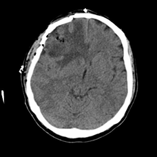
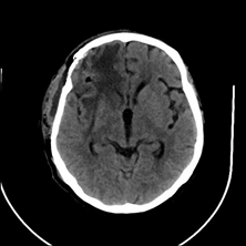
57 years old man without any comorbidities presented to the E.R. with the complaints of headache for the last 15 days and confusion. At the time of presentation GCS of the patient was 13. In the Cranial MRI study, there was a 5 x 6 cm subcortical mass lesion showing irregular nodular contrast enhancement causing cingulate herniation located in right middle frontal gyrus. The patient was operated after 2 days of ICU follow-up with anti-edema treatment. The resection was gross-total. In the postoperative neurological examination GCS was 15 without any neurological deficits. There was no complications postoperatively. The histopathology reported as glioblastoma multiforme. After the first month postoperatively, radiotherapy and chemotherapy were applied. During the routine follow-up, a relapsing lesion at the site of operation was identified 7 months postoperatively. The patient was reoperated. The resection was gross-total. GCS was 15 and there was no neurological deficits, without any complications postoperatively. The pathology was glioblastoma multiforme from the second operation. At postoperative 28th month after the first operation, there was a 4 x 5 cm new mass lesion on right orbital gyrus and right inferior frontal gyrus showing irregular nodular contrast enhancement corresponds to a glial lesion. The patients was reoperated. Postoperative GCS was 15 and there was no neurological deficits. Pathology was reported as glioblastoma multiforme. The patietn underwent 3 surgeries due to GBM and took radiotherapy and chemotherapy. The survival after the first operation was 35 months.
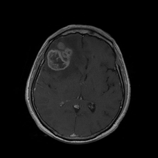
Figure 1: Preoperative MRI: 5 x 6 cm subcortical mass lesion showing irregular nodular contrast enhancement located in right middle frontal gyrus.
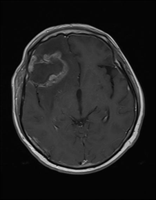
Figure 3: Preoperative MRI: Mass lesion in right middle frontal gyrus corresponds to relapse or radiation necrosis.
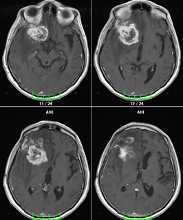
Figure 5: Preoperative MRI: 4 x 5 cm subcortical mass lesion in right orbital gyrus and right inferior frontal gyrus.
Dıscussıon
The location and rate of progression of the tumor amd edema related with tumor are responsible from the signs and symptoms. The main prognostic factors are age, Karnofsky Performance Status Scale and pathology. In general patients with older age, worse Karnofsky Performance Status Scale score and higher grade glial tumors show a shorter duration of survival.
In the last 30 years the treatment of high-grade glial tumors advanced however three main principles remained same: maximum surgical resection, radiotherapy and chemotherapy. Fractioned localized radiotherapy is a part of the standart treatment for all high-grade glial tumors. Besides, chemotherapy became an indespensable portion of the treatment in high-grade glial tumors including glioblastoma and anaplastic oligodendroglioma. The mean survival is increased under favor of including chemotherapy in the first-line treatment of these two tumors. Although these three treatment became main principles the mean survival of a patient with GBM is about 14 months. [4-5]
Pinsker and Lumenta showed survival after reoperation in patients with GBM as 5,75 moths abd overall survival as 14,3 months. [7] Tugcu and colleagues found that the survival after reoperation was 6.86 months in a study including 50 patients with GBM. [8] In a study including 365 cases conducted by Chaichana and colleagues the longest duration of survival was 26,6 months belongs a patient whom underwent 4 surgeries. [9] Helseth and colleagues found the mean survival as 5,9 months after reoperation in a study including 65 cases with GBM. [10]
Canada National Institute of Cancer compared the affect of radiotherapy alone or radiotherapy combined with chemotherapy on mean surival in a study including 573 patients recently diagnosed with malign glioma. The mean survival was 12,1 months and 14,6 months respectively. It’s found that the mean survival was 4,1 months longer in patients whom underwent gross-total surgical resection. Therefore, according to this study extensive surgery may aid to the effect of chemoradiotherapy. [11]
Conclusıon
In this case, a gross-total surgery was performed and followed by chemoradiotherapy. The patient was reoperated because of relapsing lesion. The patient was reoperated for the second time due to a new lesion in postoperative 28th month. The survival was 35 months after the first operation and without any neurological deficits. This result considered as a significant finding when compared with previous findings in the literature in terms of the duration of survival and lack of any neurological deficits.
The initial treatment of malignant glial tumors require a gross-total surgical resection within safe margins. The findings show that as a significant prognostiv factor the extend of the resection has an impact on overall survival. To prevent local invasions and relapsing disease adjuvant treatment protocols are needed. [12]
The indications for reoperation are; prescence of recent neurological deficits, increased mass-effect of the tumor causing signs and symptoms of increased intracranial pressure, inceresed frequency of seizures and radiological evidence of tumor progression. Recent findings state that the age should not be considered as an absolute contraindication. In those patients, if there is a chance of reoperation, this should be considered before the adjuvant therapy. Given that the effectiveness of the adjuvant therapy is increased in cases with wider surgical resection. [11] With advancing technology the molecular markers become more important in terms of completion of diagnosis, pathological grading and evaluation of prognosis.
References
- Louis DN., et al. “The 2007 WHO classification of tumours of the central nervous system”. Acta Neuropathologica 114.4 (2007): 97-109.
- Smith AA., et al. “A novel approach to the discovery of survival biomarkers in glioblastoma using a joint analysis of DNA methylation and gene expression”. Epigenetics (2014).
- Stupp R., et al. “High-grade malignant glioma: ESMO Clinical Practice Guidelines for diagnosis, treatment and follow-up”. Annals of Oncology 21.5 (2010): 190-193.
- Gilbert MR., et al. “Dose-densetemozolomidefornewlydiagnosed glioblastoma: a randomized phase III clinical trial”. Journal of Clinical Oncology 31.32 (2013): 4085Y4091.
- van den Bent MJ., et al. “Adjuvant procarbazine, lomustine, and vincristine chemotherapy in newly diagnosed anaplastic oligodendroglioma: long-term follow-up of EORTC brain tumor group study 26951”. Journal of Clinical Oncology 31.3 (2013): 344Y350.
- Gondi V., et al. “Primary intracranial neoplasms. In: C. A., Perez, L.W., Brady, E. C., Halperin, & D. E., Wazer, editors. Perez and Brady`s principles and practice of radiation oncology (6th ed.). Philadelphia (PA): Lippincott Willwams & Wilkins. 660. (2013).
- Pinsker M and Lumenta C. “Experiences with reoperation on recurrent glioblastoma multiforme”. Zentralbl Neurochir 62.2 (2001): 43-47.
- Tugcu B., et al. “Efficacy of clinical prognosticfactorsonsurvivalinpatientswithglioblastoma”. TurkNeurosurg 20.2 (2010): 117-125.
- Chaichana KL., et al. “Multiple resections for patients with glioblastoma: prolonging survival”. Journal of Neurosurgery 118.4 (2013): 812-820.
- Helseth R., et al. “Overall survival, prognostic factors, and repeated surgery in a consecutive series of 516 patients with glioblastoma multiforme”. Acta Neurologica Scandinavica 122.3 (2010): 159-167.
- Stupp R., et al. “Effects of radiotherapy with concomitant and adjuvant temozolomide versus radiotherapy alone on survival in glioblastoma in a randomised phase III study: 5-year analysis of the EORTC-NCIC trial”. The Lancet Oncology 10.5 (2009): 459-466.
- Bloch O., et al. “Impact of extent of resection for recurrent glioblastoma on overall survival: clinical article”. Journal of Neurosurgery 117.6 (2012): 1032-1038.
Citation:
Kaptan H and Özyörük Ş. “Unexpected Outcome In Case Of GBM”. Chronicle of Medicine and Surgery 2.6 (2018): 269-273.
Copyright: © 2018 Kaptan H and Özyörük Ş. This is an open-access article distributed under the terms of the Creative Commons Attribution License, which permits unrestricted use, distribution, and reproduction in any medium, provided the original author and source are credited.


 Scientia Ricerca is licensed and content of this site is available under a Creative Commons Attribution 4.0 International License.