Research Article
Volume 3 Issue 1 - 2019
The Value of Excision and Rotation Skin Flap for the Treatment of Plantar wart in Comparison to other Surgical Approaches
1Assistant Professor of General surgery, Faculty of Medicine, Zagazig University, Zagazig, Egypt
2Lecturer of General surgery, Faculty of Medicine, Zagazig University, Zagazig, Egypt
3Lecturer of Pathology, Faculty of Medicine, Zagazig University, Zagazig, Egypt
2Lecturer of General surgery, Faculty of Medicine, Zagazig University, Zagazig, Egypt
3Lecturer of Pathology, Faculty of Medicine, Zagazig University, Zagazig, Egypt
*Corresponding Author: Loay M. Gertallah, Lecturer of General surgery, Faculty of Medicine, Zagazig University, Zagazig, Egypt.
Received: December 21, 2018; Published: January 05, 2019
Abstract
Aim: we aimed to assess the value of excision of plantar wart and rotation skin flap closure for its management in comparison to other surgical approaches like minor surgery (electro cautery) which involves cutting or destroying the wart by using an electric needle under anesthesia with reference to patient satisfaction, patient outcomes and complication rates.
Method: 30 patients are included in the current study, 15 of them had undergone plantar lesion excision with rotation skin flap between and 15 of them are managed by electro cautery and debridement. Then we have assessed outcomes, complications and patients' satisfaction.
Results: we have found that treatment of plantar skin lesions by excision and rotation skin flap closure is better than the other minor surgical approaches and there is a statistically significant difference between both groups regarding occurrence of pain (p=0.048), post-operative recurrence (p=0.033), presence of painful ugly scar (p= 0.023) superficial ischemia (p= 0.049) and patient satisfaction (p= 0.032)
Conclusion: the current study has demonstrated that excision and rotation skin flap closure is an effective and alternative safe surgical procedure for management of intractable huge plantar warts.
Keyword: Plantar wart; Excision; Rotation skin flap closure; Recurrence
Introduction
Plantar skin lesions such as intractable plantar keratosis (IPK) which might be called plantar warts are considered a common reason for occurrence of forefoot pain [1]. IPK might be complicated by a deep central nucleus pressing into the underlying dermis. This pathological skin changes although it is not fatal but it might cause an alteration of patient’s gait resulting in disability [2]. Moreover external stress which is applied to the plantar aspect of the foot might result in an excessive growth of epithelium as a protective mechanism against extrinsic pressure and trauma [3]. Additionally, the presence of internal bony prominences might contribute to their occurrence [4].
Treatment approaches of IPKs could be divided into two categories, nonsurgical and surgical approaches. The non-surgical modalities include local debridement, topical salicylic acid ointments, and cryo-therapy [5]. If such conservative treatment failed, the clinician might consider surgical strategies which includes isolated lesion excision, electrosurgical excision, skin biopsy and metatarsal osteotomy [1]. Previous research, focused on the clinical outcomes following lesion excision in combination with performing rotational skin flap. [1]. As there is little data available regarding the success rates of surgical approaches of management of plantar wart so we have conducted such study to assess the value of excision of plantar wart and rotation skin flap closure for its management in comparison to other surgical approaches like minor surgery (electro cautery) which involves cutting or destroying the wart by using an electric needle under anesthesia with reference to patient satisfaction, patient outcomes and complication rates.
Patients and Methods
Study design
Study design
- This is a prospective cohort study that was done in department of General Surgery, Faculty of Medicine Zagazig University and Department of Pathology, Faculty of Medicine Zagazig University.
- Sample size: our study included 30 patients with plantar wart. We divided them into 2 groups; group A and group B each group contain 15 patients each group includes 6 males and 9 females. All patients accepted to perform surgical excision of the lesion after failure of other non-surgical methods of management. We performed excision and rotation skin flap closure for surgical management of the group A and minor surgery (electro cautery) or debridement for surgical management of group B.
- Patient's data :
- We included 12 male patients and 18 female patients with plantar wart
- Our patients age range from 25–74 years old.
- All the 30 patients are diagnosed with plantar wart with failed non-surgical management and agreed to perform surgical excision of the lesion.
Preoperative preparation
All patients were subjected to the following:
All patients were subjected to the following:
- Full history taking.
- Complete local and general clinical examination.
- Preoperative investigations :
- C.B.C.
- Live & renal function tests
- Plain X-ray chest
- Breast mammography
Surgical technique
Group: (A)
The Schroder’s type 1 rotation skin flap was performed in patients of group (A). The technique was as described by Dockery., et al. [6].
Group: (A)
The Schroder’s type 1 rotation skin flap was performed in patients of group (A). The technique was as described by Dockery., et al. [6].
The procedure itself is composed of five steps.
- Planning the site of incision by a sterile surgical marker to encircle the lesion and plan the excision and the flap design.
- The flap must be smaller in size than the excised lesion or smaller than the defect.
- We must avoid edges beveling when excision is performed.
- We have undermined the wound margins.
- We have made flap rotation and closure.
We must keep a 90° angle at the base of the flap to allow an easier flap rotation and to decrease incidence of dog-ear formation [Dockery., et al. [6]. Following excision of the plantar wart, a secondary incision, which is smaller than the defect is made and form about 75% the size of the defect as described by Bouche., et al. [7], and to allow easier flap mobilization. According to Tschoi., et al. [8], the wound margins are undermined to simplify skin rotation over the wound and to reduce skin tension. Finally we have closed the wound with 3.0 or 4.0 Prolene and dressed with Jelonet, sterile gauze and crepe bandage. Patients were placed in an orthopedic wedge trauma shoe postoperatively, heel weight bearing for 3 weeks with the support of crutches. We have removed sutures at 3 weeks and supplied each patient with a silicone based scar dressing to use for three months. A final review for complications, recurrence and patient’s satisfaction was scheduled for 6 months post operation.
Group: B
For management of plantar warts of patients included in group (B) we removed them under local an aesthetic by minor surgical procedures like shave, laser ablation, and curette and electro surgery or full-thickness excision. A Special small surgical instrument called a “curette” is used to shell out each wart and separates the wart tissue from the healthy tissue surrounding it. Also we cauterized the base or bottom of the lesion electronically or chemically to discourage re growth. Since plantar warts often arise on load-bearing areas, it is needed to keep weight off the area of excision following the procedure which may cause reduced mobility and interference with daily living or work commitments.
For management of plantar warts of patients included in group (B) we removed them under local an aesthetic by minor surgical procedures like shave, laser ablation, and curette and electro surgery or full-thickness excision. A Special small surgical instrument called a “curette” is used to shell out each wart and separates the wart tissue from the healthy tissue surrounding it. Also we cauterized the base or bottom of the lesion electronically or chemically to discourage re growth. Since plantar warts often arise on load-bearing areas, it is needed to keep weight off the area of excision following the procedure which may cause reduced mobility and interference with daily living or work commitments.
Postoperative follow up
We followed our patients in postoperatively for the following:
We followed our patients in postoperatively for the following:
- Wound infection seroma or Ischemia.
- Post-operative pain which was done by assessment of the average consumption of analgesics given on patient demand during the first week.
- All patients were discharged immediately after the operation.
- We have sent excised specimen to the Pathology department for histopathology.
- The patients were followed-up postoperatively 2 times in the week in the outpatient clinic for the first 2 weeks, then once monthly, and finally every 6 month.
- Postoperative pain, skin complication and patient satisfaction were recorded at every visit.
Statistical analysis
All patients’ data were computerized and analyzed statistically using Statistical Package for Social Science program. We have used Chi square test (χ2) and Fisher exact to calculate difference between variables. We have considered a P-value ≤ 0.05 significant, p < 0.001 highly significant and P > 0.05 as non-significant.
All patients’ data were computerized and analyzed statistically using Statistical Package for Social Science program. We have used Chi square test (χ2) and Fisher exact to calculate difference between variables. We have considered a P-value ≤ 0.05 significant, p < 0.001 highly significant and P > 0.05 as non-significant.
Results
Preoperative and operative results; table 1
This study was performed on 12 male patients and 18 female patients suffering from plantar wart we performed excision and rotation skin flap closure for surgical management of the group A and minor surgery (electro cautery) or debridement for surgical management of group B.
This study was performed on 12 male patients and 18 female patients suffering from plantar wart we performed excision and rotation skin flap closure for surgical management of the group A and minor surgery (electro cautery) or debridement for surgical management of group B.
No statistically significant differences between both groups regarding the age or sex of the patient. No statistically significant differences between both groups regarding the operative time duration. No intraoperative complications occurred.
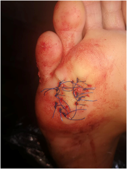
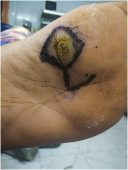
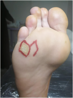
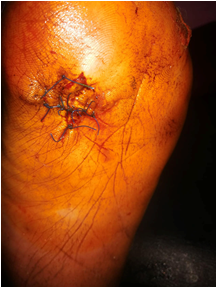
Postoperative results; table 2, Figure 1
The excision and rotation skin flap closure is better than the other surgical approaches and there is a statistically significant difference between both groups regarding occurrence of pain (p = 0.048), post-operative recurrence (p=0.033), superficial ischemia (p = 0.049) and presence of painful ugly scar (p = 0.023). Patients in the first group that was managed by excision and rotation skin flap closure are more clinically and cosmetically satisfied with the results than patients in the second group (p= 0.032).
The excision and rotation skin flap closure is better than the other surgical approaches and there is a statistically significant difference between both groups regarding occurrence of pain (p = 0.048), post-operative recurrence (p=0.033), superficial ischemia (p = 0.049) and presence of painful ugly scar (p = 0.023). Patients in the first group that was managed by excision and rotation skin flap closure are more clinically and cosmetically satisfied with the results than patients in the second group (p= 0.032).
| Surgical Procedure | P | |||||
| minor surgery (electrocautery) or debridement: N = 15 | excision and rotation skin flap closure N = 15 | |||||
| Age, Years* | Mean ± SD | 32.13 ± 9.51 | 33.47 ± 7.56 | 0.674 | ||
| N | % | N | % | |||
| Sex | M | 6 | 36.7% | 6 | 36.7% | 1 |
| F | 9 | 63.3% | 9 | 63.3% | ||
| Comorbid Condition | No | 14 | 80.0% | 13 | 66.7% | 0.409 |
| Yes | 1 | 20.0% | 2 | 33.3% | ||
Chi-square X2 test except (*) Independent T
Table 1: Demographic data data between both groups.
Table 1: Demographic data data between both groups.
| Surgical Procedure | P | |||||
| minor surgery (electrocautery) or debridement: N = 15 | excision and rotation skin flap closure N = 15 | |||||
| N | % | N | % | |||
| Complications | No | 11 | 73.3% | 15 | 100.0% | 0.032 |
| Yes | 4 | 26.7% | 0 | 0.0% | ||
| Pain | No | 13 | 86.7% | 15 | 100.0% | 0.048 |
| Yes | 2 | 13.3% | 0 | 0.0% | ||
| Recurrence | No | 13 | 86.7% | 15 | 100.0% | 0.033 |
| Yes | 2 | 13.3% | 0 | 0.0% | ||
| Superficial Ischemia | No | 13 | 86.7% | 15 | 100.0% | 0.049 |
| Yes | 2 | 13.3% | 0 | 0.0% | ||
| Painful Ugly Scar | No | 13 | 86.7% | 15 | 100.0% | 0.023 |
| Yes | 2 | 13.3% | 0 | 0.0% | ||
| Technical& Clinical Success (Complete Cure Without Complications | No | 2 | 13.3% | 0 | 0.0% | 0.039 |
| Yes | 13 | 86.7% | 15 | 100.0% | ||
| Patient Cosmotic Satisfaction | No | 4 | 26.7% | 0 | 0.0% | 0.032 |
| Yes | 11 | 73.3% | 15 | 100.0% | ||
Chi-square X2 test except (*) Independent T
Table 2: Comparison of both gropups regarding operative and post-operative results.
Table 2: Comparison of both gropups regarding operative and post-operative results.

Figure: 1D
Figure 1: excision and rotation skin flap for the treatment of plantar wart; (A) Large planter wart, (B) Romboid rotational flap was designed, (C)Excision of the wart and covering the raw area with flap, (D)Suturing skin only with 2/0 proline sutures
Figure 1: excision and rotation skin flap for the treatment of plantar wart; (A) Large planter wart, (B) Romboid rotational flap was designed, (C)Excision of the wart and covering the raw area with flap, (D)Suturing skin only with 2/0 proline sutures
Discussion
There several variable approaches of management of IPK as local excision, single- or double lobed flaps, electro-surgery and techniques of punch biopsy [6]. But it was found that there is no gold standard or definitive procedure for the most adequate line of management [1].In our study we highlighted that performing excision and rotation skin flap closure is better than the other surgical approaches and there is a statistically significant difference between groups regarding occurrence of pain, recurrence, presence of painful ugly scar and better patient satisfaction and our results were similar to results of Saipoor., et al. [1].
Moreover numbers of patients with full satisfaction and clinical recovery following such technique were similar to those reported byLopez and Kilmartin [9]. But, about half of patients that were included in Lopez and Kilmartin [9] were found to have recurrence as in contrast to the present study where it was found that 44.5% of patients suffered from recurrent lesions or callus formation, which could indicate that lesions with a viral origin might respond better to excision however the cohort was too small draw statistical inference.
Lopez and Kilmartin [9]. Discussed the efficacy of full thickness surgical excision and biopsy without wound closure in forty three patients. All biopsies were sent for histopathological examination and twenty two cases were confirmed as to be of viral origin which was near results of the current study. Satterfield and Jolly, [10] reported post-operative outcomes in 13 feet of 12 patients which underwent rotation skin flap following excision of painful plantar warts, and they reported plantar scarring in two patients (3 feet), inclusion cyst in two patients and a para-keratotic nodule in one patient. But, in our study no patients experienced recurrence of the lesion.
A prospective study which has been conducted by Chandorkar and Gaidole [11] and they have compared the outcomes of three treatment approaches in 90 patients over a period of two years. Each group was composed of 30 patients who were assigned to either wart excision with primary closure, wart excision then it was left to heal by secondary intention or by application of 40% salicylic acid ointment. Chandorkar and Gaidole, [11],found that after 6 months from warts excision with primary closure three patients had lesion recurrence. Moreover 5 patients have wart recurrence at 6 months in the excision which was left to heal by secondary intention.
Additionally, in the salicylic acid group 25 patients experienced complete cure, with only one patient was found to have recurrence after 6 months. Similar to our results it was found that performing the technique of rotation skin flap allows preservation of plantar skin sensation and it also could reduce risk of ischemia due to preservation of the epidermal venous junction [10]. Moreover such technique has a positive effect on healing following foot ulcer excision in diabetic patients [12]. Another advantage of such procedure is that it allows surgeons to add a second lobe to cover bigger defects when needed [6,14], such management might be needed to avoid increased skin tension if there are difficulties during closing the wound.
The fusiform or semi-elliptical excision is one of the alternative techniques which were used for repair of small defects or ulcers [15]. It is beneficial for smaller defects due to the increased risk of necrosis of wound edges which was caused by excessive skin tension when employed in larger defects [6]. But, the skin ellipse is reported as not being an appropriate approach for excision of plantar circular lesions [6,14] and elasticity [15]. Electro-cautery treatment has been reported by Winfield., et al. [16], to have 70% efficacy as a management strategy for plantar warts, with expected period of healing ranged from 6–8 weeks [4]. Winfield AL, Forster MSK [16], studied the effectiveness of electro surgery in treating plantar warts and they found 83% of patients were pleased with the outcome, 3% were dissatisfied and 13% give unsure results. Similarly, Bevans and Bosson [17], compared between electrosurgical excision and sharp debridement in the management of plantar warts and they found resolution of lesions. Both studies suggested that electro-surgery may be a good option for treatment of painful plantar warts when non-surgical treatments fail. However, it is not an appropriate technique if an underlying bony prominence or soft tissue abnormality is suspected [17]. Our approach avoids such drawbacks as it will be suitable for management of plantar warts in any location even if there is an underlying bony prominence or soft tissue abnormality is present.
Conclusion
Our study proved that lesion excision with insertion of a rotation skin flap might be an ideal treatment for painful intractable plantar wart. And also our results demonstrated that performing rotation skin flap is an effective and safe surgical procedure for the treatment of both IPK and chronic viral warts. However, further studies are needed to investigate the long-term outcomes of surgical treatment of intractable plantar keratosis.
Authors declared no conflict of interest
References
- Saipoor A., et al. “A retrospective audit of lesion excision and rotation skin flap for the treatment of intractable plantar keratosis”. The Foot 34 (2018): 23-27.
- Spink MJ., et al. “Distribution and correlates of plantar hyperkeratotic lesions in older people”. Journal of Foot and Ankle Research 2 (2009): 8.
- Freeman DB. “Corns and calluses resulting from mechanical hyperkeratosis”. American Family Physician 65.11 (2002): 2277-2280.
- Anderson JM., et al. “A small-scale study to determine the clinical effectiveness of electrosurgery in the treatment of chronic helomata (corns)”. The Foot 11.4 (2001): 189–98.
- Grouios G. “Corns and calluses in athletes’ feet: a cause for concern”. Foot 14.4 (2004): 175–84.
- Dockery G. Chapter 17—advancement and rotational flaps. Low Extremity Soft Tissue Cutan Plast Surg (2012): 177–93.
- Bouche RT., et al. “Unilobed and bilobed skin flaps. Detailed surgical technique for plantar lesions”. J Am Podiatr Med Assoc 85 (1995): 41–8.
- Tschoi M., et al. “Skin flaps”. Clin Plast Surg 32 (2005): 261–73.
- Lopez FM, Kilmartin TE. Corn cutting in the 21st century. Pod Now 19 (2016): 24–7.
- Satterfield VK and Jolly GP. “A new method of excision of painful plantar forefoot lesions using a rotation advancement flap”. J Foot Ankle Surg 33 (1994): 129–34.
- Chandorkar Shubha S and Gaidole Rachana. “To study and analyze various methods for treatment of planter corn”. Indian J Appl Res 5.2 (2015): 866–7.
- Merrill TJ., et al. “Relationship between smoking and plantar callus formation of the foot”. Pod Inst Updat (2012): 197–9.
- Boffeli TJ and Hyllengren SB. “Unilobed rotational flap for plantar hallux interphalangeal joint ulceration complicated by osteomyelitis”. J Foot Ankle Surg 54 (2015): 1166–71.
- Blume PA., et al. “The role of plastic surgery for soft tissue coverage of the diabetic foot and ankle”. Clin Podiatr Med Surg 31.1 (2014): 127–50.
- Chang TJ., et al. “Plastic repair techniques: skin plasties and local flaps”. Pod Inst Updat (1993): 52–65.
- Whinfield AL and Forster MSK. “Effect of electrodesiccation on pain intensity associated with chronic heloma durum”. Foot 7 (1997): 224–8.
- Bevans JS and Bosson G. “A comparison of electrosurgery and sharp debridement in the treatment of chronic neurovascular, neurofibrous and hard corns. A pragmatic randomised controlled trial”. Foot 20.1 (2010): 12–7.
Citation:
Loay M. Gertallah., et al. “The Value of Excision and Rotation Skin Flap for the Treatment of Plantar wart in Comparison to other Surgical Approaches”. Chronicle of Medicine and Surgery 3.1 (2019): 310-316.
Copyright: © 2019 Loay M. Gertallah., et al. This is an open-access article distributed under the terms of the Creative Commons Attribution License, which permits unrestricted use, distribution, and reproduction in any medium, provided the original author and source are credited.


 Scientia Ricerca is licensed and content of this site is available under a Creative Commons Attribution 4.0 International License.