Research Article
Volume 1 Issue 2 - 2018
Antidiabetic Potential of Aqueous Extract of Catharanthus Roseus
Department of Biochemistry, Adekunle Ajasin University, P.M.B. 001, Akungba Akoko, Ondo state, Nigeria
*Corresponding Author: Jamiyu Ayodeji Saliu, Department of Biochemistry, Adekunle Ajasin University, P.M.B. 001, Akungba Akoko, Ondo state, Nigeria.
Received: April 25, 2018; Published: May 11, 2018
Abstract
Aim: Catharanthus roseus, among many plants has been used from time immemorial in old folk lore medicine in many ancient countries such as India, China and Malaysia to treat diseases. This present study reports the in-vitro antioxidant and antidiabetic potential of this plant.
Material and Methods: The assays carried out were α-amylase, α –glucosidase inhibitory assays, total phenol, total flavonoids, ferric reducing antioxidant property FRAP, 2,2-diphenyl -1-picryl hydrazyl (DPPH), and iron (Fe2+) chelation for the determination of antioxidant and antidiabetic potential of this plant.
Results:The result revealed that α-amylase and α-glucosidase activities showed increase in the percentage inhibition in a dose-dependent manner. The aqueous extract of the plant also demonstrated high scavenging ability of DPPH, Fe2+-chelating ability, and inhibitory action against lipid peroxidation in pancreas.
Conclusion:Therefore, this plant may be promising therapy in the management of diabetes mellitus.
Keywords: Catharanthus roseus; Antidiabetic; Antioxidant; Phytochemicals
Introduction
Diabetes mellitus is one of the commonest chronic illnesses to human beings. It is characterized by deleterious hyperglycaemia and it is one of the leading diseases in the world (Shukla., et al. 2000). The World Health Organization (WHO 1999) estimates that by the year 2030, the number of people with diabetes would have reached 370 million. There is a high level of treatment failures and unpleasant side effectsassociated with oral anti diabetic drugs generating an urgent need and desire for alternative treatment by the use of plants based products are becoming popular in the management of diabetes.
Catharanthus roseus is an ornamental plant belonging to the family Apocynaceae. C. roseus is also called Madagascar periwinkle because it originated from Madagascar but it is widely distributed around the world countries including Nigeria due to its high survivability in different habitat. This plant grows 7 to 24 inches tall and wide, forming colorful flowers like white, pink, or purple on brittle stems. This plant has a long history as a folk medicine in many countries such as South Africa, China, India, Mexico (Patel., et al. 2012) and Malaysia (Ong., et al. 2011), where it is utilized as a remedy to alleviate diabetes complications.
Some phytochemical which have been known to exert the antioxidant and antidiabetic effect have been isolated in this plant. They are vindoline, vindolicine and vindolinine (Bisby., et al. 2007). Antioxidant activity was evaluated with the use of 2, 2-diphenyl-1-picrylhydrazyl (DPPH) and ferric reducing antioxidant power (FRAP) assays. The antidiabetic activity was carried out using α-glucosidase and α-amylase assays. Other assays such as the lipid peroxidation ability and the iron chelating ability were carried out.
Materials and Method
Chemicals
DPPH, Griess reagent [1% sulfanilamide, 0.1% of Naphthyl ethylene diaminedihydrochloride and 2% of Hydrogen phosphate(H3P04), Potassium dihydrogen Phosphate(KH2PO4)], 3, 5-dinitrosalicylic acid (DNSA), Porcine pancreatic alpha-amylase, Sodium potassium tartarate, sodium chloride, sodium hydroxide, starch, sodium dihydrogen phosphate, disodium hydrogen phosphate.
DPPH, Griess reagent [1% sulfanilamide, 0.1% of Naphthyl ethylene diaminedihydrochloride and 2% of Hydrogen phosphate(H3P04), Potassium dihydrogen Phosphate(KH2PO4)], 3, 5-dinitrosalicylic acid (DNSA), Porcine pancreatic alpha-amylase, Sodium potassium tartarate, sodium chloride, sodium hydroxide, starch, sodium dihydrogen phosphate, disodium hydrogen phosphate.
Collection of samples
Fresh samples of Catharanthus roseus were gotten from the Botanical garden at Adekunle Ajasin University, Akungba Akoko. These samples were identified in the Department of Plant Science and Biotechnology.
Fresh samples of Catharanthus roseus were gotten from the Botanical garden at Adekunle Ajasin University, Akungba Akoko. These samples were identified in the Department of Plant Science and Biotechnology.
Plant extraction
The samples were air-dried and blended after drying. 50g blended sample was poured into 1 litre distilled water for 48 hours and stirred at 4 hours interval. After 48 hours, it was decantated and the supernatant was concentrated using rotary evaporator, after which it was freeze-dried and kept in the refrigerator for further use.
The samples were air-dried and blended after drying. 50g blended sample was poured into 1 litre distilled water for 48 hours and stirred at 4 hours interval. After 48 hours, it was decantated and the supernatant was concentrated using rotary evaporator, after which it was freeze-dried and kept in the refrigerator for further use.
Pancrease homogenates preparation
The rat was dislocated in the cervical (neck) and dissected and the pancreas excised into normal saline and the weight was recorded. The tissue was subsequently homogenized in cold saline (1/10 w/v) up and down strokes at approximately 1200 rev/minutes in a Teflon glass homogenizer, the tissue was properly disrupted. The homogenates were centrifuged for 10 minutes at 4000 X g to yield a pellet that was discarded, and a low speed supernatant was kept for subsequent analysis of lipid peroxidation assay.
The rat was dislocated in the cervical (neck) and dissected and the pancreas excised into normal saline and the weight was recorded. The tissue was subsequently homogenized in cold saline (1/10 w/v) up and down strokes at approximately 1200 rev/minutes in a Teflon glass homogenizer, the tissue was properly disrupted. The homogenates were centrifuged for 10 minutes at 4000 X g to yield a pellet that was discarded, and a low speed supernatant was kept for subsequent analysis of lipid peroxidation assay.
Enzyme Inhibition Assays
α - amylase inhibition assay
The α-amylase inhibitory activity was expressed as percentage inhibition. 50 mls enzyme, 50-200 mls sample, 0-250mls of 0.02M Sodium phosphate buffer (pH 6.9) with 0.006M NaCl were incubated for 10mins at 25°C. 50 ml starch solution was added then incubated again for 5 minutes at 100-degree census. 200ml of dinitrosalicyclic acid (DNSA) was added and incubated again for 5minutes and 2 ml H20 added and read at 540 nm with spectrophotometer.
[(Absref–Abssample)/Absref] × 100
Where: Absref - Absorbance of the reference (reacting mixture without the test sample) and,
Abssample - Absorbance of reacting mixture with the test sample
Abssample - Absorbance of reacting mixture with the test sample
α - Glucosidase Inhibition Assay
The α-glucosidase inhibitory activity was expressed as percentage inhibition. 0.5mls of the enzyme, 0.15mls of glutathione (GSH), 50-200 mls sample was added into a test tube and incubated for 10 minutes at 30°C. 40mls 4-Nitrophenyl-beta-D-glucopy (PNPG) was added and incubated for 37°C for 20minutes, 1% of 2mls sodium carbonate was added and read with a spectrophotometer at 400nm.
[(Absref – Abssample)/ Absref] × 100
Where Absref-Absorbance of the reference (reacting mixture without the test sample) and, Abssample-Absorbance of reacting mixture with the test sample.
Determination of total flavonoid content
The total flavonoid content was determined using a slightly modified method reported by Meda., et al. (2005). Briefly 0.5ml of appropriate diluted sample was mixed with 0.5ml methanol, 50ul 10% AlCl3, 50μl 1M potassium acetate and 1.4ml water, and allowed to incubate at room temperature for 30minutes. The absorbance of the reaction mixture was measured at 415nm in UV-visible spectrophotometer, total flavonoid content of the extract was calculated with quercetin as standard.
Total phenol = (Abssam X Conc.std)/Absstd X Conc.sam
Determination of FRAP
The reducing property of aqueous extracts was determined by assessing the ability of the extract to reduce FeCl3 solution as described by Oyaizu (1986). A 2.5 ml aliquot was mixed with 2.5 ml of 200 mM sodium phosphate buffer (pH 6.6) and 2.5 ml of 1% potassium ferricyanide. The mixture was incubated at 50°C for 20 min and then 2.5 ml of 10% trichloroacetic acid was added and centrifuged at 3000 rpm for 10 minutes. The supernatant was mixed with an equal volume of water and 1 ml of 0.1% ferric chloride. The absorbance was measured at 700 nm and ferric reducing power was subsequently calculated using ascorbic acid equivalent.
The reducing property of aqueous extracts was determined by assessing the ability of the extract to reduce FeCl3 solution as described by Oyaizu (1986). A 2.5 ml aliquot was mixed with 2.5 ml of 200 mM sodium phosphate buffer (pH 6.6) and 2.5 ml of 1% potassium ferricyanide. The mixture was incubated at 50°C for 20 min and then 2.5 ml of 10% trichloroacetic acid was added and centrifuged at 3000 rpm for 10 minutes. The supernatant was mixed with an equal volume of water and 1 ml of 0.1% ferric chloride. The absorbance was measured at 700 nm and ferric reducing power was subsequently calculated using ascorbic acid equivalent.
DPPH Free Radical Scavenging Assay.
The free radical scavenging ability of the extracts against DPPH (2, 2-diphenyl-1- picryl hydrazyl) free radical was evaluated using a modified method described by Gyamfi., et al. 1999. Briefly, 3ml of the extract was mixed with 1 ml of the 0.1 mM methanolic solution of DPPH, the mixture was left in the dark for 30 min and the absorbance was taken at 516nm. The DPPH free radical scavenging ability was subsequently calculated.
The free radical scavenging ability of the extracts against DPPH (2, 2-diphenyl-1- picryl hydrazyl) free radical was evaluated using a modified method described by Gyamfi., et al. 1999. Briefly, 3ml of the extract was mixed with 1 ml of the 0.1 mM methanolic solution of DPPH, the mixture was left in the dark for 30 min and the absorbance was taken at 516nm. The DPPH free radical scavenging ability was subsequently calculated.
Fe2+ Chelation Assay
The ability of the extract to chelate Fe2+ was determined using a modified method described by Puntel (Puntel., et al. 2005). 150 μl of freshly prepared 500 μM FeSO4 was added to reaction mixture containing 168μl of 0.1M Tris-HCl (pH 7.4), 218 μl of 0.85% saline and the reconstituted extract (0-500 μl). The reaction mixture was subjected to incubation for 5 minutes before the addition of 13 μl of 0.25% 1, 10-phenanthroline (W/W). The absorbance was subsequently measured at 510nm and Fe2+ chelating ability was then calculated.
The ability of the extract to chelate Fe2+ was determined using a modified method described by Puntel (Puntel., et al. 2005). 150 μl of freshly prepared 500 μM FeSO4 was added to reaction mixture containing 168μl of 0.1M Tris-HCl (pH 7.4), 218 μl of 0.85% saline and the reconstituted extract (0-500 μl). The reaction mixture was subjected to incubation for 5 minutes before the addition of 13 μl of 0.25% 1, 10-phenanthroline (W/W). The absorbance was subsequently measured at 510nm and Fe2+ chelating ability was then calculated.
% Fe2+ chelating ability = (Absref - Abssamp)/Absref X 100
Results
Enzyme – Inhibition Assays
Alpha-Amylase Inhibition Assay
Alpha-Amylase Inhibition Assay
Total Phenol, Total Flavonoid and Ferric Reducing Antioxidant Property
The results for the total phenol, flavonoids content and ferric reducing antioxidant properties of the aqueous extracts of Catharanthus roseus are presented in the table below.
The results for the total phenol, flavonoids content and ferric reducing antioxidant properties of the aqueous extracts of Catharanthus roseus are presented in the table below.
| Total Phenol (mg/ml) | Flavonoid (mg/ml) | Frap (mg/ml) |
| 0.75 ± 0.007 | 1.29 ± 0.007 | 0.75 ± 0.042 |
Table 1:
Dpph Scavenging Activity
Fe2+ Chelation
Lipid Peroxidation
| Assays | IC50 (mg/ml) |
| Alpha-amylase | 0.0025 |
| Alpha-glucosidase | 0.0014 |
| Lipid peroxidation | 0.0130 |
| Iron chelation | 0.0027 |
| DPPH | 0.0181 |
Table 2: IC50 (mg/ml) of Catharanthus Roseus.
Discussion
Recent advances in understanding the activity of intestinal enzymes (α-amylase and α- glucosidase) which are important in carbohydrate digestion and glucose absorption have led to the development of newer pharmacological agents. A high postprandial glucose responses associated with complication in diabetes and is more strongly associated with the risk for cardiovascular disease than are fasting blood glucose (Mahesh, 2011).
Post prandial hyperglyceamia (PPHG) could induce the non-enzymatic glycosylation of various proteins resulting in the development of chronic complication. Thus inhibition of enzymes involved in the metabolism of carbohydrate such as α-amylase and α-glucosidase are important therapeutic approaches for reducing postprandial hyperglyceamia (Shim., et al. 2003).
α-amylase and α-glucosidase enzymes in the intestinal lumen in brush border membrane play major role in carbohydrate digestion to degrade starch and oligosachharide to monosaccharide before they can be absorbed. It was proposed that suppression of activity of such digestive enzymes would delay the degradation of starch and oligosaccharide which would in turn cause a decrease in the absorption of glucose on consequently the reduction of the postprandial blood glucose level elevation (Puls., et al. 1997).
α - amylase and α-glucosidase inhibitor disrupt the digestion of carbohydrate and slows down its absorption. They do these by inhibiting the enzymes (i.e. α-amylase and α-glucosidase) catalyzing carbohydrate degradation. Hence one of the therapeutic approaches for reducing postprandial blood glucose level in patient with diabetes mellitus is to prevent absorption of carbohydrate after food intake. Inhibition of these enzymes (α-amylase and α-glucosidase) reduces the high postprandial blood glucose peaks in diabetes (Conforti., et al. 2005). Acarbose is one of the synthetic amylase inhibitor that has been used therapeutically to control postprandial hyperglyceamia (Wikipedia encyclopedia, 2010). Acarbose is a complex oligosaccharide that delays the digestion of carbohydrate. It inhibits the action of pancreatic amylase in breakdown of starch. Synthetic inhibitor cause side effect such as abdominal pain diarrhea and soft feaces in the colon (Mahesh, 2011). So in order to overcome these, there is need to focus on the scientific exploration of herbal drugs having fewer side effects.
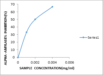
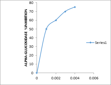
The ability of Catharanthus roseus extract to inhibit α-amylase and α-glucosidase activity in-vitro was investigated. The two enzymes inhibition assays showed increase in the percentage inhibition in a dose-dependent manner. In (figure 1), as the dose of the plant extract increased from (0.001 - 0.004) mg/ml, there was concomitant increase in the alpha-amylase inhibition from 50 to 66.6%. Similarly in (figure 2) as the dose of the plant extract increase from 0.001 to 0.004mg/ml, there was a resultant increase in the α-glucosidase inhibition from 33.3% to 50%. It is note-worthy that the level of enzyme inhibition of the plant extracts was due to the low IC50 which were 0.0025mg/ml for alpha-amylase and 0.0014mg/ml for alpha-glucosidase.
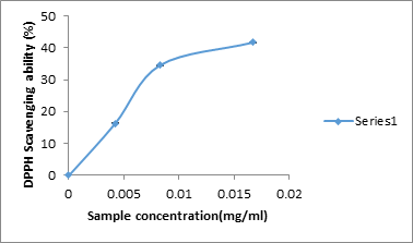
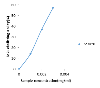
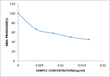
The DPPH scavenging activity of aqueous extract of Catharanthus roseus reveals that the scavenging activity of DPPH is dose dependent (0.0042 – 0.0167 mg/ml). The higher the dose the higher the plant extracts scavenge. The Fe2+ chelating activity of aqueous extract of Catharanthus roseus reveals that Fe2+ chelation is dose dependent (0.001 – 0.003 mg/ml). At dose 0.001mg/ml the chelating power was 14.3%. But respective dose increase to 0.003, the chelating power increased to 57% i.e. the higher the dose the higher the chelating power. The lipid peroxidation activity of aqueous extract of Catharanthus roseus reveals that peroxidation is dose dependent (0.001 –0.004 mg/ml). It reduces down the trend.
Conclusion
Catharanthus roseus is a promising plant that may be used in the management of diabetes mellitus because it shows positive results in all the assays conducted. However, further study is needed to isolate the active principle(s) in this plant which is responsible for its activity.
Acknowledgements
Nil
Nil
Competing interests
Authors have declared that no competing interests exist.
Authors have declared that no competing interests exist.
References
- Bisby RH., et al. “Effect of antioxidant oxidation potential in the oxygen radical absorption capacity (ORAC) assay”. Food Chemistry 108.3 (2008): 1002-1007.
- Conforti F., et al. “Biological and pharmaceutical Bulletin”. 28.6 (2005): 1098-1102.
- Gyamfi MA., et al. “Free-radical scavenging action of medicinal herbs from Ghana: Thonningia sanguinea on experimentally-induced liver injuries”. General Pharmacology 32.6 (1999): 661-667.
- Li WL., et al. “Natural medicines used in the traditional Chinese medical system for therapy of diabetes mellitus”. Journal of Ethnopharmacology 92.1 (2004): 1-21.
- Meda A., et al. “Determination of the total phenolic, flavonoid and proline contents in Burkina Faso honey, as well as their radical scavenging activity”. Food Chemistry 91.3 (2005): 571-577.
- Mahesh Bhanudas. “Investigation of in vitro α-amylase and α-Glucosidase inhibitory activity of polyherbal extract”. International Journal of Pharmaceutical Research 3.8 (2011): 97-103.
- Minotti G and Aust S. “An investigation into the mechanism of citrate- Fe2+ dependent lipid peroxidation”. Free Radical Biology and Medicine 3.6 (1987): 379-387.
- Ong HC., et al. “Traditional medicinal plants used by the temuan villagers in Kampung Tering, Negeri Sembilan, Malaysia”. Studies on Ethno-Medicine 5.3 (2011): 169-173.
- Oyaizo M. “Studies on products of browning reaction: ant oxidative activity of products of browning reaction prepared from glucosamine”. Japanese Journal of Nutrition 44 (1986): 307-315.
- Patel D.K., et al. “Natural medicines from plant source used for therapy of diabetes mellitus: An overview of its pharmacological aspects”. Asian Pacific Journal of Tropical Medicine 2.3 (2012): 239-250.
- Puls W., et al. “Inhibition of the rate of carbohydrate and lipid absorption by the intestine”. International Journal of Obesity 8.1 (1984): 181-190.
- Puntel RL., et al. “Krebs cycle intermediates modulate thiobarbituric reactive species (TBARS) production in Rat Brain In-viro”. Neurochemical Research 30.2 (2005): 225-235.
- Shim YJ., et al. “Inhibitory effect of aqeous extract from the gal of Rhuz chinesis on alpha-glucosidase activities and post prandia blood glucose”. Journal of Enthopharmacology 85.2 (2003): 283-287.
- Shukla R., et al. 14. “Medicinal plants for treatment of diabetes mellitus”. Indian Journal of Clinical Biochemistry 15.1 (2000): 169-177.
- Wikipedia the free encyclopedia (2010).
Citation:
Jamiyu Ayodeji Saliu., et al. “Antidiabetic Potential of Aqueous Extract of Catharanthus Roseus”. Archives of Endocrinology
and Diabetes Care 1.2 (2018): 81-87.
Copyright: © 2018 Jamiyu Ayodeji Saliu., et al. This is an open-access article distributed under the terms of the Creative Commons Attribution License, which permits unrestricted use, distribution, and reproduction in any medium, provided the original author and source are credited.


 Scientia Ricerca is licensed and content of this site is available under a Creative Commons Attribution 4.0 International License.