Research Article
Volume 1 Issue 3 - 2017
Anatomical Study of the Trachea of an Adult Donkey (Equus asinus): A Histomorphometric Study
1Department of Veterinary Anatomy, Usmanu Danfodiyo University, Sokoto
2Department of Veterinary Anatomy, Ahmadu Bello University Zaria
3Department of Theriogenology and Animal production, Usmanu Danfodiyo University, Sokoto
4Department of Microbiology, Faculty of Science, Usmanu Danfodiyo University, Sokoto
5Veterinary Teaching Hospital, Usmanu Danfodiyo University, Sokoto
*Corresponding Author: Dr. A Bello: Department of Veterinary Anatomy, Usmanu Danfodiyo University, Sokoto.
Received: July 19, 2017; Published: August 14, 2017
Abstract
The study aimed at investigating the histomorphometric characteristics of the trachea of donkey (Equus asinus) using standard morphometrical techniques. In this study three donkeys were used (two male and one female). The age of the donkeys was estimated using the knowledge of teeth eruption and wearing of milk and permanent teeth. The shape, location, size and relation of the trachea were observed. The length, width, diameter, volume and number of tracheal rings were also observed in this study. Histological characteristic of the trachea was studied in detail. Result shown that there was no obvious difference between the trachea of horse, sheep, goat, dog and camel histologically except in the location and size of the tracheal gland, basal cells and lymphatic tissue. Based on the above finding it was suggested that more research should be conducted using special staining technique and electron microscopy in order to finalize on the finding.
Keywords: Anatomy; Biometry; Donkey; Histology; Morphometry; Trachea
Introduction
Equine species (Equus asinus) are important domestic animals in the tropic and play a vital role in the society especially in the rural area in African countries. In northern Nigeria, donkeys are kept for transportation purposes and have a population of 41.5 million worldwide (Desalgn., et al. 2011). Nigeria is one of the countries with relatively large (800000) populations of donkeys (Mabayoje and Ademiluyu, 2004). Which are relatively concentrated in the northern state considering their climatic and vegetation influence, the Savannah type of vegetation and the fewer disease vectors such as tsetse fly (Rim, 1992). In the southern Nigeria, donkeys are used as a source of meat and about 16000 donkeys are transported annually from the northern state for this purpose (Atnesa, 1997; Blench, 2004).
Moreover, donkeys are used by Veterinary institute for practical model, for teaching gross anatomy to the undergraduate students (Blench, 2004). Donkeys originate in Nigeria started with the introduction of different breed through the Trans Sahara caravan trade across Nile to Sudan and Chad (Felding and Starkey, 2004). However, in 1970s, the population of donkey drastically reduced due to oil exploration and trade (Blench., et al. 1990).
Trachea is a walled cartilaginous tube extending from the coracoid cartilage of the larynx to the root of the lung where it bifurcates to form the right and left bronchia (Getty; 1975). The most distinctive feature of the trachea is the cartilage that forms U-shape. The trachea muscles are attached to the perichondrium on the external surface of the rings (Dallman and Brown 1984). The number of cartilage ring is not constant in all species and even within the same species (Dallman and Brown 1984).
The Pig has tracheal rings ranges from 32 to 35 and tracheal length of 15 to 20 cm (Dyce., et al. 2002). For example Goat has tracheal length of 230 to 257 mm tracheal rings from 35 to 57 (Byanet., et al. 2014). Tracheal morphometry is of significance because of the dimensions of the trachea from the conducting portion of the respiratory tract (Hyatt., et al. 1980).
Many factors affect the dimension of the trachea such as tracheal collapse (Dallman and Brown 1984). Disease like hyperplasia (Van and Pelt 1998) and tracheal collapse (Buback., et al. 1983-1993). Anatomical structure of trachea has been reported in horse, camel, dog and goat (Byanet., et al. 2014). The trachea is one of the components of respiratory system that aid in conducting air and respiratory functions as well.
Moreover, the entire trachea must be flexible to allow the movement of head, neck and larynx. This flexibility is possible due to the cartilages which form individual rings connected by fibro elastic ligaments (Ettinger and Feldman 2004). In histological terms; the trachea is composed of layers: mucosa, submucosa, layer of hyaline cartilage and adventitia. The mucosa consists of respiratory epithelia, lamina propria and elastic lamina. Respiratory epithelia is a pseudo stratified cylindrical ciliated and presents several cell types as calceiform cells (Ross., et al. 1993).
Although the anatomical structures of domestic animal are basically the same except for minor differences, there is still the need to study the various part of individual animal to note their normal and abnormal, similarities and difference, and more information on the choice of appropriate endotracheal tube, and tracheotomy tube for anaesthesia and emergency procedures.
Materials and Methods
Study Area
The study was conducted in Sokoto metropolis; the capital of Sokoto state of Nigeria. Sokoto state geographically was located on 12°15N and 05°E latitude, and 38m above sea level. The state has savannah vegetation of short grasses in the south and thorn in the north. Sokoto was bounded by Zamfara state to the east, Niger Republic to the north and Kebbi state to the west and southwest (Sokoto, 2001).
The study was conducted in Sokoto metropolis; the capital of Sokoto state of Nigeria. Sokoto state geographically was located on 12°15N and 05°E latitude, and 38m above sea level. The state has savannah vegetation of short grasses in the south and thorn in the north. Sokoto was bounded by Zamfara state to the east, Niger Republic to the north and Kebbi state to the west and southwest (Sokoto, 2001).
The state was ranked second in the nation livestock population with an estimated number of 34,532 horses, 51,388 donkeys, (Soko to 2003), 2 million cattle, 1.85 million sheep, 2 million goats, 0.8million camels, 4 million chickens and 3.2 million poultry (Buhari, 2008).
Animal Source
Tracheas from a three (3) apparently healthy adult donkeys were used in this study. The donkey’s lower respiratory tract were purchased from the Sokoto mami market at Dange-Shuni Local Government; and transported by road to the Gross Anatomy Laboratory of the Department of Veterinary Anatomy, Usmanu Danfodiyo University, Sokoto, where they are washed and used in gross anatomy unit.
Tracheas from a three (3) apparently healthy adult donkeys were used in this study. The donkey’s lower respiratory tract were purchased from the Sokoto mami market at Dange-Shuni Local Government; and transported by road to the Gross Anatomy Laboratory of the Department of Veterinary Anatomy, Usmanu Danfodiyo University, Sokoto, where they are washed and used in gross anatomy unit.
Sample Processing and Dissection
The animals were observed before slaughtering to clear respiratory problem. The lower respiratory tract was dissected out from the laryngeal region of the neck to the thoracic inlet up to the thoracic cavity. The whole trachea was then removed from the whole lower respiratory tract (trachea, bronchus, and lungs).
The animals were observed before slaughtering to clear respiratory problem. The lower respiratory tract was dissected out from the laryngeal region of the neck to the thoracic inlet up to the thoracic cavity. The whole trachea was then removed from the whole lower respiratory tract (trachea, bronchus, and lungs).
Gross Observation
The trachea samples were observed grossly for shape, size, and number of rings in relation to other species.
The trachea samples were observed grossly for shape, size, and number of rings in relation to other species.
Morphometry
The length of the trachea was measured by twine and meter rule from the cranial border of the first tracheal ring (larynx) to the bifurcation of tracheal rings as tracheal length (TL). The numbers of the tracheal rings (TR) were counted and observed. Measurement from the first tracheal ring to the last rings before thoracic inlet was considered as the cervical region (CVR) and from that point (thoracic inlet) to it bifurcate thoracic region (TR). The length from tracheal bifurcation to the hilus of the lungs was also measured as length of right bronchus (LR) and left bronchus (LB). The diameter of the trachea and bronchus in all the two parts (cervical and thoracic parts) were taken as distance from the two edges. The width of the trachea and bronchus was taken as the thickness of the tracheal wall in all two parts (cervical and thoracic parts) using a digitalized venire calliper. The volume of the trachea and bronchus were taken using Archimedes principle (water displacement technique).
The length of the trachea was measured by twine and meter rule from the cranial border of the first tracheal ring (larynx) to the bifurcation of tracheal rings as tracheal length (TL). The numbers of the tracheal rings (TR) were counted and observed. Measurement from the first tracheal ring to the last rings before thoracic inlet was considered as the cervical region (CVR) and from that point (thoracic inlet) to it bifurcate thoracic region (TR). The length from tracheal bifurcation to the hilus of the lungs was also measured as length of right bronchus (LR) and left bronchus (LB). The diameter of the trachea and bronchus in all the two parts (cervical and thoracic parts) were taken as distance from the two edges. The width of the trachea and bronchus was taken as the thickness of the tracheal wall in all two parts (cervical and thoracic parts) using a digitalized venire calliper. The volume of the trachea and bronchus were taken using Archimedes principle (water displacement technique).
Aging in Equine
From the knowledge of Jeffrey (1996) equine age determination was carried out using knowledge of eruption and wearing of milk teeth and permanent teeth. As well as other anatomical features such as increase in interdental space, change in shape of teeth and the dental star which appear in the central incisors at 10 years of age, intermediates at12 years and corners at 15 years.
From the knowledge of Jeffrey (1996) equine age determination was carried out using knowledge of eruption and wearing of milk teeth and permanent teeth. As well as other anatomical features such as increase in interdental space, change in shape of teeth and the dental star which appear in the central incisors at 10 years of age, intermediates at12 years and corners at 15 years.
Histological Techniques
Tissue samples were taken from the cervical, middle and thoracic regions of the trachea and were fixed in 10% neutral buffered formalin for 7 days. The tissue was then process as per method adapted by (Bello., et al. 2012). The paraffin blocks so formed were sectioned into 5 µm thickness and stained using haematoxylin and eosin (H&E) for proper morphological viewing (Bello., et al. 2014).
Tissue samples were taken from the cervical, middle and thoracic regions of the trachea and were fixed in 10% neutral buffered formalin for 7 days. The tissue was then process as per method adapted by (Bello., et al. 2012). The paraffin blocks so formed were sectioned into 5 µm thickness and stained using haematoxylin and eosin (H&E) for proper morphological viewing (Bello., et al. 2014).
Photomicrography
The sections were examined using a light microscope and micrographs were taken using a motic camera with 12.1 mega pixels. Data obtained were processed and presented in mean ± standard error of mean (mean ± S.E.M). Using Microsoft excel software.
The sections were examined using a light microscope and micrographs were taken using a motic camera with 12.1 mega pixels. Data obtained were processed and presented in mean ± standard error of mean (mean ± S.E.M). Using Microsoft excel software.
Results and Discussion
Gross Observation
From the three (3) samples used for the study, two were male and one female. There was no sex consideration while conducting the study.
From the three (3) samples used for the study, two were male and one female. There was no sex consideration while conducting the study.
The trachea was observed to extend from the coracoid cartilage of the larynx to the root of the lungs. It was located on the ventro-median aspect of the neck, with its distal part usually displaced to the right side (lateral deviation). This is in accordance with the reports of Smuts and Bezuindenhout, (1987); Dyce, (1995); Abdalla., et al. (1974); Salehi., et al. (2012); Bello., et al. (2012); Bello., et al. (2013).
Tracheal ring fusion with adjacent rings was observed within all different tracheal regions. Although, the number of the tracheal ring fusions was not consistent in the specimens examined, fusion of the tracheal rings occurred mostly in the cranial cervical region. It has been suggested that tracheal rings of this region are most affected by neck movements resulting in its fusion over time (Salehi., et al. 2012)
Grossly, the anatomical features of the trachea were observed to have prominent C- shape, tracheal cartilage; extensive dorsal trachealis muscle, covered with skeletal muscle and the colour was seen to be reddish in appearance. This finding is in accordance with the reports of Abdalla., et al. (1974); Salehi., et al. (2012); Bello., et al. (2012); Bello., et al. (2013). Studies have shown that, there was no cartilage overlapping in relation to the tracheal rings.
Grossly, the anatomical features of the trachea were observed to have prominent tube like appearance, cervical part is lengthier than the thoracic part, tracheal cartilage; extensive dorsal trachealis muscle, covered with skeletal muscle and the colour was seen to be reddish in appearance. There was no cartilage overlapping in relation to the tracheal rings observed. The trachea was located on a dorso-ventro lateral aspect of the neck.
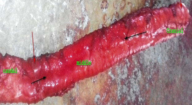
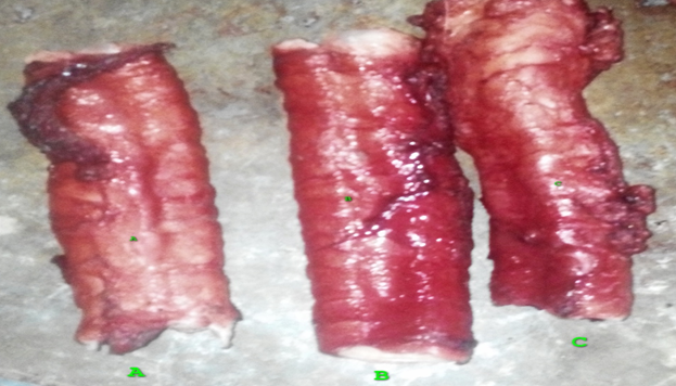
Figure 1: Tracheal tissue showing cranial, middle and caudal part of the trachea with tracheal ring (black arrow).
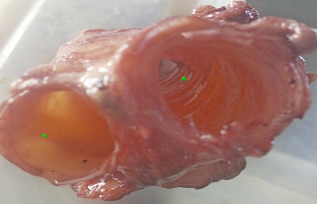
Figure 3: Tracheal tissue showing area of bifurcation attachment with the lungs
(A &B) with tracheal diameter (black arrow) and width (white arrow).
Biometric Observation
Biometrically, the weight, length, width and volume of the samples were observed and analyzed as mean ± S.E.M as shown in table 1. The tracheal cartilage were counted, observed and represented as mean ± S.E.M too as shown in table 1
Biometrically, the weight, length, width and volume of the samples were observed and analyzed as mean ± S.E.M as shown in table 1. The tracheal cartilage were counted, observed and represented as mean ± S.E.M too as shown in table 1
| Variables | Trachea | Bronchus Left Right | |
| Weight (g ± SEM) | 250.00 ± 5.00 | 55.45 ± 3.50 | 53.50 ± 2.50 |
| Length( mm ± SEM) | 205.00 ± 4.00 | 80.00 ± 3.00 | 75.00 ± 2.50 |
| Diameter (mm ± SEM) | 35.50 ± 3.50 | 2.80 ± 0.00 | 2.50 ± 0.50 |
| Width( mm ± SEM) | 3.50 ± 0.50 | 24.00 ± 2.50 | 23.00 ± 1.50 |
| Volume( cm3 ± SEM) | 20.50 ± 3.00 | 5.50 ± 0.50 | 5.00 ± 0.50 |
| Trachea rings | 55.00 ± 2.00 | ----------- | ------------- |
Table 1: The biometrics parameters of the trachea and bronchus of the donkeys.
The number of the tracheal rings varied from 52-60 with a mean value of 55.00 ± 2.00. Similar values were reported previously for the adult Indian camels (Kumar., et al. 1992). The variation in numbers of tracheal rings between specimens was due to individual anatomical variations. The average tracheal length from the first to the last tracheal ring was 87 ± 0.83. We are the first to report the tracheal length in donkey in this area.
Histological Observation
Histological observation of the organ revealed that the trachea appeared as a tubular organ with four (4) basic layers vie tunica mucosa, tunica sub mucosa, layer of hyaline cartilage and tunica adventitia as shown in figure 1-7. This is in accordance with the reports of Smuts and Bezuindenhout, (1987); Bello., et al. (2013a); Dyce, (1995); Abdalla., et al. (1974); Caccamo., et al. (2007) Salehi., et al. (2012); Bello., et al. (2012 a); Bello., et al. (2013 b).
Histological observation of the organ revealed that the trachea appeared as a tubular organ with four (4) basic layers vie tunica mucosa, tunica sub mucosa, layer of hyaline cartilage and tunica adventitia as shown in figure 1-7. This is in accordance with the reports of Smuts and Bezuindenhout, (1987); Bello., et al. (2013a); Dyce, (1995); Abdalla., et al. (1974); Caccamo., et al. (2007) Salehi., et al. (2012); Bello., et al. (2012 a); Bello., et al. (2013 b).
The tunica mucosa consist of three (3) basic layers viz lamina epithelial mucosa, lamina propria mucosa and lamina muscularis mucosa as shown in figure 1-4. Observation under light microscope showed that tracheal epithelium of donkey is ciliated pseudostratified columnar epithelium with goblet cells and basal cells as shown in figure 5-9. Similar features were also reported previously by Choi and Finkbeiner, (2000). The tracheal muscle was smooth and lied internal to the open end of the horseshoe-shaped hyaline cartilage as seen in other ruminants. It is noteworthy that tracheal muscle lies external to the cartilages in the carnivores (Nickel and Schumer, 1979). This is in accordance with the reports of Dyce, (1995); Abdalla., et al. (1974); Dabanoglu., et al. (2001); Bello., et al. (2016); Caccamo., et al. (2007) Salehi., et al. (2012); Bello., et al. (2012a); Bello., et al. (2013b).

Figure 4: Photomicrograph of adult donkey trachea showing Epithelium (Tmuc)
with well-developed cilia (Black arrow), Submucosa (Tsub) layer connective tissues
(Ct) with prominent large blood vessels (Red arrow), layer of hyaline cartilage
(Lhyl) with well-developed chondrocyte (Yellow arrow) and H&E x200.
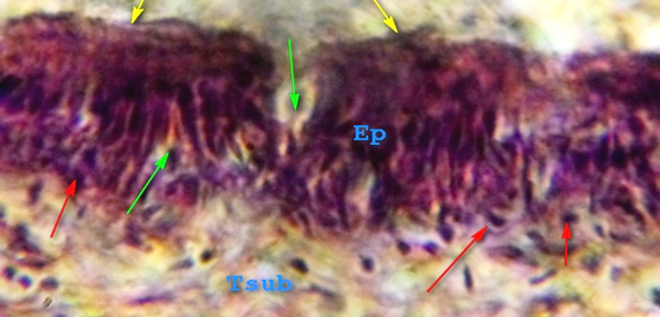
Figure 5: Photomicrograph of adult donkey trachea showing Epithelium (Ep)with
prominent cilia (Yellow arrow), functional goblet cells (green arrow), well arranged
basal cells (red arrow) and Submucosa layer (Tsub) H&E x250
In contrast, goblet cells in goat produce acidic mucosubstances, which is observed by Kahwa and Purton, (1996). The goblet cells showed appositive reaction toward PAS stain and revealed purple color due to mucopolysaccharide contents (Figure 4). Similar finding was observed by Raji and Naserpour, (2007). The mucous produced by goblet cell act as a protective barrier for the epithelium by lubricating, insulating and providing an appropriate condition for mucociliary clearance (Buchner and Maxwell, 1993). Lamina propria was loose connective tissue with prominent collagen and elastic fibers, blood vessels and lymphatic vessels (Figure 4). These features are similar to the histological features of cats and goats (William, 1990). The muscularis mucosa was very thin layer consist of few smooth muscle fibers (Figure 3) and such result comparable with the those found in cat Nasser (2012).
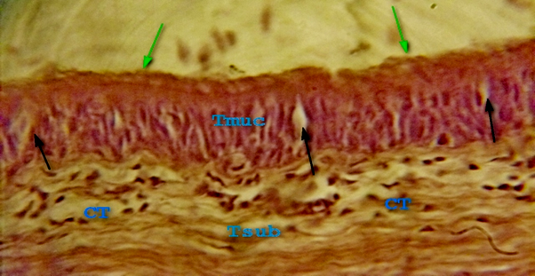
Figure 6: Photomicrograph of adult donkey trachea showing Epithelium (Tmuc)with
prominent cilia (green arrow), goblet cells (black arrow), Submucosa layer (Tsub) with
different connective tissues fibres (CT) and prominent blood vessels H&E x250.
The tunica submucosa appears as a layer of loose connective tissue contains different connective tissue cells, lymphocytes, monocytes, macrophage and plasma cells, blood vessels and the submucosal glands were very few in number, small in size and appeared as tubulo-acinar mucus type glands that were appositively reacted with PAS (Figure 4). The glands opened into the lumen of trachea by a slit shaped duct (Figure 4). Similar features were also reported previously by Choi and Finkbeiner, (2000).
The tracheal muscle was smooth and lied internal to the open end of the horseshoe-shaped hyaline cartilage as seen in other ruminants. It is noteworthy that tracheal muscle lies external to the cartilages in the carnivores (Nickel and Schumer, 1979) (Figure 4). The hyaline cartilage layer was surrounded by perichondriun with the dense fibroblastic tissue present between the cartilaginous rings, it contain the chondrocyte inside the lacuna within an amorphous matrix (Figure 2). The adventitia was consisted of connective tissue with numerous elastic fibers that are similar to cat (William, 1990).
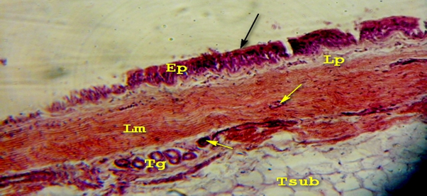
Figure 7: Photomicrograph of adult donkey trachea showing Epithelium (Ep)with prominent
cilia (black arrow), goblet cells, lamina propria mucosa (Lp) with different connective tissues
fibres, lamina muscularis mucosa (Lm) with well-developed blood vessels(Yellow arrow) and
well-arranged tracheal gland (Tg) with prominent blood vessels H&E x200.
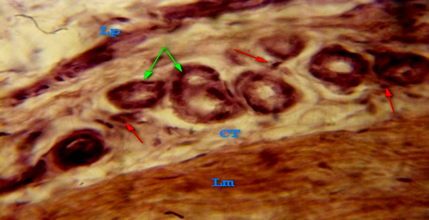
Figure 8: Photomicrograph of adult donkey trachea showing well arranged tracheal
gland (green arrow) lined by simple cuboidal epithelium with prominent blood vessels
(red arrow), lamina propria mucosa (Lp) with different connective tissues fibres (CT),
and lamina muscularis mucosa (Lm) H&E x250.
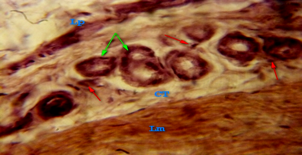
Figure 9: Photomicrograph of adult donkey trachea showing Epithelium (Ep)with prominent
cilia (green arrow), goblet cells, lamina propria mucosa (Lp) with different connective
tissues fibres (CT), lamina muscularis mucosa (Lm) with well developed blood vessels and
well arranged tracheal gland (black arrow) with prominent blood vessels H&E x200.
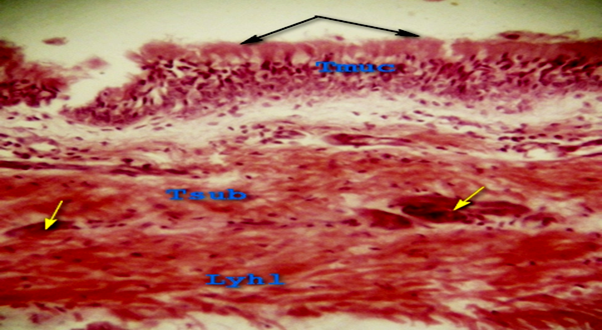
Figure 10: Photomicrograph of adult donkey trachea showing Epithelium (Tmuc)
with well-developed cilia (Black arrow), Submucosa (Tsub) layer connective
tissues with prominent large blood vessels, layer of hyaline cartilage (Lhyl) with
well-developed tracheal gland (Yellow arrow) H&E x200.
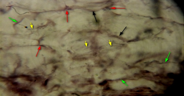
Figure 11: Photomicrograph of adult donkey trachea showing Submucosa layer (Tsub)
with different connective tissues fibres (CT); collagen (black arrow) and elastic (yellow
arrow), connective tissues cells (red arrow) and prominent blood vessels H&E x250.
The tunica sub mucosa consist of different connective tissues fibre (collagen, reticular and elastic connective tissue), with blood vessels and well-arranged tracheal glands figure lined by simple cuboidal epithelium with prominent blood vessels (4,5-6). This is in accordance with the reports of Smuts and Bezuindenhout, (1987); Dyce, (1995); Abdalla., et al. (1974); Caccamo., et al. (2007) Salehi., et al. (2012; Bello., et al. (2013a)); Bello., et al. (2012c).
The trachea submucosa layer with different connective tissues fibre, collagen (black arrow) and elastic connective tissue cell (figure 6-8) and prominent blood vessels. The of layer hyaline cartilage shown to have numerous chondrocyte, inner fibro elastic coat, outer fibro elastic coat with no blood vessels in all the section as shown in (fig 6-10). The hyaline cartilage layer was surrounded by perichondriun with the dense fibroblastic tissue present between the cartilaginous rings, it contain the chondrocytes inside the lacuna within an amorphous matrix. Within the inner fibro-elastic coat, observation showed numerous aggregate of lymphatic nodules as shown in figure 6 and 8. This is in accordance with the reports of Hyatt., et al. (1980); Dyce, (1995); Abdalla., et al. (1974); Caccamo., et al. (2007) Salehi., et al. (2012); Bello., et al. (2012c); Ibe., et al. (2011).
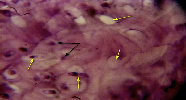
Figure 12: Photomicrograph of adult donkey trachea showing layer of hyaline cartilage (Lhyl) with well-developed connective tissues fibres; collagen (black arrow), chondrocyte within the lacunae (yellow arrow) and prominent blood vessels H&E x250.
From the result observed in this study, the shape, location, size, and relation of the trachea were observed. The length, width, diameter, volume and number of tracheal rings were also observed in this study. Histological characteristic of the trachea was studied in detail. Result shown that, there was no obvious difference between the trachea of horse, sheep, goat, dog and camel (Grossman., et al. 1953; Bello., et al. 2013a) histologically, except in the location and size of tracheal gland, basal cells and lymphatic tissue.
Conclusion and Recommendation
Based on the above finding, it was recommended that more research should be conducted using special staining technique and at ultra-structural level in order to finalize on the findings.
References
- Al Abasi., et al. “Anatomical and Histological Study on trachea and lung of one-humped camel in middle of Iraq”. M.Sc.Thesis Veterinary Medicine College, University of Baghdad (2001).
- Aspinall V., et al. “Textbook of introduction to Veterinary Anatomy and Physiology 2ndediton”. Butter worth Heiman ELSEVIER (2009): 79-96.
- ATNESA. “Improving donkey utilization and management”. Report of the International ATNESIA workshop 5-9th May (1997).
- Bello., et al. “The Prenatal Development of Thyroid Gland in one Humped Camel (Camelus dromedarius): Histomorphological Study”. JBR Journal of Clinical Diagnosis and Research2.1 (2014): 1-3.
- Bello A., et al. “Histomorphometric Study of the Prenatal Development of the Circumvallate Papillae of One-Humped Camel (Camelus Dromedarius)”. Anatomy and Physiology 4 (2014): 168.
- Bello A., et al. “Prenatal Development of Yankasa Sheep (Ovis aries) Kidney: A Histomorphometric Study”. Journal of Kidney 1.4 (2016): 126.
- Bello A., et al. “Gross Embryonic Diffrentiation of the Stomach of the One Humped Camel (Camelus dromedarius)”. Anatomy and Physiology 4.1 (2014): 131.
- Bello A., et al. “Histomorphological Studies of the Prestiatal Development of Oesophagus one hump camel (Camelus) dromedaries)”. Scientific journal of Agricultural 1.14 (2012): 100- 104.
- Bello A., et al. “A biometric study of the digestive tract of one-humped camel (Camelus dromedarius) foetus”. Science journal Zoology 1.1(2012): 11-16.
- Bello A., et al. “Prenatal Development of the Kidney of One-Humped Camel (Camelus dromedarius) – A Histomorphometric Study”. African Journal of Biomedical Research 16.1 (2013): 31-37.
- Bello A., et al. “Age estimation of camel in Nigeria using rostral dentition”. Scientific Journal of Animal Science 2.1 (2013): 34-40.
- Brown RE., et al. “The elephants Respiratory System: Adaptations to gravitational stress”. Respiration and Physiology109.2 (1997): 177-194.
- Buback JL., et al. “Surgical treatment of tracheal collapse in dogs: 90 cases”. Journal of American Veterinary Medical Association208.3 (1993):380-384.
- Caccamo R., et al. “Endoscopic. Bronchial Anatomy in the cat”.Journal of Feline Medicine & Surgery9.2 (2007):140-149.
- Dabanoglu I., et al. “A quantitative Study on Trachea of Dog”. Anatomy, Histology, Embryology 30.1 (2001): 57-59.
- Dabanoglu I., et al. “Quantitative Study on the Trachea of the Dog”. Anatomia, Histologia, Embryologia30.1 (2001): 57-59.
- Dallman MJ and Brown EM. “Statement Analysis of Selected Trachea Measurement in Normal Dogs and Dogs with Collapsed Trachea”. American Journal of Veterinary Research 49.5 (1984): 1033-1037.
- Desalegne A., et al. “Status of Parasitial in Donkey Project and Control Areas in Central Region of Ethiopia: A comparative Study”. Ethiopian Veterinary Journal15.2 (2011).
- Dyce KM., et al. “Textbook of Veterinary Anatomy Elsevier|Saunders, Philadelphia, P.a USA, 3rd edition”. (2002).
- Ethinger and Feldman. “Tratado de medicine Inter Veterinarian 3rd Edition”. Rio deFenaio G Guanabara Kaogan (2004): 21-56.
- Fielding D and Starkey P. “A Resources book of the Animal Traction Network for Eastern and Southern African (ATNESA)”. (2004): 92- 211.
- Getty R. ”The Anatomy of Domestic Animals 5th edition”. 1 and 2. W. B. Saunders Company (1973).
- Grossman JP and Sisson S. “The Respiratory System” In Anatomy of Domestic Animals”. W. B. Saunders Philadelphia, USA 4thedition (1953): 517-563.
- Hyatt RE., et al. “Prediction of maximal expiratory of local in excised human lungs”. Journal of applied physiology48.6(1980):991-998.
- Ibe CS., et al. “Anatomy of Lower Respiratory System of Africa giant Pouches rat (Cricetomysa anater louse 1840)”. International journal of Morphology 29.1 (2011): 27-33.
- Jeffrey D. “Horse Dentistry, the theory and Practice of Equine Dental Maintenance Norfolk, Nebraska”. Norfolk Primary Company (1996).
- Mabayoje Al and Ademiluyi VA. ”A Role on Animal Power and Donkey Utilization In Nigeria”. Animal traction Network for Eastern and Southern Africa (Atnes) (2004): 2-7.
- Perelman MI. “Surgery of Trachea”. MIR Publisher, Mucous, Edition 49.55 (1976): 97.
- Rim. “Nigeria National Livestock Resources Survey”. (V. Vola) Report by Resources Inventory and management Limited (RIM) to Federal Department Livestock and Pest Control Services (FDL & PCS) Abuja, Nigeria (1992).
- Sokoto BA. “The Study of Local Geography of Nigerian Secondary School”. First edition, Ministry of education Sokoto (2001): 104-110.
Citation:
Bello A., et al. “Anatomical Study of the Trachea of an Adult Donkey (Equus asinus): A Histomorphometric Study”. Multidisciplinary
Advances in Veterinary Science 1.3 (2017): 89-99.
Copyright: © 2017 Bello A., et al. This is an open-access article distributed under the terms of the Creative Commons Attribution License, which permits unrestricted use, distribution, and reproduction in any medium, provided the original author and source are credited.


 Scientia Ricerca is licensed and content of this site is available under a Creative Commons Attribution 4.0 International License.