Case Report
Volume 1 Issue 1 - 2016
Traumatic iliopsoas hematoma: A case report
Department of Orthopaedic Surgery, G. Gennimatas, Hospital, Thessaloniki, Greece
*Corresponding Author: NK Sferopoulos, Department of Orthopaedic Surgery, G. Gennimatas, Hospital, Thessaloniki, Greece.
Received: May 30, 2016; Published: June 10, 2016
Abstract
A 67-year-old man presented with a painful swelling of the right groin following a fall from a height a month ago. A palpable tender mass was felt in the right iliac fossa next to the iliac crest. There was a flexion contracture of the right hip, a restriction of the right hip joint movements and he was unable to bear weight on his right limb. Radiographs were not indicative of a fracture, but revealed right hip osteoarthritis. Further imaging examination excluded skeletal injuries and vascular disorders. It indicated a chronic hematoma in resolution in the area of the right iliopsoas muscle. Bed rest and non-weight bearing crutch walking for a month, followed by partial weight bearing for another month was recommended. Considerable improvement of the clinical and imaging findings of the hematoma was detected 3 months post-injury. Six months after injury, the hematoma had completely resolved. There was no recurrence within 12 months.
Introduction
Most reported cases of iliopsoas hematoma are associated with bleeding disorders more likely due to anticoagulant treatment. Iliopsoas hematoma following a traumatic injury is very rare. Most post-traumatic cases have previously been reported in young patients aged between 12 and 24 years and the vast majority resulted from a sports injury. Case reports of a traumatic iliopsoas hematoma referring to middle-aged or older adults, with no bleeding disorder, are scarce in the literature [1-3].
A post-traumatic hematoma of the iliopsoas muscle in a 67-year-old man that resolved following conservative management is presented. The current literature is reviewed indicating the importance of clinical awareness of this lesion, the difficulty of clinical differential diagnosis in older adult patients also suffering from osteoarthritis of the ipsilateral hip and the value of imaging evaluation to delineate the lesion and exclude an occult bone injury.
Case report
A 67-year-old man was admitted with complaints of a painful groin swelling on the right side that prevented him from bearing weight on the right limb. The patient reported a traumatic injury that involved a fall from his tractor a month ago. There was no evidence of fracture in the initial plain radiographs. He was treated with bed rest and ambulation with a walker to weight off his right limb. He was referred for further evaluation, since there was no improvement of his symptoms and signs. He reported morning stiffness and right groin pain following heavy agricultural activities of 2 years duration. He reported no significant medical history, such as hypertension, diabetes mellitus, vascular disease or peripheral neuropathy and clotting disorders. He was on no regular medication and also denied receiving any aspirin-based tablet or anticoagulant agents in the past.
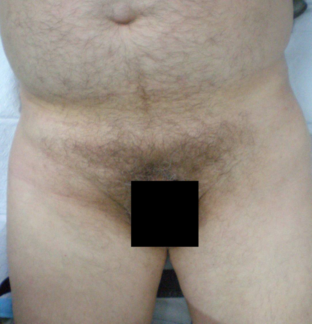
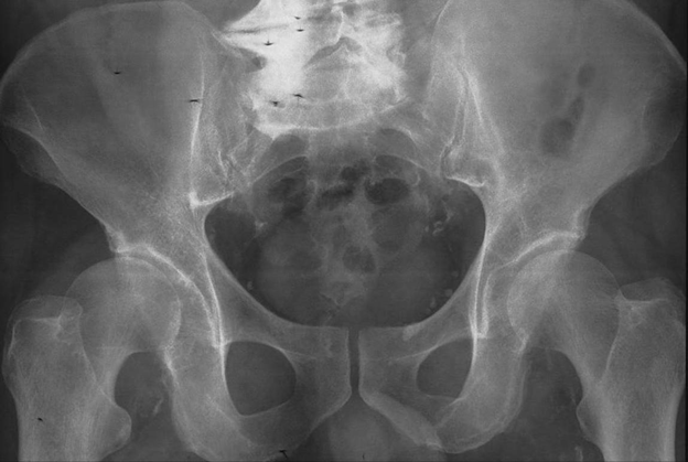
Physical examination revealed a painful right groin swelling Figure: 1. A palpable tender mass was felt in the right iliac fossa next to the iliac crest. There was a 20 degrees flexion contracture of the right hip and a diminished hip range of motion in 2 planes, including flexion and internal rotation. Pain was severe with weight bearing. When in bed the hip was held in lateral rotation. Surgical and genitourinary evaluation revealed no evidence of a hernia or any other disorder. Neurological examination revealed no abnormal findings. Manual muscle testing as well as pinprick and light touch sensation of the lower limbs were normal. Plain radiographs indicated findings of degenerative osteoarthritis of the hip, but no fracture line Figure: 2. Laboratory test results were within normal limits. The patient’s coagulation profile and platelet counts were normal.

Figure 1: Clinical presentation of a 67-year-old patient with a painful swelling of the right groin following a fall from a height a month ago.

Figure 2: Anteroposterior radiograph of the pelvis and hips reveals no evidence of a fracture line, but degenerative changes of the lumbar spine and of the right hip as well as extensive calcification of the femoral arteries.
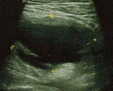
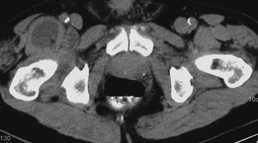
The clinical suspicion of an occult hip fracture necessitated further imaging of the patient. Imaging evaluation included ultrasonography, scintigraphy, computed tomography (CT) and magnetic resonance imaging (MRI). Ultrasonography showed no findings suggesting vascular disorders. Echogenic atherosclerotic plaques were evident on the arteries of the lower extremities. A fluid-signal, avascular muscle lesion in the area of the right iliopsoas muscle was diagnosed Figure: 3. A conventional 3-phase bone scintigraphy (300 MBq) with technetium-99m HDP indicated increased uptake of the soft tissues around the right hip joint in the early phase, as well as increased uptake of the upper femur consistent with degenerative arthritis or fracture. Contrast-enhanced CT of the abdomen revealed a soft tissue lesion consistent with a chronic hematoma in resolution, depicting fluid signal intensity and a slightly thick wall, in the area of the right iliopsoas muscle. There was no evidence of a fracture. There was also no relation of the lesion with the right femoral vessels Figure 4. A MRI of his pelvis and hips demonstrated the right iliopsoas muscle hematoma. No fracture or bone marrow edema was detected. No underlying vascular lesion was identified. The calculated size of the lesion in all imaging modalities was approximately 10.3 x 4.7 x 3.9 cm.

Figure 3: Ultrasonography indicates a fluid-signal, avascular muscle lesion of the right iliopsoas muscle.

Figure 4: Contrast-enhanced axial computed tomography of the abdomen shows the soft tissue lesion with fluid-signal density in the area of the right iliopsoas muscle.
It was considered best not to attempt percutaneous drainage. He was restricted from bearing weight on the affected limb when using crutches for a month, and was kept on partial weight bearing for another month. Local symptoms and signs showed notable improvement 3 months post-injury. A new MRI performed at 3 months after injury indicated considerable reduction in the size of the hematoma. Full weight bearing was then allowed. The local symptoms and signs that were due to the hematoma, including the palpable mass in the iliac fossa, pain on ambulation, flexion contracture and supine hip external rotation, were completely relieved after 6 months. Symptoms that were due to the right hip osteoarthritis, including morning stiffness, exertional hip pain and limited hip flexion and internal rotation range of motion, were still evident. There was no recurrence of the lesion within 12 months.

Figure 5: (a) Sagittal T1-weighted after gadolinium injection, coronal T2-weighted (b) and short tau inversion recovery (c) magnetic resonance images demonstrate a chronic hematoma of the right iliopsoas muscle.
Discussion
Intramuscular hematoma of the iliopsoas muscle may be either spontaneous or post-traumatic. The latter etiology is questioned, since a direct external force does not seem sufficient to produce a hematoma due to the deep, protected, intrapelvic location of the iliopsoas muscle [4]. Spontaneous lesions are usually seen in patients receiving anticoagulant agents [5-9], or suffering from clotting disorders and vascular malformations [4, 10-13], but may also be evident in otherwise healthy patients [14]. Post-traumatic lesions may be cases of minor or overuse trauma [15,16], indoors injuries [3], as well as following sport injuries [17-20].
Intramuscular hematomas of the iliopsoas muscle may be clinically evident as a painful swelling of the groin. The symptoms may be nonspecific like abdominal or flank pain radiating to the inguinal region, labia, or scrotum. Compression of the femoral nerve by the hematoma may be associated with neurologic findings including paresthesia or weakness of the thigh and leg. Paralysis of the quadriceps muscle due to compression of the femoral nerve may follow the increase of the intracompartmental pressure in the case of an intramuscular hematoma. It may be more likely associated with anticoagulant therapy induced hematomas than post-traumatic lesions [2,8, 21-29].
Imaging studies, including ultrasonography, CT and MRI are usually needed to delineate iliopsoas compartment injuries [30-32].
The lesion usually disappears by absorption without sequelae, so treatment is usually conservative, while in patients under anticoagulant therapy it focuses on the correction of the coagulation profile [14,33,34]. However, a few persist and can undergo organization into slow-growing lesions, mimicking neoplasm. They are described as chronic expanding hematomas [4,27]. Extensive calcification and ossification of the hematoma has also been reported [16]. Surgical treatment may be required in hemodynamically unstable patients [25,35] and in chronic encapsulated [4,8] or radiographically sclerotic lesions [16].
The patient presented in this report was an older adult who suffered a painful groin swelling following a traumatic injury. The differential diagnosis of such a lesion may be very wide to include soft tissue hematoma, hernias, vascular or genitourinary disorders, infectious or inflammatory processes, and neoplasms [28,29, 36-38]. The clinical detection of a tender mass in the mid-inguinal area or in the iliac fossa next to the iliac crest is usually diagnostic. The lesion may not be palpable in patients examined soon after injury because hematoma takes some time to reach its full size or due to tenderness and ‘guarding’ [39]. The symptoms and signs of a post-traumatic painful hip joint in the presented patient necessitated imaging evaluation to exclude an occult bone injury or other hip joint pathology. The right hip osteoarthritis further complicated the clinical differential diagnosis. Flexion contracture of the hip may appear as a consequence of both hip osteoarthrosis and iliacus hematoma. In the latter, other hip movements are present, if hip extension is avoided, which is the only clinical sign to distinguish iliacus hematoma from hemarthrosis or acute suppurative arthritis of the hip [39-41]. Further imaging clearly demonstrated the soft tissue lesion and excluded any bone pathology. Conservative treatment including rest and ambulation on crutches with no weight bearing on the involved side for a sufficient period led to an uncomplicated resorption of the hematoma.
Conflict of interest statement
The author certifies that he has no commercial associations (such as consultancies, stock ownership, equity interest, patent/licensing arrangements, etc) that might pose a conflict of interest in connection with the submitted article. The author received no financial support for this study.
The author certifies that he has no commercial associations (such as consultancies, stock ownership, equity interest, patent/licensing arrangements, etc) that might pose a conflict of interest in connection with the submitted article. The author received no financial support for this study.
References
- Guivarc'h M. “Hematoma of the iliac psoas muscle. 29 cases”. Journal de Chirurgie 134.9-10 (1997): 382-389.
- Murray IR., et al. “Femoral nerve palsy secondary to traumatic iliacus haematoma-a young athlete's injury”. BMJ Case Reports 22 (2010): pii: bcr0520103045.
- Smuczyński W., et al. “Pain caused by the iliopsoas muscle injury. Case study”. Medical and Biological Sciences 27.4 (2013): 59-61.
- Yamasaki T., et al. “Chronic expanding hematoma in the psoas muscle”. International Journal of Urology12.12 (2005): 1063-1065.
- Ozkan OF., et al. “Iliopsoas haematoma: a rare complication of warfarin therapy”. Journal of the College of Physicians and Surgeons 22.10 (2012): 673-674.
- Ho KJ., et al. “Psoas haematoma and femoral neuropathy associated with enoxaparin therapy”. International Journal of Clinical Practice 57.6 (2003): 553-554.
- Casoni P and Dalla Valle R. “Femoral neuropathy due to a spontaneous hematoma of the iliopsoas muscle during therapy with heparin-calcium”. Acta Biomed Ateneo Parmense65.5-6 (1994): 289-296.
- Yegen SF., et al. “Spontaneous iliopsoas haematoma presenting with groin pain under warfarin therapy”. Pain Physician 18.6 (2015): E1145-E1146.
- Sasson Z., et al. “Spontaneous iliopsoas hematoma in patients with unstable coronary syndromes receiving intravenous heparin in therapeutic doses”. Canadian Journal of Cardiology412.5 (1996): 490-494.
- Bauduceau O., et al. “Spontaneous hematoma of the iliac psoas muscle in chronic myeloid leukemia. A case report”. Annales de Médecine Interne 154.4 (2003): 183-186.
- Flipo RM., et al. “Spontaneous hematoma of the psoas in Gaucher's disease”. Revue de Médecine Interne 13.4 (1992): 293-295.
- Uncini A., et al. “Femoral neuropathy from iliac muscle hematoma induced by oral anticoagulation therapy. Report of three cases with CT demonstration”. Journal of Neurology 226.2 (1981): 137-141.
- Dauty M., et al. “Iliopsoas hematoma in patients with hemophilia: a single-center study”. Joint Bone Spine 74.2 (2007): 179-183.
- Marquardt G., et al. “Spontaneous haematoma of the iliac psoas muscle: a case report and review of the literature”. Archives of Orthopaedic and Trauma Surgery 122.2 (2002): 109-111.
- Giuliani G., et al.“CT scan and surgical treatment of traumatic iliacus hematoma with femoral neuropathy: case report”. Journal of Trauma 30.2 (1990): 229-231.
- Petropoulos A and Sferopoulos N. "Post traumatic myositis ossificans of the iliopsoas muscle. Apropos of a case with review of the literature". Rev Chir Orthop Reparatrice Appareil Mot 83.8 (1997):747-751.
- Maffulli N., et al. “Iliopsoas haematoma in an adolescent Taekwondo player”. Knee Surgery, Sports Traumatology, and Arthroscopy 3.4 (1996): 230-233.
- Morelli V and Smith V. “Groin injuries in athletes”. American Family Physician 64.8 (2001): 1405-1414.
- Mozes M., et al. “Iliopsoas injury in soccer players”. British Journal of Sports Medicine 19.3 (1985): 168-170.
- Chambers S., et al. “Iliacus haematoma causing femoral nerve palsy: an unusual trampolining injury”. BMJ Case Reports 27 (2015): pii: bcr2014208758.
- Niakan E., et al. “iliopsoas hematoma and femoral nerve compression”. American journal of Family Physician44.6 (1991): 2100-21002.
- Sreenivas VI., et al. “Femoral neuropathy secondary to anticoagulant therapy”. International Surgery 65.3 (1980): 279-281.
- Piazza I., et al. “Femoral nerve palsy secondary to anticoagulant induced iliacus hematoma. A case report”. International Angiology9.2 (1990): 125-126.
- Tamai K., et al. “Complete paralysis of the quadriceps muscle caused by traumatic iliacus hematoma: a case report”. Journal of Orthopaedic Science7.6 (2002): 713-716.
- Kumar S., et al. “Posttraumatic hematoma of iliacus muscle with paralysis of the femoral nerve”. Journal of Orthopaedic Trauma 6.1 (1992): 110-112.
- Berlusconi M and Capitani D. “Post-traumatic hematoma of the iliopsoas muscle with femoral nerve entrapment: description of a rare occurrence in a professional cyclist”. Italian journal of orthopaedics and traumatology 17.4 (1991): 563-566.
- Reid JD., et al. “Chronic expanding hematomas. A clinicopathologic entity”. The Journal of the American Medical Association 244.21 (1980): 2441-2442.
- LeBlanc KE., et al. “Inguinal Hernias: Diagnosis and Management”. American Family Physician15.87 (2012): 844-848.
- Apostolidis S., et al. “Groin swelling, the anatomic way out of abdominal haematomas: a case report and explicative literature review”. Acta chirurgica Belgica 108.2 (2008): 251-253.
- Shabshin N., et al. “MR imaging of iliopsoas musculotendinous injuries”. Magnetic Resonance Imaging Clinics of North America 13.4 (2005): 705-716.
- Lenchik L., et al. “CT of the iliopsoas compartment: value in differentiating tumor, abscess, and hematoma”. American Journal of Roentgenology162.1 (1994): 83-86.
- Zago G., et al. “Iliopsoas muscle hematoma during treatment with warfarin”. Arquivos Brasileiros de Cardiologia 94.1 (2010): 1-3.
- Fealy S and Paletta GA. “Femoral nerve palsy secondary to traumatic iliacus muscle hematoma: course after nonoperative manage- ment”. Journal of Trauma47.6 (1999): 1150-1152.
- Patel A., et al. “Non-operative management of femoral neuropathy secondary to a traumatic iliacus haematoma in an adolescent”. Journal of Bone and Joint Surgery 90.10 (2008): 1380-1381.
- Takami H., et al. “Traumatic rupture of iliacus muscle with femoral nerve paralysis”. Journal of trauma23.3 (1983): 253-254.
- Malietzis G., et al. “When a groin lump is more than a simple groin lump”. BMJ Case Reports 15 (2011) pii: bcr1220103607.
- Shadbolt CL., et al.“Imaging of Groin Masses: inguinal anatomy and pathologic conditions revisited”. RadioGraphics (2001): s261-271.
- Sandler MA., et al. “Inflammatory lesions of the groin: ultrasonic evaluation”. Radiology151.3 (1984): 747-750.
- Goodfellow J., et al. “Iliacus haematoma. A common complication of haemophilia”. Journal of Bone Joint Surgery 49.4 (1967): 748-756.
- Cibere J., et al. “Reliability of the hip examination in osteoarthritis: effect of standardization”. Arthritis and Rheumatology 59.3 (2008): 373-381.
- Sniegowski M., et al. “Roentgenometric assessment of spine shape in patients with a flexion contracture of hip as a consequence of hip osteoarthrosis”. Chir Narzadow Ruchu Ortop Pol76.5 (2011): 266-273.
Citation:
NK Sferopoulos. “Traumatic iliopsoas hematoma: A case report”. Orthopaedic Surgery and Traumatology 1.1 (2016): 1-6.
Copyright: © 2016 NK Sferopoulos. This is an open-access article distributed under the terms of the Creative Commons Attribution License, which permits unrestricted use, distribution, and reproduction in any medium, provided the original author and source are credited.


 Scientia Ricerca is licensed and content of this site is available under a Creative Commons Attribution 4.0 International License.