Case Report
Volume 2 Issue 1 - 2018
Tuberculosis of Sacral Spine: A Case Report
1Department of Orthopaedics and Trauma Surgery, Suri Seri Begawan Hospital, Kuala Belait, Brunei
2Department of Orthopaedics and Trauma Surgery, RIPAS Hospital, Bandar Seri Begawan, Brunei
2Department of Orthopaedics and Trauma Surgery, RIPAS Hospital, Bandar Seri Begawan, Brunei
*Corresponding Author: Pramod Devkota, Department of Orthopaedics and Trauma Surgery, Suri Seri Begawan Hospital, Kuala Belait, Brunei.
Received: January 29, 2018; Published: March 27, 2018
Abstract
Tuberculosis (TB) is one of the common public health problems in the developing world. Spinal tuberculosis accounts for 50% of all musculoskeletal tuberculosis and 95% of spinal TB presented on dorsolumbar junction. The tuberculosis of sacral spine is rare. We present a case of sacral spine tuberculosis which was presented with low back pain and swelling on the gluteal region. Computerized tomography (CT) of the sacral spine and histopathology confirmed the TB and treated with anti-tuberculosis drugs for 18 months.
Keywords: Sacral Spine; Spinal Tuberculosis; Low Back Pain
Introduction
The incidence of vertebral tuberculosis (TB) accounts for to all TB cases varied from 1% to 5% [1]. TB is one of the common musculoskeletal problems in the developing world, also increasing in the developed world. Spine is one of the common places for the TB incidence and about 95% incidence at dorsolumbar junction [2]. The incidence of sacral spine TB is extremely rare and only reported as cases in the literatures. [3-5]. We a case of sacral spine TB, which was managed successfully with anti-tuberculosis treatment (ATT)
Case Report
A 54 years old female was referred to Orthopaedic clinic for low back pain which she was having for the past three months. She gave no history of trauma, injury and a having Diabetes mellitus (DM), hypertension (HTN) medicine. Her physical examination was unremarkable and routine bloods, x-ray of pelvic, lumbosacral (LS) spine were done. X-rays were unremarkable and bloods showed slightly raised erythrocyte sedimentation rate (ESR), C-reactive protein (CRP). She was advised for physiotherapy and some analgesics prescribed.
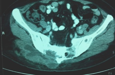
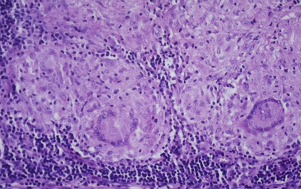
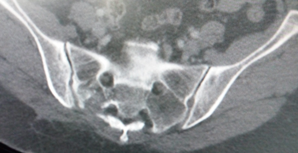
After one moth she was reviewed. Back pain was still present and she was having some swelling on right gluteal region. Ultrasound was done and suggestive of blood collection formation. Aspiration of the swelling done which showed the frank pus and computerized tomography (CT) of the pelvic done which showed suggestive of sacral vertebral osteomyelitis (Figure 1). CT guided biopsy done and confirmed the tuberculosis on histopathology report (Figure 2). She was referred to TB clinic and ATT was started. After 18 months of ATT, the CT showed (Figure 3) regeneration of the right sacral ala with much smaller lytic lesion. Clinically also her general condition improved a lot.

Figure 1: CT scan of sacral spine showing lytic lesion involving the body
and processes of 2nd and 3rd sacral vertebrae, suggestive of infective lesion.

Figure 2: Histopathology study of (HE x 40) showing the multiple
giant cells and inflammatory cells, confirming the TB.

Figure 3: After 18 months of ATT the CT scan shows the regeneration of the right
sacral ala with much smaller lytic lesion. Evidence of the healing of the TB lesion.
Discussion
Spinal TB is a common disease in the developing countries [6,7]. Dorsolumbar spine is the common place of TB Spine and the incidence on this place is about 95% and incidence on the cervical spine is 5% [8]. Sacral spine TB is rare and large series has not been reported in the literature. The diagnosis of the sacral spine TB is challenging for physicians, radiologists and spine surgeons. Clinical assessment of the patients should start with a careful history and a detailed physical examination. The clinical presentation can vary according to the age, health status, and site of infection, stage of the disease and the absence or presence of abscesses, sinus tracts or neurological status [5].
The common presenting symptoms of the sacral TB are non-specific pain and swelling. Sometime, the skeletal tuberculosis frequently mimics neoplasia like chordoma and osteoclastoma leading to incorrect initial diagnosis and delay in the institution of treatment [9]. CT and Magnetic Resonance Imaging (MRI) scans are the choice of radiological investigation. CT can reveal destructive lesions in the sacrum and the sacrum cortex may be diffusely destroyed secondary to the lesion expansion. And the MRI of the sacrum usually has a severe replacement of bone marrow, presenting with a low signal intensity mass on T1-weighted images patients and on T2-weighted images on spin echo sequences [10].
In this case, the patient presented with low back pain (LBP) and aspiration confirmed the cold abscess. The CT scan suggested the TB and the CT guided biopsy of the lesion proved the sacral spine TB. The patient was referred to the TB clinic for ATT. After 18 months of ATT the repeat CT of the sacral spine showed the healed lesion. The general condition of the patients also improved a lot. The sacral spine TB was managed successfully. Other literature also showed diagnosis by biopsy and successful treatment of the sacral spine TB with ATT [3,4,5 and 9]. Isolated sacral tuberculosis is rare but high degree of suspicion is needed in the presence of atypical clinical symptoms like LBP, gluteal region swelling and patient suffering from DM.
Conclusion
Isolated sacral tuberculosis is not common. Literature reported cases only. High degree of suspicion is needed in the presence of atypical clinical symptoms like swelling on the gluteal region, LBP with increased inflammatory markers.
References
- Fuentes Ferrer M., et al. “Tuberculosis of the spine. A systematic review of case series”. International Orthopaedics 36.2 (2012): 221-231.
- Coffman GJ. “Presentation of a rare sacral tuberculosis in an otherwise healthy patient: diagnostic challenge and review of treatment”. Journal of the Louisiana State Medical Society 164.2 (2012): 67-69.
- Wellons JC., et al. “Sacral tuberculosis: a case report and review of the literature”. Surgical Neurology International 61.2 (2004): 136-141.
- Oniankitan O., et al. “Sacrum Pott’s disease: A rare location of spine tuberculosis”. The Egyptian Rheumatologist 36.4 (2014): 209–211.
- Cavalcante RAC., et al. “Sacral Tuberculosis in the Elderly: An Unusual Cause of Refractory Low Back Pain”. Journal Brasileiro de Nefrologia 24 (2013): 335-339.
- Godlwana L., et al. “Incidence and profile of spinal tuberculosis in patients at the only public hospital admitting such patients in KwaZuluNatal”. Spinal Cord 46.5 (2008): 372-374.
- Oniankitan O., et al. “Spondylodiscitis at a hospital outpatient clinic in Lome, Togo”. Medecine Tropicale 69.5 (2009): 581-582.
- Maftah M., et al. “Pott’s disease about 320 cases”. Medecine du Maghreb 90 (2001):19–22.
- Shantanu K., et al. “Tuberculosis of sacrum mimicking as malignancy”. BMJ Case Reports 27 (2012): 2012.
- Patankar T., et al. “Imaging in isolated sacral Tuberculosis: a review of 15 cases”. Skeletal Radiology 29.7 (2000): 392-396.
Citation:
Pramod Devkota., et al. “Tuberculosis of Sacral Spine: A Case Report”. Orthopaedic Surgery and Traumatology 2.1 (2018):
286-289.
Copyright: © 2018 Pramod Devkota., et al. This is an open-access article distributed under the terms of the Creative Commons Attribution License, which permits unrestricted use, distribution, and reproduction in any medium, provided the original author and source are credited.


 Scientia Ricerca is licensed and content of this site is available under a Creative Commons Attribution 4.0 International License.