Case Report
Volume 2 Issue 3 - 2018
Malignant Transformation of Chronic Limb Wounds. Four Cases Reported
Department of Orthopedics Traumatology of the University Hospital of Treichville (CHUT), 05 BP 2836 Abidjan 05, Cote d’Ivoire
*Corresponding Author: Gogoua Dallo R, Department of Orthopedics Traumatology of the University Hospital of Treichville (CHUT),
05 BP 2836 Abidjan 05, Cote d’Ivoire.
Received: May 23, 2018; Published: July 07, 2018
Abstract
Introduction: Neoplastic transformation of chronic wounds was described by Marjolin J. N. in 1928, which later gave the name "Marjolin ulcer" to any secondary cancer arising from chronic ulceration. In this text, the authors described 4 cases of malignant transformation and list the epidemiological characteristics of these lesions. They also evoke the technique of early diagnosis that can avoid radical treatment to the patient.
Cases Report: 3 cases were from old scars and one from phagedenic ulcer. The characteristic of our cases was the long occurrence time preceding the transformation. In three cases this malignant transformation occurred in a period over15 years, which is the mean usual time according literature. One case appeared 30 years later. Then a common characteristic was the precarious socioeconomic origin of our patients.
Conclusion: These case reports show that old scars are possibly at risk of malignant transformation and any ulceration of such scar should be suspected and managed aggressively. It's the only way for the patients to receive a less mutilating treatment.
Keywords: Chronic wound; Marjolin ulcer; Spinocellular epithelioma
Introduction
Neoplastic transformation of chronic wounds was described by Marjolin J. N. in 1928, which later gave the name "Marjolin ulcer" to any secondary cancer arising from chronic ulceration. The development of these lesions is slow and the healing is ephemeral [1].
Their evolution is unpredictable [2]. The authors of this article report four cases of malignant transformation of chronic limb ulcers. The therapeutic treatment applied to those patients was the amputation of the limb concerned.
Case Reports
Case 1
A 64-year-old farmer was admitted to hospital in January 2003 for right leg absolute functional impotence. Examination showed on the antero-medial aspect of the leg, a large wound (8 X 7 cm) with a raised edge, which had been developing for about 15 years. That wound was the result of an insect bite. Its outcome was characterized by infection episodes. The patient's general condition was good but he was pale. The X-ray of the leg showed a fracture of the middle 1/3 of the fibula and particularly a total lysis of the diaphysis of the tibia (Figure 1).
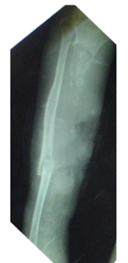
Figure 1: X-ray (diagonal view because of the patient’s height) of a total lysis of the
diaphysis of the tibia and of a Spinocellular epithelioma on a chronic leg wound.
In view of this result, we decided to offer an amputation and histopathologic exam. The preoperative work- up showed Anaemia with a haemoglobin of 7g/dl. After the correction of this Anaemia, an above-knee amputation was done with histopathology of the skin, muscle, and bone tissue as well as that of the ipsilateral inguinal lymph glands. The postoperative period was uneventful and the wound healed totally. Sutures were removed on the 16th postoperative day. Histopathology of the operative specimen showed a differentiated Spinocellular epithelioma.
Case 2
A 27-year-old woman from the rural area had chronic ulceration of the elbow on the scar of a thermal burn (hot water) which occurred in the patient's childhood when she was 5 years old. This wound healed completely. 22 years after, an ulceration appeared on the outer aspect of the elbow. This ulceration was due to a lesion following scratching. Different local treatment did not result in healing.
A 27-year-old woman from the rural area had chronic ulceration of the elbow on the scar of a thermal burn (hot water) which occurred in the patient's childhood when she was 5 years old. This wound healed completely. 22 years after, an ulceration appeared on the outer aspect of the elbow. This ulceration was due to a lesion following scratching. Different local treatment did not result in healing.
That wound was bleeding at the least contact and gave off a foul smell. It was purulent in places. The patient's general condition was good. The X-rays of the arm and the right forearm showed lysis of the top extremity of the ulna and the radius associated with infiltration of soft tissues. Biopsy with histopathologic examination was done. It showed the diagnosis of a differentiated Spinocellular epithelioma. Then an above-elbow amputation was performed.
Case 3
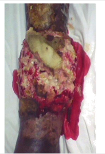
A 29-year-old man from the rural area had an old scar on the right leg for 3 years. This wound was due to a thermal burn. The ulceration of that old scar was the result of a fall which happened 6 months before presentation. The wound did not heal and bled at the least contact and gave off a foul smell. It was purulent in places (Figure 2). The patient's general condition was good. A biopsy with histological analysis confirmed the diagnosis of differentiated Spinocellular epithelioma (Figure 3). The treatment consisted in an amputation and the immediate and secondary postoperative outcome was simple.
A 29-year-old man from the rural area had an old scar on the right leg for 3 years. This wound was due to a thermal burn. The ulceration of that old scar was the result of a fall which happened 6 months before presentation. The wound did not heal and bled at the least contact and gave off a foul smell. It was purulent in places (Figure 2). The patient's general condition was good. A biopsy with histological analysis confirmed the diagnosis of differentiated Spinocellular epithelioma (Figure 3). The treatment consisted in an amputation and the immediate and secondary postoperative outcome was simple.
Case 4
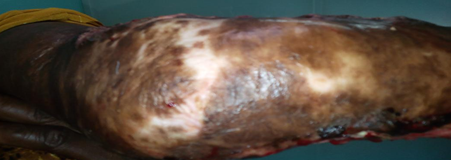
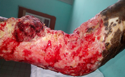
A 39-year-old woman from a modest socio-economic condition, with no particular medical history, presented at 8 years of age a thermal burn by boiling water of the right elbow (3rd degree). This wound required a pellet graft was completely healed (Figure 4). 31 years later appeared persistent itching causing scratching lesions. This persistent itching was complicated by massive skin, ulcerations, budding, hemorrhagic at the slightest contact (Figure 5) requiring a biopsy and a histological analysis of the operative specimen. Histological examination confirmed the diagnosis of a differentiated Spinocellular epithelioma. The treatment consisted in an amputation and the immediate and secondary post-operative outcome was simple.
A 39-year-old woman from a modest socio-economic condition, with no particular medical history, presented at 8 years of age a thermal burn by boiling water of the right elbow (3rd degree). This wound required a pellet graft was completely healed (Figure 4). 31 years later appeared persistent itching causing scratching lesions. This persistent itching was complicated by massive skin, ulcerations, budding, hemorrhagic at the slightest contact (Figure 5) requiring a biopsy and a histological analysis of the operative specimen. Histological examination confirmed the diagnosis of a differentiated Spinocellular epithelioma. The treatment consisted in an amputation and the immediate and secondary post-operative outcome was simple.
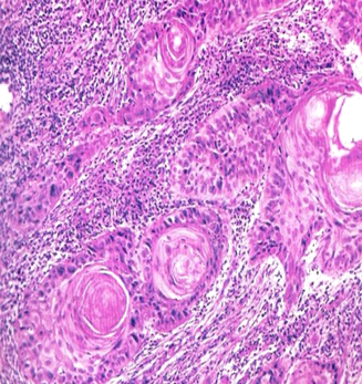
Figure 5: Spinocellular epitelioma H&E stain magnification
X10. Invasive malignant tumor proliferation of malphigian
cells, some of which surround horny globes. Cytonuclear
abnormalities exist. The stroma is inflammatory.
Discussion
These 4 cases reports concerned chronic limb wounds. They had three common characteristics: malignant transformation; the long time between the initial wound and this malignant transformation and the precarious social condition of the patients. The outcome of these chronic wounds is known since Marjolin's pieces of work. The proportion of transformation risk would be in the region of 0.33% according to Barrière [3] and Maréchal [4].
Seventy to 99% of the cases are differentiated epidermoid- Spinocellular epithelioma [1-3]. Rarely is it transformed into fusocellular carcinoma or Spinocellular epithelioma with malignant fibrohystiocytoma as reported by Touré [2]. Transformation into basal cell carcinoma has been described; but Walden [5] and other authors think that this basal cell carcinoma would be a primitive carcinoma. In our case reports, all the transformations were differential Spinocellular epithelioma.
The second characteristic of our cases was the long occurrence time preceding the transformation. In three cases this malignant transformation occurred in a period over15 years, which is the mean usual time according literature [2-4]. One case appeared 30 years later. The third common characteristic was the precarious socioeconomic origin of our patients. This notion is confirmed by the data of the literature [2]. A Spinocellular epithelioma can then occur on a healed wound, whether that healing is natural or the result of a skin grafting [6].
The prognosis of this cancer is alarming because of the local muscular and bone invasion as well as the metastatic diffusion. This metastatic diffusion could appear in the proportion of 50% according to Robinson [7]. The physiopathology of this malignant transformation is not yet totally elucidated and some hypothesizes are put forward about their genesis: they are of infectious, traumatic or immunologic nature. These hypothesizes impose a rigorous good and continuous surveillance of any chronic wound or of any skin scar.
The treatment of epithelioma is surgical in the first intention [2,4]. The diagnosis must be made early to make this treatment less mutilating. This is the reason why Moh's technique is very important. This technique consists of performing regular biopsies and histologic examinations. This allows the detection malignant transformation of wounds early.
Conclusion
These case reports show that old scars are possibly at risk of malignant transformation and any ulceration of such scar should be suspected and managed aggressively. It's the only way for the patients to receive a less mutilating treatment.
References
- Marjolin JN. “Ulcer”. Medical dictionary 21. 1828 : 31-50.
- Touré M., et al. “Malignant transformation of Phagedenic ulcers of the leg. A study of 473 cases”. Masson éd., Chirurgie, Paris (1988): 103-110.
- Barrière H., et al. “Carcinomatous transformation of chronic ulcers of the leg. 9 cases reported”. Annales de Dermatologie et de Vénéréologie 104 (1977): 6-7.
- Maréchal V., et al. “Carcinomatous transformation of chronic ulcers of the leg. 9 cases reported”. Annales Médicales de Nancy et de lorraine 38 (1999): 239-241.
- Walden V and Black M. “Basal cell carcinomatous changes on the lower leg: an association with chronic venous stasis”. Histopathology 7.2 (1981): 219-227.
- Voisard JJ., et al. “Leg ulcers and cancer. 6 Cases reported”. Journal of vascular diseases 26.2 (2001): 85-91.
- Robinson DC and Hay RJ. “Tropical ulcer in Zambia”. Transactions of the Royal Society of Tropical Medicine and Hygiene 80.1 (1986): 132-137.
Citation:
Gogoua Dallo R., et al. “Malignant Transformation of Chronic Limb Wounds. Four Cases Reported”. Orthopaedic Surgery and
Traumatology 2.3 (2018): 338-342.
Copyright: © 2018 Gogoua Dallo R., et al. This is an open-access article distributed under the terms of the Creative Commons Attribution License, which permits unrestricted use, distribution, and reproduction in any medium, provided the original author and source are credited.


 Scientia Ricerca is licensed and content of this site is available under a Creative Commons Attribution 4.0 International License.