Case Report
Volume 2 Issue 4 - 2018
Two Simultaneous Stress Fractures in Pubic Branches in Female Military: A Case Study
1Centro Universitario Lusiada/ FCMS - Unilus
2Universidade Metropolitana de Santos - Unimes
3Universidade Federal de São Paulo - Unifesp
4Universidade Federal do Estado do Rio de Janeiro - UniRio
5Pontifícia Universidade Católica do Paraná - PUCPR
6Hospital Santa Casa de Santos – Orthopedics and Traumatology Department
2Universidade Metropolitana de Santos - Unimes
3Universidade Federal de São Paulo - Unifesp
4Universidade Federal do Estado do Rio de Janeiro - UniRio
5Pontifícia Universidade Católica do Paraná - PUCPR
6Hospital Santa Casa de Santos – Orthopedics and Traumatology Department
*Corresponding Author: Áquilla dos Anjos Couto, Medicine resident at the Hospital of the University Federal in São Paulo, Napoleão de Barros St., 715 - Vila Clementino, São Paulo city; São Paulo state; CEP: 04024-002, Brazil.
Received: July 26, 2018; Published: August 08, 2018
Abstract
Stress fracture can be associated with physical exercise overload with inefficient recovery time. The main group at risk are military, dancers and runners. The presence of injury leads to withdrawal from regular activities, absenteeism of physical activities, complementary examinations and drug treatment. In this report will be disclosed the case of a 22-year-old female military without previous diseases, who developed two simultaneous pelvic fractures in the ischio-pubic branches.
Keywords: Stress fracture; Military training; Prevention; Pelvis fracture; Military
Introduction
Stress fracture is based on the triad: repetitive movements, exercise overload and inadequate recovery time [1]. The groups considered at risk are race athletes, dancers, cyclists and military [1-3]. The most affected bones are the tibia, fibula, metatarsal, femoral neck and pelvis [4], which shows lower incidence than the others [4]. Women are the most affected group in which etiology may be related to hormonal changes due to the high quantity and intensity of the training [5,6].
It can be divided into two main groups: (I) Fatigue resulting from abnormal stress applied to an apparently normal bone (i.e. this military case report); (II)insufficiency of abnormal muscular force in addition to bone with poor mass composition [7].
The treatment is based on the removal of the causative agent, reduction of new lesions and recovery of previously affected areas [8] and gradual return to exercising after any local pain episode [22]. We present a case report of a female military with two simultaneous fractures due to pelvic stress.
Case Report
A 22-year-old white female patient was admitted to military training in January 2017. She seeked for medical advice after a month of training, complaining of pain in the proximal right thigh, medial face, which started insidiously without correlating to an event and no previous history of local trauma. The patient did not present previous musculoskeletal lesions (according to her military admission examination). The patient also denied: the use of medication, smoking or alcoholic habits, systemic arterial hypertension, and diabetes mellitus. The patient reported the use of combined oral contraceptive for 2 years and absence of sexual activity in the last 6 months.
During examination, pain was reported on the palpation of the right thigh adductors and on the adduction of the limb against resistance pressure, thus being initially diagnosed as tendonitis of adductors. The patient was medicated with analgesia, and radiography was scheduled. Two days later, the patient seeked for emergency services referring to worsening of the pain in the region of the proximal insertion of the adductors of the right thigh during training, progressing towards incapacitation of movements.
During physical examination, pain was identified through palpation in the tendon region of the adductors and, when performing passive, active adduction and left thigh resistance test, the patient reported increased pain. Further to that, bilateral Grava test was also positive. The radiograph results showed fracture in the ischium-pubic branch with the presence of bone callus. Based on physical exams and image diagnosis, provided treatment was interruption of physical exercises for 15 days, use of non-steroidal anti-inflammatory drugs (NSAIDs) combined with four weeks of physiotherapy. After the treatment period, the patient showed clinical improvement and returned to sports activities without restrictions.
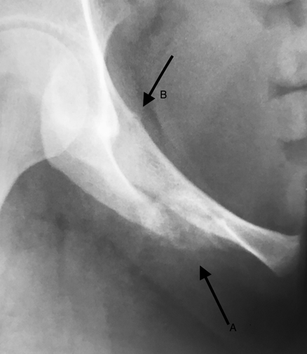
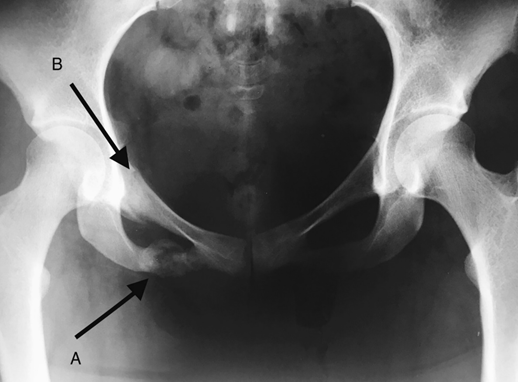
After two months of regular activities, the patient returned to the emergency department with similar complaints to previous situation. Physical examination was repeated and suggested hypothesis of a new fracture. A new radiography was performed and presented a hypodense line 01 cm proximal to the inferior branch bone callus and a hypodense line in the upper pubic branch, confirming a new fracture.

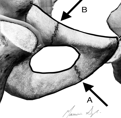
Image 1: Graphic representation of both fractures of the case report.
A – Medial fracture of inferior pubic branch (most common fracture among pelvic stress fractures).
B – Uncommon site of superior pubic branch.
A – Medial fracture of inferior pubic branch (most common fracture among pelvic stress fractures).
B – Uncommon site of superior pubic branch.

Image 2: “In let view” incidence.
A – Inferior pubic branch fracture.
B- Superior pubic branch fracture.
A – Inferior pubic branch fracture.
B- Superior pubic branch fracture.

Image 3: Radiography in pelvic AP.
A- Inferior pubic branch fracture.
B- Superior pubic branch fracture.
A- Inferior pubic branch fracture.
B- Superior pubic branch fracture.
Discussion
The importance of this report is based on the unusual presentation of two simultaneous stress fractures in pubic branches. It also highlights the need of including this diagnostic hypothesis in cases of pain in the groin area and thigh of athletes, especially military soldiers. It is estimated that women, as showed in this report, represent 16% of the Brazilian Air Force's personnel [9].
The incidence of stress fracture in extremities during military training is around 0.8%-6.9% for men and 3.4%-21.0% for women [11]. In military recruits it is 18 times higher than cases reported in veteran military [12]. A study conducted in India by Dash., et al. 2012 showed an incidence of around 10% in military training centers [13]. In the pelvis, this presentation is uncommon and represents 1.6 to 7.1% of all of stress fractures [14].
Similar to that, as described by Cosman., et al. 2013 [3], the diagnosis of the current case was delayed due to vague clinical manifestations. Initially there were no radiological signs as the appearance of these occurs in average of four weeks [1,3]. The magnetic resonance imaging (MRI) could have demonstrated the lesion at its initial stage. However the high cost and non-availability in various health care units of such diagnostic tool can be a barrier to the large population scale [15].
Stress fractures are generally seen in the medial portion or at the junction between the lower pubic branch and the ischium [16] (Image 1). They may occur due to tension in the adductor muscle [6], insufficient muscular training of overload exercises, characteristic of military training, which leads to bone overload [17]. As previously mentioned, we emphasize that females have a higher prevalence of such fracture and may be correlated with hormonal and metabolic alterations [18].
The combination of hormonal changes and training overload suggest imbalance between bone production and resorption, enabling calcium removal from the bone and leading to bone fracture [1]. Among extrinsic risk factors the association between the low level of physical conditioning and the large volume of training, as well as the inadequate rehabilitation time of previous injuries are the most frequent ones [20].
Early diagnosis is fundamental for prognosis and efficient treatment [19]. The literature emphasizes symptom control in young athletes diagnosed before three weeks of symptoms presentation. Athletes with early diagnoses return to activities in 10.4 weeks as opposed to late diagnoses with a return period of 18.4 weeks [19].
Conclusion
The prognosis and treatment of stress fracture is established according to some variables: duration of symptoms, fracture site, risk and pseudoarthrosis [21]. The main treatment is developed with prevention of new episodes and recovery of the injured area. The main treatment is based on prevention of new episodes and recovery of affected area. Rehabilitation time may require from 8 to 12 weeks [8] and clinical follow up is necessary to prevent functional loss and new fractures.
References
- Astur DC., et al. "Stress fractures: definition, diagnosis and treatment". Revista brasileira de ortopedia 51.1 (2016): 3-10.
- Kucera KL., et al. "Association of injury history and incident injury in cadet basic military training". Medicine and science in sports and exercise 48.6 (2016): 1053.
- Cosman F., et al. "Determinants of stress fracture risk in United States Military Academy cadets". Bone 55.2 (2013): 359-366.
- De Souza Laurino CF. “Atualização Em Ortopedia E Traumatologia Do Esporte”.
- Castelo-Branco C., et al. Menstrual history as a determinant of current bone density in young hirsute women. Metabolism-Clinical and Experimental 45.4 (1996): 515-518.
- Moreira CA and Bilezikian JP. "Stress fractures: concepts and therapeutics". The Journal of Clinical Endocrinology & Metabolism 102.2 (2016): 525-534.
- Talbot J., et al. "Femoral neck stress fractures in military personnel–a case series". Journal of the Royal Army Medical Corps 154.1 (2008): 47-50.
- Raasch WG and Hergan DJ. "Treatment of stress fractures: the fundamentals". Clinics in Sports Medicine 25.1 (2006): 29-36.
- Santos MML and Rocha-Coutinho ML. "Mulheres Na Força Aérea Brasileira: um estudo sobre as primeiras oficiais aviadoras". Estudos de Psicologia 15.3 (2010):259-267.
- Alessi A., et al. "I Brazilian position paper on prehypertension, white coat hypertension and masked hypertension: diagnosis and management". Arquivos brasileiros de cardiologia 102.2 (2014): 110-119.
- Knapik J., et al. "Stress fracture risk factors in basic combat training". International Journal of Sports Medicine 33.11 (2012): 940-946.
- Lee D. "Stress fractures, active component, US Armed Forces, 2004-2010". MSMR 18.5 (2011): 8-11.
- Dash N and Kushwaha A. "Stress fractures—a prospective study amongst recruits". Medical Journal Armed Forces India 68.2 (2012): 118-122.
- Behrens SB., et al. "Stress fractures of the pelvis and legs in athletes: a review". Sports Health 5.2 (2013): 165-174.
- Lee SW and Lee CH. "Fatigue stress fractures of the pubic ramus in the army: imaging features with radiographic, scintigraphic and MR imaging findings". Korean Journal of Radiology 6.1 (2005): 47-51.
- Pavlov H., et al. "Stress fractures of the pubic ramus. A report of twelve cases". The Journal of bone and joint surgery American volume 64.7 (1982): 1020-1025.
- Buist I., et al. "Predictors of running-related injuries in novice runners enrolled in a systematic training program: a prospective cohort study". The American Journal of Sports Medicine 38.2 (2010): 273-280.
- Brukner P., et al. "Stress fractures: a review of 180 cases". Clinical Journal of Sport Medicine 6.2 (1996): 85-89.
- Ohta-Fukushima M., et al. "Characteristics of stress fractures in young athletes under 20 years". Journal Of Sports Medicine and Physical Fitness 42.2 (2002): 198-206.
- Hosey RG., et al. "Evaluation and management of stress fractures of the pelvis and sacrum". Orthopedics 31.4 (2008).
- DeFroda SF., et al. "Bone stress injuries in the military: diagnosis, management, and prevention". American Journal of Orthopedics 46.4 (2017): 176-183.
- Matheson G., et al. "Stress fractures in athletes: a study of 320 cases". The American Journal of Sports Medicine 15.1 (1987): 46-58.
- Bertolini FM., et al. "Pubis stress fracture in a 15-year-old soccer player". Revista Brasileira de Ortopedia 46.4 (2011): 464-467.
Citation:
Áquilla dos Anjos Couto., et al. “Two Simultaneous Stress Fractures in Pubic Branches in Female Military: A Case Study”.
Orthopaedic Surgery and Traumatology 2.4 (2018): 351-355.
Copyright: © 2018 Áquilla dos Anjos Couto., et al. This is an open-access article distributed under the terms of the Creative Commons Attribution License, which permits unrestricted use, distribution, and reproduction in any medium, provided the original author and source are credited.


 Scientia Ricerca is licensed and content of this site is available under a Creative Commons Attribution 4.0 International License.