Case Report
Volume 1 Issue 2 - 2017
Ovarian Yolk Sac Tumour in a 47 Years Old Patient: A Case Report
1Department of Clinical Biology, School of Medicine and Pharmacy, University of Rwanda, Kigali, Rwanda
2Department of Pathology, University Teaching Hospital of Kigali, Kigali, Rwanda
3Department of Internal Medicine, School of Medicine and Pharmacy, University of Rwanda, Kigali, Rwanda
4Department of Radiology, University Teaching Hospital of Kigali, Kigali, Rwanda
2Department of Pathology, University Teaching Hospital of Kigali, Kigali, Rwanda
3Department of Internal Medicine, School of Medicine and Pharmacy, University of Rwanda, Kigali, Rwanda
4Department of Radiology, University Teaching Hospital of Kigali, Kigali, Rwanda
*Corresponding Author: Dr. Belson Rugwizangoga, Lecturer, Department of Clinical Biology, School of Medicine and Pharmacy, University of Rwanda and Pathologist, Department of Pathology, University Teaching Hospital of Kigali, Rwanda.
Received: December 16, 2017; Published: December 28, 2017
Abstract
Yolk sac tumour, also known as Endodermal Sinus Tumour, is a malignant germ cell tumour that most frequently occurs in the testis, ovary, and sacrococcygeal areas in children and young individuals. Yolk sac tumours are highly aggressive and because of the early metastatic or invasive pattern, their prognosis has been poor. We report a unique case of ovarian yolk sac tumour diagnosed in a 47-year-old patient. With this case, the elderly should not preclude a clinical diagnosis of ovarian yolk sac tumour.
Keywords: Elderly; Endodermal Sinus Tumour; Ovary; Rwanda; Yolk sac tumour
Abbreviations: AFP: Alpha-Fetoprotein; CA-125: Cancer Antigen 125; CD: Cluster of Differentiation; CT: Computerized Tomography; H&E: Hematoxylin and Eosin
Introduction
Ovarian yolk sac tumours are rare malignant germ cell tumours generally affecting children and young adults with a median age of 19 years [1,2]. Yolk sac tumour is the second most common malignant ovarian germ cell tumour [2,3]. It is a highly malignant tumour that metastasizes early and invades the intraabdominal structures [2]. Reticular, endodermal sinus, polyvesicular-vitelline, hepatoid, and glandular architectures are the main morphologic patterns [1,4]. Other important features include Schiller-Duval bodies and hyaline globules [1,4].
Serologic and immunohistochemical α-fetoprotein (AFP) expression characterize this tumour [1,5]. AFP is also useful for monitoring the recurrence of ovarian yolk sac tumour [5]; elevated serum AFP levels during chemotherapy indicate a poor prognosis [2,6]. This report presents a unique case of ovarian yolk sac tumour diagnosed in a 47-year-old patient.
Case Presentation
A 47-year-old female patient with obstetric history of nine gestations with eight term pregnancies, consulted for an 11-month history of progressive abdominal distension with sensation of a mass. A previous biopsy of the mass performed in a peripheral hospital was diagnosed as a clear cell adenocarcinoma. Therefore, chemotherapy was prescribed prior to debulking surgery. At the teaching hospital, physical examination revealed a firm, mobile and flat mass in the Douglas pouch.
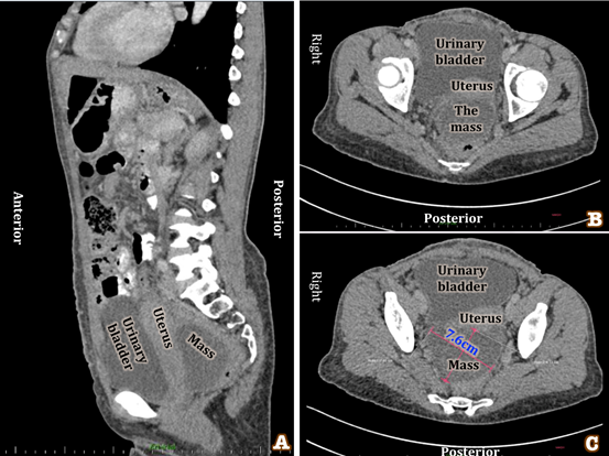
Examination under ultrasound (images not shown) revealed an irregular mass measuring 7 x 4 cm behind the uterus; there was a free fluid in the Douglas. Computerized tomography (CT) scans (Figure 1 A-C) showed an irregular, slightly complex and cystic mass measuring 7.6 x 6.3 cm, located posterior to uterus, suggesting an ovarian origin. Differential diagnoses include a complex cyst versus a low malignant potential neoplasm. Small amount of ascites was also seen.
Figure 1: CT scan images. All these images show a mass located in the posterior pelvis, behind the uterus.
A. Post contrast arterial sagittal image
B. Post contrast arterial axial image
C. Post contrast venous axial image
A. Post contrast arterial sagittal image
B. Post contrast arterial axial image
C. Post contrast venous axial image
Intraoperative findings are a multifocal tumour with a 3-cm mass in anterior cul-de-sac, a 4-cm mass in the left pelvic omentum and multiple nodules in the Douglas pouch, liver and diaphragm. Total abdominal hysterectomy with bilateral salpingo-oophorectomy and left pelvic mass resection were performed. The tumour was glistening on the surface, while hemorrhagic on cut sections.
Microscopic examination showed a right ovary reticular tumour with labyrinth of primitive cells forming micro cysts (Figure 2A) and frequent Schiller–Duval bodies (Figure 2B), consistent with a yolk sac tumour. The morphology showed that the tumour also involved the left pelvic omentum and the cervix. Immunohistochemistry showed focal moderate to strong cytoplasmic reactivity to alpha-fetoprotein (AFP, Figure 2C and 2D). The tumour showed no response to the prior chemotherapy. The patient had ascites and metastatic lesions in the lungs with respiratory difficulties. She received palliative care, but deceased two months after the diagnosis of yolk sac tumour.
Figure 2: Photomicrographs of Yolk Sac Tumour.
A. Reticular or micro cystic patterns formed by a loose network of flat/cuboidal cells, haematoxylin and eosin stain (H&E, 40X)
B. Schiller-Duval body: central blood vessel enveloped by germ cells within a space similarly lined by germ cells, resembles glomerulus (H&E, 200X)
C and D: Immunoperoxidase stain to AFP shows cytoplasmic positivity (AFP, 40X and 200X, respectively).
Discussion and Conclusion
Ovarian yolk sack tumour is a type of malignant ovarian germ cell tumours and is generally rare [1-3]. The peak incidence is found in young women or adolescent girls, the median age at diagnosis being 19 years [2]. We report an exceptional case of ovarian yolk sac tumour diagnosed in an elderly patient; rare similar cases were previously reported in post-menopausal patients [7-9]. Thus, the old age should not preclude a clinical diagnosis of ovarian yolk sac tumour.
Yolk sac tumour is thought to be caused by the hyper methylation of the RUNX3 gene promoter [10], and the overexpression of the transcription factor GATA-4 gene which normally regulates the differentiation of yolk sac endoderm [11]. The majority of cases present with abdominal pain and swelling as symptoms at the time of diagnosis [12], as it was in this case.
The tumour is grossly glistening on the surface while cystic and hemorrhagic on cut sections. Histologically, the pathognomonic morphological findings are the Schiller-Duval body, consisting central blood vessel surrounded by germ cells within a space lined by germ cells mimicking glomeruli. The histological patterns of yolk sac tumour are reticular or macro cystic, polyvesicular vitelline, microcytic, endodermal sinus or festoon or micro papillary, alveolar-glandular (with endometrioid variant), solid, hepatoid and myxoid patterns [8,12].
The tumour may show pure yolk sac tumour features or in combination with features of other germ cell tumours [12]. Clear cell carcinoma shares many of the morphological features of yolk sac tumour [8], which may explain the reason of the prior diagnosis in this case. Tumour markers such as AFP and CA-125 are elevated; while the immunohistochemistry antibodies to confirm yolk sac tumour include AFP, CD10 and alpha-1-antitrypsin [13]. AFP immunosperoxidase stain was strongly positive in tumour cells in this case.
The poor prognostic factors include tumour size > 2 cm, stage > I and presence or amount of ascites [2,12,14]. Yolk sac tumour diagnosed in early stages could be successfully treated with surgery combined with bleomycin, etoposide and cisplatin chemotherapy which are known to exhibit good outcomes in ovarian yolk sac tumours [6]. This case was treated using palliative therapy because the disease was very advanced. Since this case had metastatic lesions, was of size greater than 2 cm and presented with ascites, the prognosis was expectedly poor [2,6,12,14].
Conflict of interest
There is no financial interest or any conflict of interest to declare.
There is no financial interest or any conflict of interest to declare.
References
- Dallenbach P., et al. “Yolk sac tumours of the ovary: an update”. European Journal of Surgical Oncology 32.10 (2006): 1063-1075.
- Umezu T., et al. “Long-term outcome and prognostic factors for yolk sac tumor of the ovary”. Nagoya Journal of Medical Science 70 (2008): 29-34.
- Dave AN., et al. “Ovarian Yolk Sac Tumor”. Indian Journal of Radiology and Imaging 15.4 (2005): 525–527.
- Nasioudis D., et al. “Management and prognosis of ovarian yolk sac tumors; an analysis of the National Cancer Data Base”. Gynecologic Oncology 147.2 (2017): 296-301.
- Wang X., et al. “Ovarian Yolk Sac Tumor: The Experience of a Regional Cancer Center”. International Journal of Gynecological Cancer 26.5 (2016): 884–891.
- T. de La Motte Rouge., et al. “Survival and reproductive function of 52 women treated with surgery and bleomycin, etoposide, cisplatin (BEP) chemotherapy for ovarian yolk sac tumor”. Annals of Oncology 19.8 (2008): 1435–1441.
- Ashihara T., et al. “Ovarian yolk sac tumor in a postmenopausal woman : case report and review of the literature”. International Cancer Conference Journal 1.2 (2012): 96–102.
- Ulbright T. “Germ cell tumors of the gonads: a selective review emphasizing problems in differential diagnosis, newly appreciated, and controversial issues”. Modern Pathology 18 (2005): 61–79.
- Conter CF., et al. “Ovarian Yolk Sac Tumors; Does Age Matter?” International Journal of Gynecological Cancer 28.1 (2018): 77-84.
- Kato N., et al. “Hyper methylation of the RUNX3 Gene Promoter in Testicular Yolk Sac Tumor of Infants”. The American Journal of Pathology 1663.2 (2003): 387–391.
- Siltanen S., et al. “Transcription Factor GATA-4 Is Expressed in Pediatric Yolk Sac Tumors”. The American Journal of Pathology 155.6 (1999): 1823–1829.
- Cicin I., et al. “Yolk sac tumours of the ovary: Evaluation of clinic pathological features and prognostic factors”. European Journal of Obstetrics & Gynecology and Reproductive Biology 146.2 (2009): 210-214.
- Ramalingam P., et al. “The use of cytokeratin 7 and EMA in differentiating ovarian yolk sac tumors from endometrioid and clear cell carcinomas”. The American Journal of Surgical Pathology 28.11(2004):1499-1505.
- Nawa A., et al. “Prognostic factors of patients with yolk sac tumors of the ovary”. American Journal of Obstetrics & Gynecology 184.6 (2001):1182–1188.
Citation:
Carine Nyampinga., et al. “Ovarian Yolk Sac Tumour in a 47 Years Old Patient: A Case Report”. Gynaecology and Perinatology
1.2 (2017): 115-119.
Copyright: © 2017 Carine Nyampinga., et al. This is an open-access article distributed under the terms of the Creative Commons Attribution License, which permits unrestricted use, distribution, and reproduction in any medium, provided the original author and source are credited.


 Scientia Ricerca is licensed and content of this site is available under a Creative Commons Attribution 4.0 International License.