Case Report
Volume 2 Issue 3 - 2018
A Rare Case of Uterine Rupture in an Unscarred Uterus
1,6,7Junior Resident, Department of obstetrics & Gynaecology, ESIC-MC & PGIMSR, Bangalore
2,3Senior Resident, Department of obstetrics & Gynaecology, ESIC-MC & PGIMSR, Bangalore
4,5Professor, Department of obstetrics & Gynaecology, ESIC-MC & PGIMSR, Bangalore
2,3Senior Resident, Department of obstetrics & Gynaecology, ESIC-MC & PGIMSR, Bangalore
4,5Professor, Department of obstetrics & Gynaecology, ESIC-MC & PGIMSR, Bangalore
*Corresponding Author: Dr. Sreelatha S, Professor, Department of obstetrics & Gynaecology, ESIC-MC & PGIMSR, Bangalore.
Received: June 10, 2018; Published: July 27, 2018
Abstract
Uterine rupture is a rare life-threatening complication. It is rarely seen in an unscarred uterus. Most of the cases in literature have been reported are with the use of labour induction agents or in a scarred uterus. We are reporting a case of uterine rupture in a multigravida patient with term gestation who was in the latent phase of labour. Patient was taken up for emergency LSCS in view of foetal distress and intra-operatively there was uterine rupture and foetal parts were seen in the peritoneal cavity. Uterine repair was done in layers. Patient received 1-pint packed cells. Postoperatively patient was stable and was discharged on post-operative day 9.
Keywords: Unscarred uterus; LSCS; latent phase of labour; Foetal distressmulti gravida
Introduction
Rupture of a pregnant uterus is one of the life-threatening complications encountered in obstetric practice. It is one of the causes of maternal and perinatal morbidity and mortality. There are several risk factors associated with rupture of uterus but the most common is a previous Caesarean section. Advanced maternal age, multiparity, placenta increta, macrosomia, shoulder dystocia and medical termination of pregnancy are some other important contributing factors to this condition. The overall incidence of rupture uterus in unscarred uterus and scarred uterus varies from 0.7 and 5.1 per 10,000 deliveries respectively [1]. Rupture of an unscarred uterus is a rare event.
Case Report
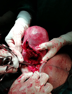
A 30-year-old patient, gravida 2 para 1, with 40 weeks period of gestation was admitted to the hospital in latent phase of labour. She had no significant past history of curettage. On examination her pulse rate was 96/min, BP was 110/70 mm of Hg. Respiratory rate was 20/min. SPO2 was 100%. Per abdomen uterus was term size, acting with 1 to 2 contractions lasting for 20 seconds in 20 minutes. Patient was taken up for emergency LSCS in view of foetal distress. On opening the abdomen, there was hemoperitoneum, and rupture of the uterus was found in the postero-lateral wall extending from fundus to cervix.
The foetal parts were outside the uterus and there was hemoperitoneum of 1.0 litres. Extracted a live male baby of weight 2.5 kg uterine repair was done in layers using No.1 vicryl. Bilateral tubes and ovaries were healthy. Bilateral tubectomy was done. The abdomen was closed with an intraperitoneal drainage tube. Patient was given adequate packed cells. Patient got discharged after 9 days of postoperative hospitalization without any complications.
Discussion
Uterine rupture is defined as a full-thickness separation of the uterine wall and the overlying serosa. It is a rare peripartum complication associated with severe maternal and neonatal morbidity and mortality [2]. According to a systematic review conducted by World Health Organization (WHO), the median incidence of uterine rupture was estimated at 5.3 per 10,000 deliveries [3]. According to literature incidence of spontaneous rupture in intact uterus is 1:15,000 [16]. Rupture is mainly due to impaired collegen synthesis due to underlying collegen deficiency. The causes for rupture of uterus maybe endometritis, previous history of curettage which leads to uterine synechite, uterine perforation results in scar which in turn leads to rupture [17].
Rupture of an unscarred uterus may be caused by trauma or congenital or acquired weakness of the myometrium. Sources of trauma include motor vehicle accidents and obstetric manoeuvres (e.g., internal or external version). The myometrium may be inherently weak because of a congenital disorder, such as Ehlers-Danlos type IV or it may become weakened from protracted labour or use of strong uterotonic drugs (e.g., misoprostol), which place prolonged stress on the myometrium [4-7]. Over distension of the uterine cavity, whether absolute or relative to the size of the cavity, may be the major physical factor provoking rupture when the myometrium is weakened for any reason.
Grand multiparty, advanced maternal age, endometriosis, arteriovenus malformation and abnormal placentation fetal macrosomia, neglected labor, malpresentation, breech extraction, and uterine instrumentation are all predisposing factors for uterine rupture [8].
Compared with spontaneous labour, combination inductions showed the highest risk of uterine rupture, followed by prostaglandin induction, and then by oxytocin induction. Lowest risk for uterine rupture comprised sweeping of membranes and trans cervical balloon catheter [9] rupture usually occur on left side if rupture occur on right side due to wrong application of forceps.
The usual site of rupture in second trimester is fundal region unlike lower uterine segment in third trimester [10]. The most consistent early indicator of uterine rupture is the onset of a prolonged, persistent, and profound foetal bradycardia. Other Clinical presentation of uterine rupture includes impaired foetal activity, vaginal bleeding, abdominal pain, maternal tachycardia and other symptoms of hypovolemia [11].
Differential diagnosis includes perforation of bowel, acute appendicitis, acute pancreatitis, rupture aortic aneurysm should also be considered and ruled out before making a definitive diagnosis [12]. Early surgical intervention is usually the key to successful treatment of uterine rupture. The therapeutic management is a total or subtotal hysterectomy. The suture can be performed and helps to preserve the reproductive function of patients who have never given birth with a recurrence risk of uterine rupture assessed between 4 and 19% at a subsequent pregnancy [13].
For this reason, it has been recommended that women with a previous uterine rupture undergo an elective Caesarean delivery as soon as foetal lung maturity can be demonstrated [14]. Uterine rupture of an unscarred uterus is associated with significant morbidity and mortality. Schrinsky and Benson [15], in their study, found a maternal and foetal mortality rate of 20.8% and 64.6%, respectively.
Conclusion
Spontaneous uterine rupture in an unscarred uterus is very rare and is an obstetric emergency which is difficult to diagnose. The maternal and foetal prognosis depends on awareness of risk factors, recognition of clinical signs and symptoms, intrapartum fatal monitoring and prompt surgical management.
References
- Abdulwahab and Dalia F., et al. “Second-trimester uterine rupture: lessons learnt.” The Malaysian Journal of Medical Sciences 21.4 (2014): 61-65.
- Ofir K., et al. “Uterine rupture: risk factors and pregnancy outcome”. American Journal of Obstetrics and Gynecology 189.4 (2003): 1042-1046.
- Hofmeyr GJ., et al. “WHO systematic review of maternal mortality and morbidity: the prevalence of uterine rupture”. BJOG: An International Journal of Obstetrics & Gynaecology 112.9 (2005): 1221–1228.
- Pepin M., et al. “Clinical and genetic features of Ehlers-Danlos syndrome type IV, the vascular type”. The New England Journal of Medicine 342.10 (2000): 673-680.
- Walsh CA., et al. “Unexplained prelabor uterine rupture in a term primigravida”. Obstetrics & Gynecology 109.2 (2007): 455.
- Taylor DJ., et al. “Ehlers-Danlos syndrome during pregnancy: a case report and review of the literature”. Obstetrical & Gynecological Survey 36.6 (1981): 277-281.
- Rudd NL., et al. “Pregnancy complications in type IV Ehlers-Danlos Syndrome”. Lancet 1.8314-1 (1983): 50-53.
- Wang PH., et al. “Posterior uterine wall rupture during labour”. Human Reproduction 15.5 (2000): 1198–1199.
- Al-Zirqi I., et al. “Uterine rupture: trends over 40 years”. BJOG: An International Journal of Obstetrics & Gynaecology 123.5 (2015): 780-787.
- Vaknin Zvi., et al. “Clinical, sonographic, and epidemiologic features of second-and early third-trimester spontaneous antepartum uterine rupture: a cohort study.” Prenatal Diagnosis 28.6 (2008): 478-484.
- Menihan CA. “Uterine rupture in women attempting a vaginal birth following prior cesarean birth”. Journal of Perinatology 18.6 (1998): 440–443.
- Hill Christina C and Jennifer Pickinpaugh. “Trauma and surgical emergencies in the obstetric patient”. Surgical Clinics of North America 88.2 (2008): 421-440.
- S. Ahmadi., et al. “Uterine rupture of the unscarred uterus. About 28 cases”. Gynécologie Obstétrique & Fertilité 31.9 (2003): 713–717.
- R. Conturso., et al. “Spontaneous uterine rupture with amniotic sac protrusion at 28 weeks subsequent to previous hysteroscopic metroplasty”. European Journal of Obstetrics Gynecology and Reproductive Biology 107.1 (2003): 98–100.
- DC Schrinsky and RC Benson. “Rupture of the pregnant uterus: a review”. Obstetrical and Gynecological Survey 33.4 (1978): 217–232.
- Siddique M and Rana Singh JS. “Spontaneous rupture of the uterus”. Journal of Clinical Anesthesia 14 (2002): 368-370.
- Walsh LA., et al. “Unexplained pre labour uterine rupture in term primie”. Obstetrics and gynaecology 108-3 (2006): 725-727.
Citation:
Sreelatha S., et al. “A Rare Case of Uterine Rupture in an Unscarred Uterus”. Gynaecology and Perinatology 2.3 (2018): 281-284.
Copyright: © 2018 Sreelatha S., et al. This is an open-access article distributed under the terms of the Creative Commons Attribution License, which permits unrestricted use, distribution, and reproduction in any medium, provided the original author and source are credited.


 Scientia Ricerca is licensed and content of this site is available under a Creative Commons Attribution 4.0 International License.