Case Report
Volume 1 Issue 1 - 2017
A Rare Case of Odontogenic Myxoma in Posterior Maxilla: A Case Report
1Reader, Department of Oral and Maxillofacial surgery, Manav Rachna Dental College, India
2Head, Department of Oral and Maxillofacial Pathology, Manav Rachna Dental College, India
3Head, Department of Oral and Maxillofacial Surgery, Manav Rachna Dental College, India
4Reader, Department of Oral and Maxillofacial Pathology, Manav Rachna Dental College, India
2Head, Department of Oral and Maxillofacial Pathology, Manav Rachna Dental College, India
3Head, Department of Oral and Maxillofacial Surgery, Manav Rachna Dental College, India
4Reader, Department of Oral and Maxillofacial Pathology, Manav Rachna Dental College, India
*Corresponding Author: Dr. Amit Mohan, Reader, Department of Oral and Maxillofacial Surgery, Manav Rachna Dental College, India.
Received: December 09, 2016; Published: January 04, 2017
Abstract
Odontogenic myxoma is a rare, benign, locally aggressive and non metastasizing neoplasm of the jaw bones. The tumour is supposed to be derived from the ectomesenchymal portion of the tooth germ. This case report presents a rare case of odontogenic myxoma occurring in a 35 year old female patient with emphasis on the management of the lesion.
Keywords: Odontogenic myxoma; Benign tumour; Jaw tumour
Clinical Relevance: The article describes a rare case of odontogenic myxoma which was treated conservatively by enucleation and curettage. The article focuses on the need for specific surgical management depending on the size and aggressive nature of the lesion especially in case of maxilla where the large resection results in defect which is difficult to reconstruct.
Introduction
Odontogenic myxomas (OMs) are benign, slow growing, locally aggressive and non metastasizing neoplasms of the jaw bone. [1] According to the World Health Organization (WHO) classification 2005, OM belongs to benign tumor of ectomesen¬chymal origin with or without odontogenic epithelium. [2] Amongst all the odontogenic tumors the incidence of OM ranges from 0.5% to 20%. [3]
Although OMs predominantly involve the mandible but the maxillary tumors are more aggressive than those in the mandible. [4] OM is more commonly seen during 2nd and 4th decades, rarely found in children and the elderly and has a slight predilection for females. [5]
Odontogenic myxomas are usually asymptomatic. Tumor is often scalloped between roots and usually cause displacement of teeth and rarely causing root resorption. [6] Lesions involving maxilla, can expand inside the maxillary sinus and are then diagnosed later only after having grown to larger sizes. [7] The histological characteristic of this tumor resembles the mesenchymal portion of a tooth in development-consists of rounded, spindled, fusiforms and star cells arranged in a loose, abundant myxoid stroma with few collagen fibrils. [8] The surgical treatment of OM, including curettage and radical resection, is controversial due to the varying recurrence rates, especially due to its gelatinous and mucous aspect and having no capsule. A long term follow up is required. [7] The present study reports the case of a female patient with a large maxillary OM in the posterior region.
Case Report
A 35 year old female reported with complain of pain and swelling in left maxillary posterior region since 6 months. The swelling was slowly growing and was not associated with any discharge. Patient gave a history of similar swelling 4 years back in the region slightly posterior to the present swelling for which she underwent surgery but lost the records. Patient also gave history of left maxillary sinusitis for which she underwent surgery 2 months back.
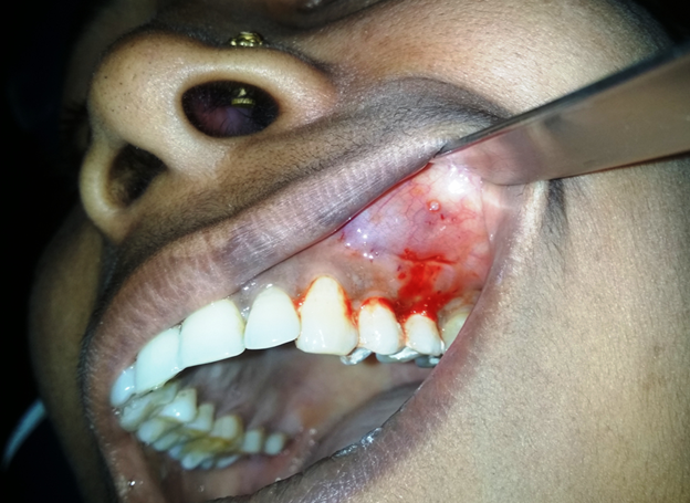
On examination, a diffuse bony hard swelling was seen in the left maxillary vestibule in the premolar region. The overlying mucosa was normal and there was no discharge seen (Figure 1). On palpation, the swelling was fixed and non compressible. Adjacent teeth (1st and 2nd premolar) were non carious, slightly tender on percussion and were non vital. On aspiration, there was no fluid collected.
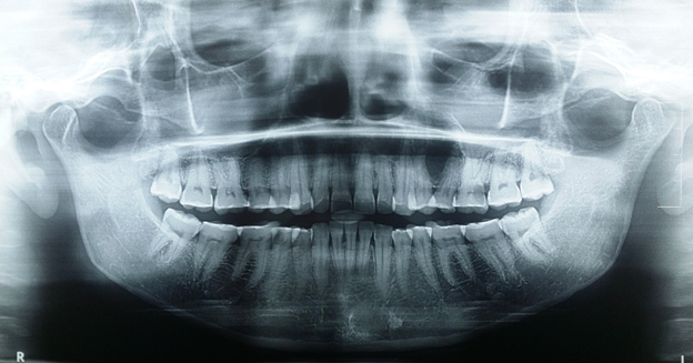
On radiographic examination, a well defined radiolucency was noted in relation to left maxillary premolars. The roots of first and second premolar were deviated without any signs of resorption (Figure 2).
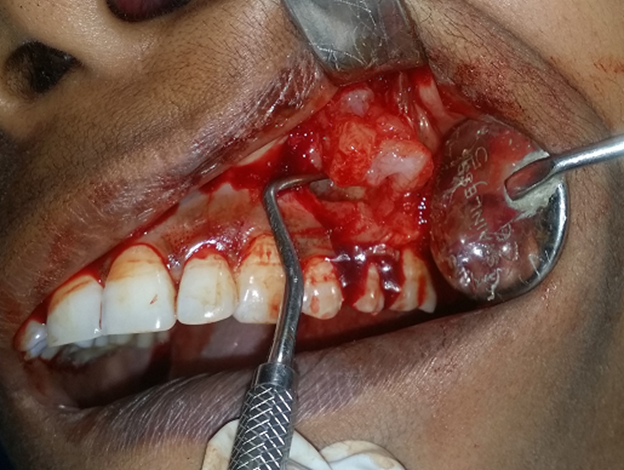
A differential diagnosis of Odontogenic myxoma, Adenomatoid Odontogenic Tumour, ossifying fibroma, fibrous dysplasia and peripheral giant cell granuloma was considered. The lesion was planned for biopsy. Both the premolars underwent endodontic treatment before the surgery. Incision was made from 1st premolar to 1st molar region and muco-periosteal flap was reflected. The overlying bone was very thin and was carefully removed with a sharp instrument to create adequate bony window. The lesion was found to be non-encapsulated and was well separated from maxillary sinus and nasal cavity. Since the lesion was well circumscribed within cortical margins, the lesion was completely curetted instead of doing an incisional biopsy to avoid second surgery (Figure 3). Hemostasis was attained and closure was done with 3-0 silk sutures.
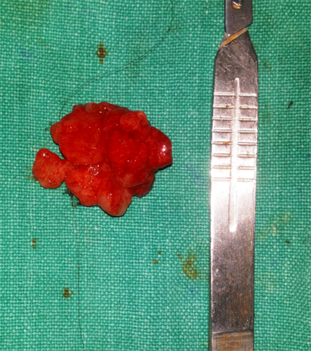
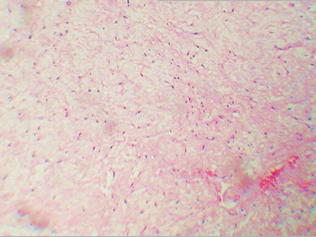
The gross specimen was soft glistening, gelatinous and non-encapsulated (Figure 4). On microscopic examination, abundant loose myxoid stroma with large number of spindle and stellate shaped cells having hyperchromatic nuclei and long anastmosing processes were seen which were suggestive of odontogenic myxoma (Figure 5). Patient was advised for regular follow up.
Discussion
OM of the maxilla was first reported by Thoma and Goldman in 1947. It is commonly seen in middle-aged individuals with predilection for females. [7] These findings were consistent with the present case.
The lesions are mostly asymptomatic apart from being a slowly growing mass within the jaw bone. When the lesion expands into the maxillary sinus, patients start experiencing symptoms of sinusitis. In the present case the patient gave history of previous sinus surgery. Although there was no obvious clinical invasion seen, possibility of the lesion affecting the maxillary sinus can be considered. Other possible features include pain, paresthesia, and ulceration. Pain is more severe in cases of soft tissue involvement as compared to intrabony lesions. [9] In the present case the patient was experiencing only mild pain due to the fact that the lesion was well encased with the bony margins.
When the cortical plates are invaded, the expanding tumor is soft on palpation. In our case the lesion was covered under the thin cortical bone without invasion into the soft tissues. Also, aspiration is non productive as seen in our case.
On radiographic examination, the tumor presents as unilocular or multilocular radiolucency with well-defined borders with fine bony trabeculae within its internal structure expressing a “honeycombed”, “soap bubble” or “tennis racket” appearance. [4,10] Root deviation or root resorption could be evident on plain radiograph. [6] In our case the unilocular radiolucent lesion was present between the roots of premolars, causing the root deviation. Other radiographic investigation modalities suggested in literature include CT, MRI and CBCT. [11,12]
The histogenesis of odontogenic myxoma is controversial. Some authors suggest that the tumor is the consequence of myxomatous degeneration of fibrous stroma while others consider it as a tumor of primitive mesenchyme, rather than a result of a secondary change in tissue. Another theory considers the myxoma cell as an aberrant development of mesodermal cells into myxoblasts which actively secrete the myxomatous ground substance. Moreover, this secretion is suggested to be a result of an inductive effect on the part of the odontogenic epithelium. [13] Microscopically, the tumor consists of rounded, spindled, and stellate cells arranged in a loose, myxoid stroma with few collagen fibrils and delicate fibrous connective tissue as seen in our case. Small islands of apparently inactive epithelial odontogenic rests may be scattered through the myxoid substance without any capsule, and they are important to establish the diagnosis [8,14] Immunohistochemistry studies suggest that the spindle-shaped cells constituting this neoplasm have a combined fibroblastic and smooth muscle typing, suggesting that it is of myofibroblastic origin. [2]
Differential diagnosis includes lesions showing typical multilocular radiolucency such as ameloblastoma, odontogenic keratocyst, central hemangioma, aneurysmal bone cyst, central giant cell granuloma, giant cell lesions of hyperparathyroidism, cherubism, and metastatic tumors in the jaws and in cases of unilocular lesions, periapical, lateral, periodontal and simple bone cysts. [7,15]
The surgical management includes-Radical (excision) or conservative (enucleation or curettage) surgery. [16] Although, complete surgical removal by conservative treatment can be difficult, because unlike most benign neoplasms, the myxoma is non-encapsulated and its myxomatous tissue infiltrates the surrounding bony tissue; [17] however, enucleation and curettage have several advantages over more radical treatments as they are substantially less invasive, can be achieved by means of an intraoral surgical approach, preserve function and aesthetics, have a shorter hospitalization time, and are more cost-effective. [18] Nonetheless, the risk of recurrence after more conservative surgery is higher. Hence a long term follow up is necessary in cases of OM. In our case conservative treatment was done as the lesion was well confined within the cortical margins without infiltration into the surrounding bone or soft tissues. Patient has shown no recurrence after 1 year follow up.
References
- Leiser Y., et al. “Odontogenic myxoma – A case series and review of the surgical management”. Journal of Cranio-Maxillo-Facial Surgery 37.4 (2009): 206-209.
- Kramer IRH., et al. “Histological typing of odontogenic tumours. 2nd edition”. Springer Verlag, Berlin, 70.12 (1992): 2988-2994.
- Barnes L., et al. “World health organization classification of tumors -Pathology and genetics of head and neck tumors”. IARC Press, Lyon, 2006.
- Deron PB., et al. “Myxoma of the maxilla: a case with extremely aggressive biologic behavior”. Head Neck 18.5 (1996): 459‑464.
- Abiose BO., et al. “Fibromyxomas of the jawbones ‑ a study of ten cases”. British Journal of Oral and Maxillofacial Surgery 25.5 (1987): 415‑421.
- Spencer KR and Smith A. “Odontogenic myxoma: Case report with reconstructive considerations”. Australian Dental Journal 43.4 (1998): 209-212.
- Carvalho de Melo AU., et al. “Martorelli Fde O. Maxillary odontogenic myxoma involving the maxillary sinus: Case report”. Brazilian Journal of Otorhinolaryngology 74.3 (2008): 472-475.
- Aquilino RN., et al. “Odontogenic myxoma in the maxilla: A case report and characteristics on CT and MR”. Oral Oncology Extra. 42.4 (2006): 133-136.
- Simon EN., et al. “Odontogenic myxoma: a clinicopathological study of 33 cases”. International Journal of Oral and Maxillofacial Surgery 33.4 (2004): 333-337.
- Sivakumar G., et al. “Odontogenic myxoma of maxilla”. Indian Journal of Dental Research 19.1 (2008): 62-65.
- Dabbaghi A., et al. “Rare appearance of an odontogenic myxoma in cone-beam computed tomography: a case report”. Journal of Dental Research, Dental Clinics, Dental Prospects 10.1(2016): 65-68.
- Kheir E., et al. “The imaging characteristics of odontogenic myxoma and a comparison of three different imaging modalities”. Oral Surgery, Oral Medicine, Oral Pathology, Oral Radiology 116.4 (2013): 492-502.
- Nitzan dw., et al. “Childhood odontogenic myxoma:report of two cases pediatric dentistry”. The American journal of Pediatric Dentistry 7.2 (1985) : 140-144.
- Nevelle BW., et al. “Oral and maxillofacial pathology. 2nd ed. Saunders, Philadelphia”, (2007): 635-637.
- Nayak MT., et al. “Maxillary odontogenic myxoma: A rarity”. International Journal of Oral and Maxillofacial Pathology 2.3 (2011): 32-35.
- Li TJ., et al. “Odontogenic myxoma: a clinicopathologic study of 25 cases”. Archives of pathology & laboratory medicine 130.12 (2006): 1799-1806.
- Lo Muzio L., et al. “Odontogenic myxoma of the jaws: a clinical, radiologic, immunohistochemical, and ultrastructural study”. Oral Surgery, Oral Medicine, Oral Pathology, Oral Radiology, and Endodontology 82.4 (1996): 426-433.
- Kawase-Koga Y,. et al “Surgical management of odontogenic myxoma: a case report and review of the literature”. BMC research notes 7:214 (2014).
Citation:
Amit Mohan., et al. “A Rare Case of Odontogenic Myxoma in Posterior Maxilla: A Case Report”. Oral Health and Dentistry 1.1 (2017): 67-71.
Copyright: © 2017 Amit Mohan., et al. This is an open-access article distributed under the terms of the Creative Commons Attribution License, which permits unrestricted use, distribution, and reproduction in any medium, provided the original author and source are credited.


 Scientia Ricerca is licensed and content of this site is available under a Creative Commons Attribution 4.0 International License.