Research Article
Volume 1 Issue 1 - 2017
Incidence of Lingual Nerve Injury during Mandibular Third Molar Surgery: A Prospective Study
1Dr. Ankur Srivastava,Senior Resident,HinduRao Hospital,New Delhi
1Dr. Ruchi Pathak Kaul,Women Scientist fellow,JPN Apex Trauma Centre,AIIMS,New Delhi
2Dr. Prachur Malhotra,Reader,K M Shah Dental College,Vadodara
3Dr. C S Ram,Professor,ITS Dental college,Ghaziabad
1Dr. Ruchi Pathak Kaul,Women Scientist fellow,JPN Apex Trauma Centre,AIIMS,New Delhi
2Dr. Prachur Malhotra,Reader,K M Shah Dental College,Vadodara
3Dr. C S Ram,Professor,ITS Dental college,Ghaziabad
*Corresponding Author: Dr. Ruchi Pathak Kaul,Women Scientist fellow,JPN Apex Trauma Centre,AIIMS,New Delhi.
Received: February 12, 2017; Published: March 11, 2017
Abstract
Introduction: Third molar removal is one of the commonest procedure performed in oral and maxillofacial surgery. Because of high incidence of short-term morbidity (pain, swelling, trismus, postoperative infection) and potential long-term complication, such as damage to the inferior alveolar and the lingual nerves one is forced to reduce the frequency of this operation. However the goal of sensory impairment is to acquire information to render a clinical diagnosis, to aid in determining meaningful prognosis, and to determine beneficial therapy for the nerve injury.
Aims and Objective: To determine the incidence of lingual nerve injury and persistent sensory disturbances of the lingual nerve after impacted mandibular third molar surgery. To modify the surgical technique to reduce lingual nerve injury.
Materials and Method: 100 patients were studied prospectively. An informed consent was signed before the study. The following test were used to determine lingual nerve injury: point reference, two point discrimination test, Electrogustometry. Measurements were recorded on VAS on 1st, 7th, 30th, 90th and 180th day post operatively.
Results: In the present study, the incidence of lingual nerve during third molar surgery after raising and retracting lingual mucoperiosteal flap with Howarth periosteal elevator is 27.3% , which is statically significant (p = .004).
Discussion: In this study, the incidence of lingual nerve injury during impacted mandibular third molar surgery is 4.0% and shows temporary effect on lingual nerve. Males are more affected with distoangular impaction (2%) followed by mesioangular (1%) and horizontal impaction (1%). The age of the patients who are suffered from lingual nerve injury above 24 yrs. are 3% and below 24 yrs. is 1%. Incidence of lingual nerve injury during lower third molar surgery seen more in cases with raising and retracting a lingual mucoperiosteal flap with Howarth periosteal elevator.
Keywords: Lingual Nerve Injury; Third Molar Surgery; Lingual Flap
Introduction
Third molar disimpaction is one of the commonest procedures performed in oral and maxillofacial surgery. Because of high incidence of short-term morbidity (pain, swelling, trismus, postoperative infection) and potential long-term complication, such as damage to the inferior alveolar nerve and the lingual nerve, one is forced to reduce the frequency of this operation. [1] The neurosensory disturbances to the inferior alveolar nerve during lower third molar removal presents paraesthesia or anaesthesia of the lower lip, chin and buccal gingival of the affected side. Its reported incidence of 1.3% to 7.8% and there is little evidence to suggest that surgical technique affects the frequency of this complication. [3-5] Lingual nerve deficits presents with numbness of ipsilateral anterior two third of the tongue and disturbed taste perception. The incidence of lingual nerve damage, as reported in the literature, during lower third molar surgery ranges from 0% to 23% and it appears likely that the surgical technique markedly affect the frequency. [6] The majority of lingual nerve injuries which occur during lower third molar surgery result in transient disturbance, with recovery of normal sensation within 4-6 months. In these cases, it is likely that the injury results from manipulation of the tissue with the Howarth's periosteal elevator on the lingual side, resulting in either crush injury or temporary conduction block. In a small proportion of patients (approximately 0.5%) the sensory disturbance is permanent, with a variable level of recovery and symptoms which can include hypoesthesia (reduced sensation), paraesthesia (abnormal sensation) or, even worse, various forms of dysesthesia (unpleasant abnormal sensation). In these cases it is likely that the injury often results from direct nerve damage by the rotating bur. It is also this group which is most distressed by the complication, report difficulties with speech and mastication. [5] The risk ratio for lingual nerve deficit with lingual flap retraction being 1.94 times more 4.
The use of lingual retractors and rotating instruments may cause lingual nerve damage and the use of a lingual nerve retractor during third molar surgery was associated with an increased incidence of temporary nerve damage, but did not influence that rate of permanent nerve damage. [7] However the goal of this study is to acquire information to render a clinical diagnosis, to aid in determining meaningful prognosis, and to determine beneficial therapy for the nerve injury. This study shall evaluate the incidence of lingual nerve injury during mandibular third molar surgery. It shall determine the incidence of persistent sensory disturbances of the lingual nerve after impacted mandibular third molar surgery. Hence this study shall provide modified operative technique which decreases the lingual nerve injury during impacted mandibular third molar surgery.
Material and Methods
This is a prospective study, with sample size of 100 patients. All patients signed an informed consent before participating in the study, which was reviewed and approved by the ethical committee of our institute All patients with unilateral or bilaterally impacted mandibular third molar teeth including both healthy male and female between the age group 18-70 years were included. The patients with fully erupted third molar, medically compromised, acute infection or swelling with respect to mandibular third molar, allergic to any drug/local anaesthesia and Inability to provide informed consent to the maxillofacial surgeon.
Evaluation of altered lingual nerve sensation after third molar surgery was done on 1st, 7th, 30th, 90th and 180th day post operatively using Point of reference, Two point discrimination, Electrogustometry, Patient subjective report (A series of specific questions designed to elicit information of various aspect of tongue sensation like touch, taste, temperature, teeth, trauma , tingling, talk).
1. Point of reference:
Point of reference was created on the tongue using a Perspex template with measured numbered holes and marked with Bonney‘s blue stain. Touch stimuli was measured and marked. Score was recorded on Visual analogue scale from 0 to 10.
Point of reference was created on the tongue using a Perspex template with measured numbered holes and marked with Bonney‘s blue stain. Touch stimuli was measured and marked. Score was recorded on Visual analogue scale from 0 to 10.
2. Two point discrimination
This was done with a modified Vernier calliper instrument with separation ranging from 2 to 20 mm at 2 mm intervals. The probe was drawn 5-10 mm across the surface of the tongue, approximately 1-2 cm from the tip. Score was recorded on Visual analogue scale from 0 to 10.
This was done with a modified Vernier calliper instrument with separation ranging from 2 to 20 mm at 2 mm intervals. The probe was drawn 5-10 mm across the surface of the tongue, approximately 1-2 cm from the tip. Score was recorded on Visual analogue scale from 0 to 10.
3. Electrogustometry
This was done with Monopolar constant- current stimuli (PHYSIOSTIM-D) of up to 7 mA with flat stainless steel electrode (diameter 5 mm). It was applied to an area about 1 cm from the tip and 1 cm from the midline of the tongue. Scores was recorded when patient indicates for tingle or metallic test on stimuli. Score was recorded on Visual analogue scale from 0 to 10 twice and taken the mean value of the score as a final score.
This was done with Monopolar constant- current stimuli (PHYSIOSTIM-D) of up to 7 mA with flat stainless steel electrode (diameter 5 mm). It was applied to an area about 1 cm from the tip and 1 cm from the midline of the tongue. Scores was recorded when patient indicates for tingle or metallic test on stimuli. Score was recorded on Visual analogue scale from 0 to 10 twice and taken the mean value of the score as a final score.
4. Patient subjective report
A series of specific question was designed to elicit information of various aspect of tongue sensation like touch, taste, temperature, teeth, trauma, tingling, and talk. Patient subjective score was recorded on visual analogue score from 0 to 10 for individual question.
A series of specific question was designed to elicit information of various aspect of tongue sensation like touch, taste, temperature, teeth, trauma, tingling, and talk. Patient subjective score was recorded on visual analogue score from 0 to 10 for individual question.
Surgical Technique
Pre-operative intra oral periapical radiographs (IOPA) or Orthopantomogram (OPG) were made to assess the type of impaction using Winter‘s classification and Pell & Gregory classification. Pederson difficulty index was applied to access the difficulty of the procedure. Surgical site was prepared and draped by aseptic technique. This is followed by intraoral irrigation with povidine iodine solution and normal saline. 2% lignocaine hydrochloride with 1:80000 adrenaline is administered using Inferior alveolar nerve block, lingual nerve block and long buccal nerve block Standard Ward‘s incision was followed in all cases. Full thickness mucoperiosteal flap was raised to expose sufficient bone on lateral and distal aspect of the impacted molar. Removal of surrounding bone is done using stainless steel burs (No.8, Rosehead bur). Constant irrigation with saline was used while removing bone to prevent thermal necrosis. Third molar is then luxated with dental elevator and extracted with molar forceps employing minimal forces. Sectioning of tooth was done to deliver it through its path of withdrawal depending upon the type of impaction. Wound Toilet and primary Closure done. Pressure pack was given. Post-operative Instructions explained and patient prescribed antibiotics and analgesics for 5 days.
Pre-operative intra oral periapical radiographs (IOPA) or Orthopantomogram (OPG) were made to assess the type of impaction using Winter‘s classification and Pell & Gregory classification. Pederson difficulty index was applied to access the difficulty of the procedure. Surgical site was prepared and draped by aseptic technique. This is followed by intraoral irrigation with povidine iodine solution and normal saline. 2% lignocaine hydrochloride with 1:80000 adrenaline is administered using Inferior alveolar nerve block, lingual nerve block and long buccal nerve block Standard Ward‘s incision was followed in all cases. Full thickness mucoperiosteal flap was raised to expose sufficient bone on lateral and distal aspect of the impacted molar. Removal of surrounding bone is done using stainless steel burs (No.8, Rosehead bur). Constant irrigation with saline was used while removing bone to prevent thermal necrosis. Third molar is then luxated with dental elevator and extracted with molar forceps employing minimal forces. Sectioning of tooth was done to deliver it through its path of withdrawal depending upon the type of impaction. Wound Toilet and primary Closure done. Pressure pack was given. Post-operative Instructions explained and patient prescribed antibiotics and analgesics for 5 days.
Results
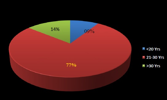
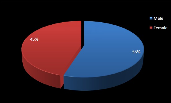
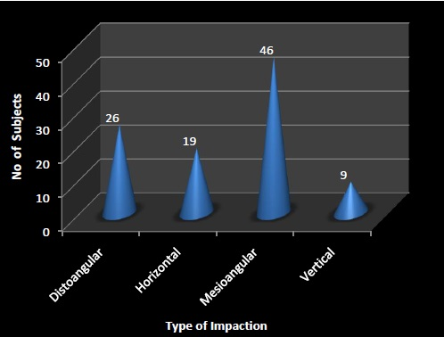
A total of 100 patients requiring unilateral or bilateral surgical extraction of impacted mandibular third molar were included in the present study. (Table 1 and Table 2) shows majority of subjects were aged between 21 to 30 years (77%). The mean age of the subjects was 26.12. The male to female ratio of study subjects was 1: 0.81. (Table 3) shows Majority (46%) of subjects had mesioangular, 26 (26%) subjects had distoangular, and 19 (19%) subjects had horizontal and 09 (09%) had vertical type of impacted tooth.
| S No | Age | No. of Patient | Percentage |
| 1 | < 20 years | 09 | 09% |
| 2 | 21-30 years | 77 | 77% |
| 3 | < 30 years | 14 | 14% |
Table 1: Age wise Distribution of Subjects.
| S No | Gender | No. of Patient | Percentage |
| 1 | Male | 55 | 55% |
| 2 | Female | 45 | 45% |
Table 2: Genderwise Distribution of Subjects.
| S No | Type of impaction | No of Patient | Percentage |
| 1 | Mesioangular | 46 | 46% |
| 2 | Distoangular | 26 | 26% |
| 3 | Horizontal | 19 | 19% |
| 4 | Verticle | 09 | 09% |
Table 3: Distribution of Subjects on the basis of type of impaction.
(Table 4) shows the distribution of subjects on the basis of lingual flap raised. In majority (89%) of subjects lingual flap is not raised. (Table 5) shows the distribution of subjects on the basis of lingual nerve injury. There were (4%) subjects had lingual nerve injury after mandibular third molar surgery. (Table 6) shows the lingual flap raised was observed in 11 subjects out of them 3 (27.3%) subjects had lingual nerve injury. Lingual nerve injury is higher in subjects in which lingual flap was raised as compare in which lingual flap was not raised this difference is significant (p = .004). (Table 7) shows according to type of impaction 24 patient with distoangular, 18 patient with horizontal, 9 patient with vertical and 9 patient with vertical impaction had undergone surgery out of which 2 patient (7.7%) with distoangular (5.3%) and horizontal (2.2%) had lingual nerve injury.
| S No | Lingual Flap | No of Patient | Percentage |
| 1 | Raised | 11 | 11% |
| 2 | Not Raised | 89 | 89% |
Table 4: Distribution of Subjects on the basis of lingual flap raised.
| S No | Lingual Nerve Injury | No. of Patient | Percentage |
| 1 | Abscent | 04 | 04% |
| 2 | Present | 96 | 96% |
Table 5: Distribution of Subjects on the basis of lingual nerve injury.
| S No | Lingual Nerve Injury | Lingualflap Raised | Percentage | |
| Abscent | Present | |||
| 1 | Abscent | 88 | 8 | 96 |
| 98.9% | 72.7% | 96% | ||
| 2 | Present | 1 | 3 |
4 |
| 1.1% | 27.3% | 4% | ||
| Total | 89 | 11 | 100 | |
| 100% | 100% | 100% | ||
Table 6: Distribution of Subjects on the basis of lingual nerve injury and lingual flap raised.
| Lingual Nerve Injury | Type of Impaction | Total | |||
| Distoangular | Horizontal | Mesioangular | Vertical | ||
| Abscent | 24 | 18 | 45 | 9 | 96 |
| 92.3% | 94.7% | 97.8% | 100% | 96% | |
| Present | 2 | 1 | 1 | 0 | 4 |
| 7.7% | 5.3% | 2.2% | 0% | 4% | |
| Total | 26 | 19 | 46 | 9 | 100 |
| 100% | 100% | 100% | 100% | 100% | |
Table 7: Distribution of Subjects on the basis of lingual nerve injury and type of impaction.
The outcome parameters were noted as altered lingual sensation, two point discrimination test, electrogustometry test and neurosensory questions. The follow up was done at 1st, 7th post-operative day, 1st month, 3rd month and 6th month post operatively. (Table 8) shows Assessment of Altered lingual sensation at different time intervals time intervals of 24 hrs, 7th day, and 1st, 3rd and 6th month. Within 24 hrs 4 subjects do not have the nerve sensation and this percentage still remains same after 1 week. After a month it is reduced to 1% and after 3 months no nerve sensation deficit was observed. (Table 9) shows Assessment of Altered lingual sensation at different time intervals. But after 1 month all the subjects achieved the score 10 on VAS. The results of the above mentioned parameters definitely signifies lingual nerve regeneration starts after 24 hrs in all cases (4%), return to all the function seem after 1 month in 3% , and after 3 months no lingual sensory deficit was observed in all cases (4%).
| S No | Altered Lingual Sensation | 24 Hrs | 7 Days | 1 Month | 3 Month | 6 Month |
| 1 | Present | 96 | 96 | 99 | 100 | 100 |
| 2 | Anscent | 04 | 04 | 01 | 0 | 0 |
Table 8: Assessment of Altered lingual sensation at different time intervals.
| Two Point Disriminate Test Score | 24 Hours | 1 Week | 1 Month | 3 Month | 6 Month | |||||
| Left | Right | Left | Right | Left | Right | Left | Right | Left | Right | |
| 6 | 0 | 1 | 0 | 0 | 0 | 0 | 0 | 0 | 0 | 0 |
| 8 | 2 | 1 | 2 | 2 | 0 | 1 | 0 | 0 | 0 | 0 |
| 10 | 98 | 98 | 98 | 98 | 100 | 99 | 100 | 100 | 100 | 100 |
Table 9: Two point discriminate test Score at different Point of time in right and left side.
| ElectrogustroMetry Test Score | 24 Hours | 1 Week | 1 Month | 3 Month | 6 Month | |||||
| Left | Right | Left | Right | Left | Right | Left | Right | Left | Right | |
| 6 | 0 | 1 | 0 | 0 | 0 | 0 | 0 | 0 | 0 | 0 |
| 8 | 2 | 1 | 2 | 2 | 0 | 1 | 0 | 0 | 0 | 0 |
| 10 | 98 | 98 | 98 | 98 | 100 | 99 | 100 | 100 | 100 | 100 |
Table 10: Electrogustrometry test Score at different Point of time in right and left side.
Statistical Tools The statistical analysis was done using SPSS (Statistical Package for Social Sciences) Version 15.0 statistical Analysis Software. The incidence of lingual nerve during third molar surgery with raising and retracting lingual mucoperiosteal flap with Howarth periosteal elevator is 27.3% , which is statically significant (p = .004).
Discussion
Disimpaction of third molar is the most common minor oral surgical procedure. The major complications following this procedure include postoperative neurosensory deficit. The thesis of Mason D.A also summarized the incidence of lingual nerve injury following lower third molar surgery [9]. Cheung L.K., et al. reported that the lingual nerve deficit, which commonly present with numbness of the ipsilateral anterior two third of tongue and taste disturbance, has an incidence of 0.2% to 22 % [1]. Hillerup and Stoltze reported the rate of temporary effects on lingual nerve after third molar surgery has 15% and permanent damage may occur in 0.3% to 0.6%. Blackburn C.W described the factors associated with lingual nerve injury during lower third molar surgery are type of anaesthesia, state of eruption of lower third molar, lingual flap retraction and distal bone removal [10]. Jerjes., et al. described that the factors found to be associated with higher incidence of lingual nerve injury included male patients, distoangular impaction and close radiographic proximity to the inferior alveolar canal [44]. Chiapasco., et al. described that incidence of lingual nerve injury is more in patient who were over 24 yrs age are more [32]. In this study the incidence of lingual nerve injury during impacted mandibular third molar surgery is 4.0% and shows temporary effect on lingual nerve. Males are more affected with distoangular impaction (2%) followed by mesioangular (1%) and horizontal impaction (1%). The age of the patients who are suffered from lingual nerve injury above 24 yrs are 3% and below 24 yrs is 1%.
Incidence of lingual nerve injury during lower third molar surgery seen more in cases with raising and retracting a lingual mucoperiosteal flap with Howarth periosteal elevator. Rood reported an initial incidence of 6.6%, Blackburn and Bramley, 11%, Von Arx and Simpson, 22% 4. Chan and To suggested that the incidence of lingual nerve injury is reduced to a very low level with the broad yet anatomically shaped retractor and the incidence of temporary lingual nerve paresthesia of 0.278%.15 In the present study the incidence of lingual nerve during third molar surgery with raising and retracting lingual mucoperiosteal flap with Howarth periosteal elevator is 27.3%, which is statically significant (p = .004). According to Gregg patients with lingual nerve injuries should be examined for lingual nerve function by noting speech and swallowing patterns and palpation of the tongue over the lingual nerve distribution [20]. Blackburn proposed the various technique to evaluate the incidence of lingual nerve injury during third molar surgery are point of reference, moving two point discrimination and patient subjective report in which a series of specific questions were designed to elicit information of various aspect of tongue sensation like touch, taste, temperature, teeth, trauma, tingling and talk.
The data obtained at 3 months was made and a score of 1-3 out of 7 was taken to indicate that a patient was likely to recover and score of 4 and above 4 was unlikely to recover fully [11]. Robinson, Loescher and Smith proposed Electrogustometry test by monopolar constant current electric stimuli of up to 7mA were applied to an area applied to an area about 1 cm from the tip and midline of tongue with stainless steel electrode. The values are recorded on VAS twice and mean value was used for statistical comparison [6]. In the present study, Lingual nerve injury was evaluated by point of reference, moving two point discrimination test, electrogustometry test and patient subjective report in which a series of specific questions were designed to elicit information of various aspect of tongue sensation like touch, taste, temperature, teeth, trauma, tingling and talk on 1st, 7th, 30th, 90th, and 180th day postoperatively and scores are recorded on visual analogue scale. Blackburn noted that the return of tongue sensation seems to start at the tip of the tongue and progress posteriorly and Robinson made similar observations and also found some transmedian innervations near the tip when the remainder of tongue was completely anaesthetic. Progressive improvement of sensation posteriorly may merely reflect the innervations density at the tip of the tongue and would not necessarily imply that axons reach the tip of the tongue first 1. Joshi and Rood reported that the return to all function can be expected in 6-12 months in less severe injuries 40. In the present study lingual nerve regeneration starts after 24 hrs in all cases (4%), return to all the function seem after 1 month in 3%, and after 3 months no lingual sensory deficit was observed in all cases (4%).
Summary and Conclusion
This study attempted to evaluate the incidence of persistent sensory disturbance of lingual nerve after impacted mandibular third molar surgery and to provide a modified technique which decreases the lingual nerve injury during impacted mandibular third molar surgery. This study showed that the clinical neurosensory testing algorithm is a reliable diagnostic test to rule in and rule out lingual nerve injuries. The tests are easy, non-invasive, inexpensive, and can be performed chairside in a short time, its routine use should be encouraged for lingual nerve injuries patients. The clinical neurosensory testing algorithm will need to be carefully looked at in the future in light of better testing methods for lingual nerve injuries. The present study may require a longer post-operative follow-up to determine the incidence of persistent sensory disturbances of the lingual nerve after impacted mandibular third molar surgery and regeneration of lingual nerve which get injured during third molar surgery.
Acknowledgement: The authors would like to express their appreciation and gratitude to Late Dr Suhas Godhi for his guidance and support in conducting this study.
References
- Robinson PP and Smith KG. “A study on the efficacy of late lingual nerve repair”. British Journal of Oral and Maxillofacial Surgery 34.1 (1996): 96-103.
- Contar CMM., et al. “Complication in third molar removal: A retrospective study of 588 patients”. Medicina Oral Patologia Oral y Cirugia Bucal 15.1 (2010): 74-78.
- Beukelaer Josephina GP. et al. “Is short-term neurosensory testing after removal of mandibular third molar efficacious?”. Oral Surgery, Oral Medicine, Oral Pathology, Oral Radiology, and Endodontology 85.4 (1998): 366-370.
- Steven B., et al. “Lingual nerve injury”. Headache 43.9 (2003): 975-983.
- Robinson PP and Smith KG. “Lingual nerve damage during lower third molar removal, a comparison of two surgical method”. British Journal of Oral and Maxillofacial Surgery 180.12 (1996): 456-461.
- Cheung LK., et al. “Incidence of neurosensory deficits and recovery after lower third molar surgery: a prospective clinical study of 4338 cases”. International Journal of Oral and Maxillofacial Surgery 39.4 (2010): 320-326.
- Hillerup S and Stoltze K. “Lingual nerve injury in third molar surgery I. Observations on recovery of sensation with spontaneous healing”. International Journal of Oral and Maxillofacial Surgery 36.10 (2007): 884-889.
- Meyer R A., et al. “The accuracy of clinical neurosensory testing for nerve injury diagnosis”. Journal of Oral and Maxillofacial Surgery 56.1 (1998): 2-8.
- Mason DA. “Lingual nerve damage following lower third molar surgery”. International Journal of Oral and Maxillofacial Surgery 17.5 (1988): 290-294.
- Blackburn CW, and Brimley PA. “Lingual nerve damage associated with the removal of lower third molar”. British Dental Journal 167.3 (1989): 103-107.
- Blackburn CW. “A method of assessment in cases of lingual nerve injury”. British Dental Journal 28.4 (1990) 238-245.
- CarmichaeL FA and Mc Gowan DA. “Incidence of nerve damage following third molar removal”. British Journal of Oral and Maxillofacial Surgery 30.2 (1992): 78-82.
- Absi EG and Shepherd JP. “A comparison of morbidity following the removal of lower third molar by the lingual spilt and surgical bur methods”. International Journal of Oral and Maxillofacial Surgery 22.31(993): 149-153.
- Greenwood M., et al. “A comparison of broad and narrow retractors for lingual nerve protection during lower third molar surgery”. British Journal of Oral and Maxillofacial Surgery 32.2 1(994): 114-117.
- E W H To and Chan FFY. “Lingual nerve retractor”. British Journal of Oral and Maxillofacial Surgery 32.21(994): 125-126.
- Walters H. “Reducing lingual nerve damage in third molar surgery: a clinical audit of 1350 cases”. British dental journal 178.4 (1995): 140-144.
- Smith KG and Robinson PP. “An experimental study on the recovery of the lingual nerve after injury with or without repair”. International Journal of Oral and Maxillofacial Surgery 24.5 (1995): 372-379.
- Sandstedt P and Sorensen S. “Neurosensory disturbances of the trigeminal nerve: A long-term follow-up of traumatic injuries”. Journal of Oral and Maxillofacial Surgery 53.5(1995): 498-505.
- Williams M. “Post-operative nerve damage and the removal of the mandibular third molar: a matter of common consent”. British Journal of Oral and Maxillofacial Surgery 34.51(996): 386-388.
- Fielding A F., et al. “Lingual nerve paraesthesia following third molar surgery”. Oral Surgery Oral Medicine Oral Pathology Oral Radiology and Endodontology 84.4 (1997): 345-348.
- Appiah AS and Appiah MG. “Protection of the lingual nerve during operations on the mandibular third molar: a simple method”. British Journal of Oral and Maxillofacial Surgery 35.3 (1997): 170-172.
- Zuniga JR., et al. “Chemosensory and somatosensory regeneration after lingual nerve repair in humans”. Journal of Oral and Maxillofacial Surgery 55.1 (1997): 2-13.
- Pichler JW and Beirne OR. “Lingual flap retraction and prevention of lingual nerve damage associated with third molar surgery: A systemic review or literature”. Oral Surgery Oral Medicine Oral Pathology Oral Radiology and Endodontology 91.4 (2001): 394-401.
- Moss CE and Wake MJC. “Lingual access for third molar surgery: a 20-year retrospective audit”. British Journal of Oral and Maxillofacial Surgery 37.4 (1999): 255–258.
- Robinson PP., et al. “The effect of surgical technique on lingual nerve damage during lower 3rd molar removal by dental students”. European Journal of Dental Education 3.21(999): 52-55.
- Brann C R., et al. “Factors influencing nerve damage during lower third molar surgery”. British dental journal 186.10 (1999): 514-516.
- Valmaseda C E., et al. “Lingual nerve damage after third lower molar surgical extraction”. Oral Surgery Oral Medicine Oral Pathology Oral Radiology and Endodontology 90.5 (2000): 567-573.
- Behnia H., et al. “An anatomic study of the lingual nerve in the third molar region”. Journal of Oral and Maxillofacial Surgery 58.6 (2000): 649-651.
- Gargallo A J., et al. “Lingual nerve protection during surgical removal of lower third molars: A prospective randomised study”. International Journal of Oral and Maxillofacial Surgery 29.4 (2000): 268-277.
- Elean Q G., et al. “Frequency and evaluation of lingual nerve lesions following lower third molar extraction”. Journal of Oral and Maxillofacial Surgery 64 (2006): 402-407.
- Batainch A B. “Sensory nerve impairment following mandibular third molar surgery”. Journal of Oral and Maxillofacial Surgery 59.9 (2001): 1012-1017.
- Renton T and McGurk M. “Evaluation of factors predictive of lingual nerve injury in third molar surgery”. British Journal of Oral and Maxillofacial Surgery 39.6 (2000): 423-428.
- Holzle FW and Wolff KD. “Anatomic position of the lingual nerve in the mandibular third molar region with special consideration of an atrophied mandibular crest: an anatomical study”. International Journal of Oral and Maxillofacial Surgery 30.4 (2001): 333-338.
- Pogrel MA., et al. “Lingual nerve damage due to inferior alveolar nerve blocks: a possible explanation”. The Journal of the American Dental Association 134.2 (2003): 195-199.
- Graff-Radford S B and Evans R W. “Lingual nerve injury”. Headache (2003): 975-983.
- Bernard G W and Victor Mintz V. “Evidence-based means of avoiding Lingual Nerve Injury following Mandibular Third Molar Extractions”. Brazilian Journal of Oral Sciences 2.5 (2003):
- Robinson PP., et al. “Current management of damage to the inferior alveolar and lingual nerves as a result of removal of third molars”. British Journal of Oral and Maxillofacial Surgery 42.4 (2004): 285-292.
- Gomes AC., et al. “Lingual nerve damage after mandibular third molar surgery: a randomized clinical trial”. Journal of Oral and Maxillofacial Surgery 63.10 (2005): 1443-1446.
- Rutner TW., et al. “Long-term outcome assessment for lingual nerve microsurgery”. Journal of Oral and Maxillofacial Surgery 63.8 (2005): 1145-1149.
- Joshi A and Rood JP. “External neurolysis of the lingual nerve”. International Journal of Oral and Maxillofacial Surgery 31.1 (2005): 40-43.
- Renton T., et al. “Objective evaluation of iatrogenic lingual nerve injuries using the jaw-opening reflex”. British Journal of Oral and Maxillofacial Surgery 43.3 (2005): 232-237.
- Hillerup S and Stoltze K. “Lingual nerve injury in third molar surgery II. Observations on recovery of sensation with spontaneous Healing”. International Journal of Oral and Maxillofacial Surgery 36.10 (2007): 1139-1145.
- Costantinides F., et al. “Abscess" as a perioperative risk factor for paresthesia after third molar extraction under general anesthesia”. Oral Surgery Oral Medicine Oral Pathology Oral Radiology and Endodontology 107.2 (2009): 8-13.
- Jerjes W., et al. “Risk factors associated with injury to the inferior alveolar and lingual nerves following third molar surgery-revisited”. Oral Surgery Oral Medicine Oral Pathology Oral Radiology and Endodontology 109.3 (2010): 335-345.
- Leung YY and Cheung LK. “Risk factors of neurosensory deficits in lower third molar surgery: a literature review of prospective studies”. International Journal of Oral and Maxillofacial Surgery 40.1 (2011): 1-10.
- Robinson PP., et al. “A prospective, quantitative study on the clinical outcome of lingual nerve repair”. British Journal of Oral and Maxillofacial Surgery 38.4 (2000): 255-263.
- Malamed SS. “handbook of local anaesthesia, 4th Edition” 1997.Hardcourt India; Mosby. Page 3-5.
- Bennett RC. “Monhemi‘s local anaesthesia and pain control in dental practice. 7th Edition 1990”. CBS Publisher and distributor. Page 32, 40-41.
Citation:
Dr. Ruchi Pathak Kaul., et al. “Incidence of Lingual Nerve Injury during Mandibular Third Molar Surgery: A Prospective Study”. Oral Health and Dentistry 1.1 (2017): 83-92.
Copyright: © 2017 Dr. Ruchi Pathak Kaul., et al. This is an open-access article distributed under the terms of the Creative Commons Attribution License, which permits unrestricted use, distribution, and reproduction in any medium, provided the original author and source are credited.


 Scientia Ricerca is licensed and content of this site is available under a Creative Commons Attribution 4.0 International License.