Case Report
Volume 1 Issue 2 - 2017
Full Mouth Implants Rehabilitation of a Patient with Ectodermal Dysplasia After 3-Ds Ridge Augmentation and
Bilateral Inferior Alveolar Nerve Transposition. A Clinical Report.
1Department of Oral and Maxillofacial Surgery, the Baruch Pade Medical Center, Poria, Tiberias, Israel
2Private dental clinic Megdal Haemek, Israel
2Private dental clinic Megdal Haemek, Israel
*Corresponding Author: Dr Fares Kablan, The Baruch Pade Medical Center, Poria, M.P. The lower Galilee, Tiberias, Israel.
Received: April 01, 2017; Published: April 18, 2017
Abstract
Ectodermal dysplasia(ED) is a hereditary disorders involving absence or Deficiency of tissues and structures derived from the embryonic ectoderm. Tooth agenesis is a common occurrence in patients with ED. Dental implants are an acceptable and safe rehabilitation solution in the adult patients, but usually need bone grafting as a first stage.
Patient and methods: A 23-year-old male presented with anodontia and severe atrophy of the alveolar ridges associated with ectodermal dysplasia. He was treated with 3Ds bone augmentation of the maxilla and the anterior mandible. Bone blocks were harvested from the anterior iliac crest. Bilateral inferior alveolar nerve transposition of the posterior mandible were performed at this surgery. Seventeen implants were placed, eight in the maxilla and nine in the mandible. A Cement-retained full mouth implants full zirconia were fabricated 4 months after the implants placement. Results: the outcome of the treatment provided the patient oral rehabilitation with fixed prosthesis over dental implants that enhanced the patients function and aesthetics. The treatment may have a great impact at the patient quality of life and the self- esteem. This patient is followed for 22 months.
Conclusions: the combination of 3Ds bone augmentation, nasal floor elevation and bilateral inferior alveolar nerve transposition can give the adult patients with ectodermal dysplasia the opportunity to have full mouth rehabilitation over dental implants.
Keywords: Ectodermal dysplasia; Anodontia; Bone augmentation; Nerve transposition; Nasal floor augmentation
Abbreviations: Ectodermal Dysplasia(ED): Three-Dimensions (3-Ds);
Introduction
Ectodermal dysplasia(ED) is a hereditary disorders involving absence or Deficiency of tissues and structures derived from the embryonic ectoderm [1]. Tooth agenesis is a common occurrence in patients with ED. The congenital absence of teeth have significant impact upon oral health-related quality of life [2]. Furthermore tooth agenesis may lead to reduced alveolar bone growth, and those cases usually have atrophied jaws.
Oral rehabilitation of ED patients has historically involved partial or complete removable prostheses supported by tissue. The development and acceptance of endosseous dental implants has provided an additional treatment modality for these patients [3]. In order to place dental implants in accurate functional and aesthetic locations, major bone augmentation procedures are usually required.
Many reconstructive augmentation procedures have been suggested for the atrophied maxillae and the atrophied mandible before implant placement [4-10]. Rehabilitation of edentulous posterior mandibular regions with severe ridge atrophy using implants is subject to anatomical, surgical, and biological difficulties, and poses a challenge to the dental team. In that situation, reconstructive options include the use of short implants, guided implants insertion, inlay/Onlay bone grafting to increase the ridge height and width, distraction osteogenesis, and inferior alveolar nerve transposition [11].
Inferior alveolar nerve transposition for endosseous implants has been used more than 30 years [12]. This procedure is sometimes the only option to rehabilitate severely atrophied posterior mandible with fixed prosthesis over implants.
The advantage of IAN transposition include the ability to place longer fixtures and to engage two cortices for initial implant stability. Apart from longer implants, IAN transposition allows first for the use of greater number of implants, which improves the overall strength of the final prosthesis [11,12].
The aim of this paper is to introduce a modality for treatment of a patient with ectodermal dysplasia that suffered of severely atrophied jaws and teeth agenesis.
Case Report
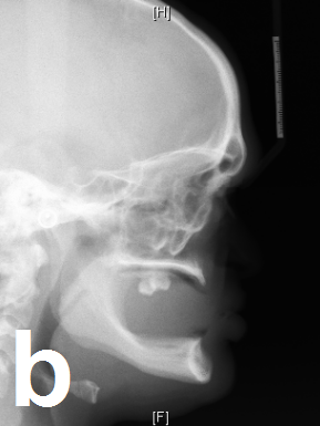
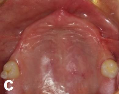
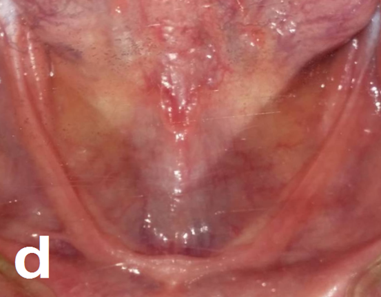
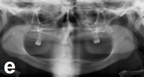
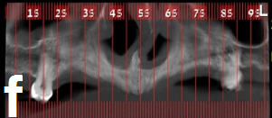
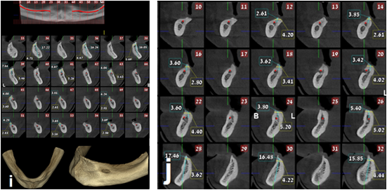
A 23-years old male was referred to our department for rehabilitation with fixed prosthesis over implants at his both jaws. The patient had tooth agenesis due to ectodermal dysplasia. Extraoral examination revealed a decreased lower face height, edentulous appearance, and protuberant lower lip (figure 1a). Phonetic problems were also obvious. Slight mandibular pragmatism was also observed (figure 1b). Intraoral examination revealed an edentulous mandible. At the maxilla two maxillary first molars were found. A prominent view of severely atrophied jaws was observed (figure 1c and 1d). On the radiographic examination, complete edentulous the both jaws except of two molars at the maxilla (figure 1e). Cone Beam Cross Sectional Computed Tomography (CBCT) was performed to assess the bone dimensions of the jaws, and revealed that the ridges in both the maxilla and the mandible were extremely atrophied horizontally and vertically with knife edge (figure 1f to 1j).
The patient treatment plan included reconstruction of the atrophied ridges in three dimensions, nasal floor elevation, and bilateral
inferior alveolar nerve transposition at the posterior mandible. Placement of dental implants in good anatomical locations to give the
ability for final rehabilitation with fixed prosthesis over the inserted implants. The treatment plan employed a staged approach and was
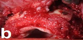
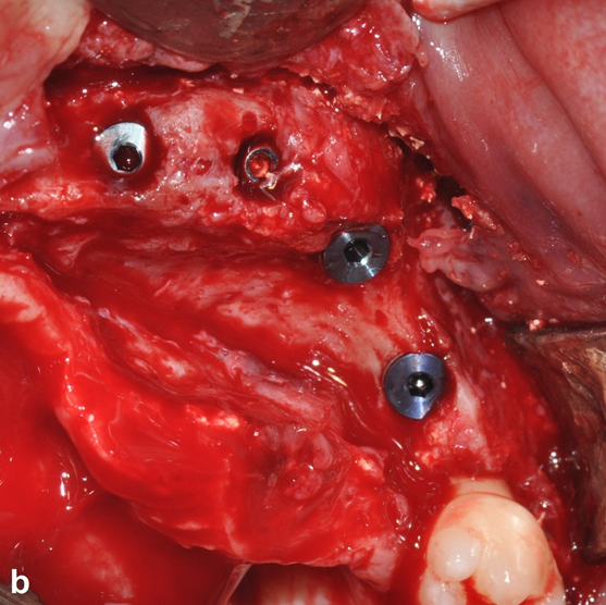
divided in three stages. At the first stage the patient underwent maxillary three dimensional bone augmentation with Onlay autogenic
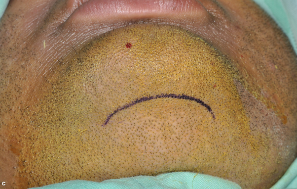
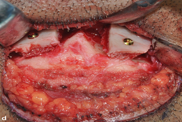
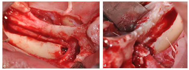
bone blocks and nasal floor elevation (figure 2a and 2b). The anterior mandible was accessed extra orally via sub mental incision and
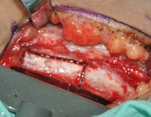
Onlay autogenic bone blocks were also used to augment the anterior mandible between the two mental foramina (figure 2c and 2d). The
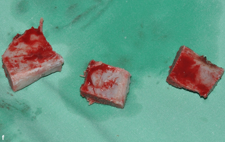
patient right anterior crest was addressed for harvesting of bone blocks (figure 2e and 2f). The radiographs obtained after this stage
demonstrated the bone grafted regions (figure 3a and 3b). Four months after the first surgery the patient sent to CBCT, that revealed a
good new bone gain at the augmented sites (figure 4a to 4f)).
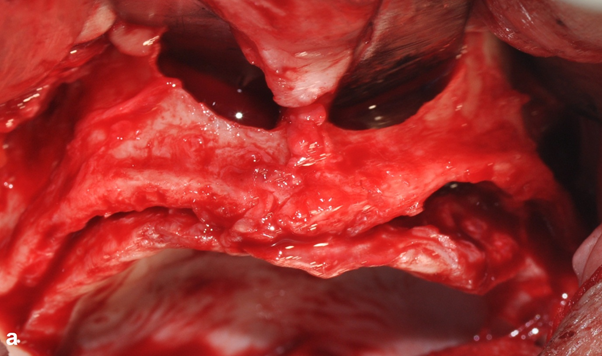
Figure 2: First Surgery; Intraoperative views.
Figure 2a: Severe atrophy of the maxilla, nasal floor was elevated.
Figure 2a: Severe atrophy of the maxilla, nasal floor was elevated.
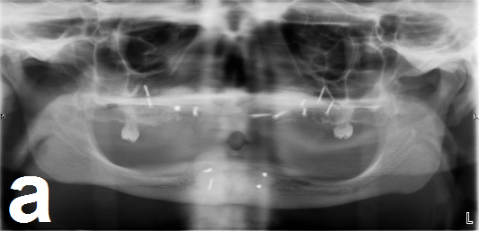
Figure 3: Radiographic views after the first surgery.
Figure 3a: Panoramic view demonstrates the augmented sites.
Figure 3a: Panoramic view demonstrates the augmented sites.
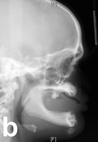
Figure 3b: Lateral cephalogram demonstrates the improvement
of the maxillary-mandibular relationship.

Figure 4: CBCT four months after the first surgery demonstrates
the excellent bone gain of the augmented regions.
Figure 4a to 4c: Maxilla.
Figure 4a to 4c: Maxilla.
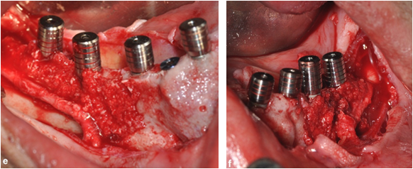
At the second stage surgery, the patient underwent placement of 8 implants between the pre-existing first molars (figure 5a and 5b).
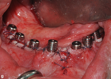
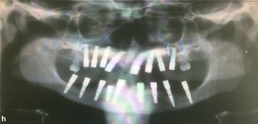
At his mandible 9 implants were placed. At the posterior sites bilateral inferior alveolar nerve transposition was performed simultaneously
with the implants insertion (figure 5c to 5j). The panoramic radiograph after the second surgery showed the satisfied implants
locations and dimensions (figure 5h). At three months after the second surgery the patient was referred to his prosthodontics for fixed
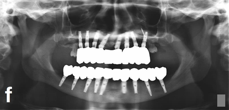
prosthesis over the implants (figure 6a to 6e). The patient is followed 22 months with excellent esthetic and functional outcomes. He
stated a huge improvement in his quality of life, he can eat, smile, and speak clearly.
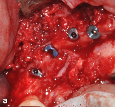
Figure 5: Intraoperative views during the second surgery.
Figure 5a: Implants insertion right maxilla.
Figure 5a: Implants insertion right maxilla.
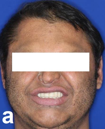
Figure 6: Full mouth rehabilitation with fixed
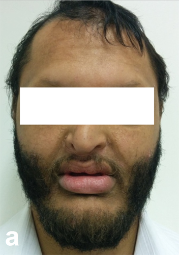
prosthesis over the implants (one year follow up).
Figure 6a: Exraoral view, harmonic facial profile.
Figure 6a: Exraoral view, harmonic facial profile.

Figure 6b to figure 6d: Fixed prosthesis over the implants, intraoral
views; Occlusion (6b), Maxillary arch (6c), Mandibular arch (6d).
Results
The recovery of the patient from the first surgery was uneventful, and excellent bone gain that was obtained. The horizontal bone gain at the maxilla and the mandible was 5-7 mm, and the nasal floor augmentation enhanced the bone height at the maxilla. The second surgical procedures healed with minor complications. The inferior alveolar nerve transposition allowed the insertion of long implants at the posterior mandible. The recovery of the nerves was completed after 3 month with complete resolution of the paraesthesia. Seventeen implants were placed in the both jaws with good dimensions (mean length 13 mm, mean width 3.75). The implants were inserted in good anatomical locations. The outcome of the treatment provided the patient oral rehabilitation with fixed prosthesis over dental implants that enhanced the patients function and aesthetics. The treatment had a great impact at the patient quality of life and the self-esteem. This patient is followed till now for 22 months.
Discussion
Dental implants are increasingly being used in the management of ED [13,14], in adult patients that complete their growth. Due to tooth agenesis, the bone height and width will not be sufficient for implant insertion without advanced surgical procedures. Our patient, has a mild form of ED with minor extra oral manifestations, He suffered from tooth agenesis, and had only two first maxillary molars. There was severe atrophy of the both jaws and edentulous appearance.
Marked atrophy of the both jaws was obvious intraoral and confirmed by the CBCT. The case required major bone augmentation procedures that were combined by special procedures. In the maxilla nasal floor elevation and augmentation were performed to improve the implants length at the anterior maxilla. At the mandible two special procedures were performed. The first was bone augmentation of the anterior mandible at the interforamine area, and this was performed via extra oral approach in order to prevent incisions inside the mouth at this region. In this way the danger of wound dehiscence over the augmented area was excluded, and the augmented bone blocks were maintained during the healing period.
The external scar was hided under the chin in a skin fold. It heals very well and left invisible. There are several reports of successful dental implants reconstruction in patients with ectodermal dysplasia. Although most of the reports have addressed implant reconstruction in the severely atrophic maxilla, few have addressed reconstruction of the both maxilla and the mandible [15,16]. Further, only a few authors report the use of implants in the treatment of adult patients with ED and reports on bone augmentation are lacking [15,17].
In this case, the severely atrophied jaws required special procedures to reconstruct both maxilla and mandible.
1- Three dimensional bone augmentation with Onlay bone blocks. The mandibular rami and chin are usually utilized as donor sites for bone blocks in bone augmentation [18,19], but in our patient those areas were not an option due to the severe atrophy of the entire mandible.
1- Three dimensional bone augmentation with Onlay bone blocks. The mandibular rami and chin are usually utilized as donor sites for bone blocks in bone augmentation [18,19], but in our patient those areas were not an option due to the severe atrophy of the entire mandible.
The donor site for the autogenous corticocancellous block graft was the anterior iliac crest and provided the desired bone volume [20]. The surgery to harvest the bone was completed with minor complications.
2- The patient had a wide nose variant. Nasal floor augmentation was performed to increase the length of the futured implants that should be inserted at the anterior maxilla. The maxillary sinuses were left intact for utilizing in case the patient will need retreatment with implants in the future.
3- The severe atrophy of his posterior mandibular ridges with mean bone height above the mandibular nerves of 2- 3 mm, the superior location of the inferior alveolar nerves, and the good mandibular height had led to the preference to do bilateral inferior alveolar nerve transposition for dental implants placement at the patient posterior mandible. This special procedure was predictable way to insert long implants (mean length 13 mm). The use of this method has not been well embraced because of the high risk of damaging the inferior alveolar nerve. In systematic review of neurosensory complications, Abayev and Juodzbaly in 2015 found that 99.47% of the cases had transient sensation changes for 1-6 months, and only 0.53% had permanent sensation disturbances [21]. In the present case the post- surgical paraesthesia was the major complication of this surgery but was completely resolved after three months.
The success of the first surgery was good and an excellent bone volume was achieved. Dental implants were placed in the second surgery with good dimensions (mean length 13 mm, mean width 3,75 mm). The final rehabilitation was performed after successful osseointegration of the implants. Two fixed prosthesis over the implants, at the both maxilla and mandible. The final outcomes were satisfied, and excellent function and esthetic were achieved.
Conclusions
The combination of 3Ds bone augmentation, nasal floor elevation and bilateral inferior alveolar nerve transposition can give the adult patients with ectodermal dysplasia the opportunity to have full mouth rehabilitation over dental implants with final outcomes that improve the patient quality of life.
Acknowledgement
To doctor Al-Keesh Kotaiba for his technical support
To doctor Al-Keesh Kotaiba for his technical support
Conflicts of interest
There are no conflicts of interest.
There are no conflicts of interest.
References
- Dorlands Illustrated Medical Dictionary, 30th edition. London: Saunders (2003).
- Wong ATY., et al. “Oral health-related quality of life and severe hypodontia”. Journal of Oral Rehabilitation 33.12 (2006): 869-873.
- Guckes AD., et al. “Using endosseous dental implants for patients with ectodermal dysplasia”. The Journal of the American Dental Association 122.10 (1991): 59-62.
- Restoy-Lozano A., et al. “Calvarial bone grafting for three-dimensional reconstruction of severe maxillary defects: a case series”. The International Journal of Oral & Maxillofacial Implants 30.4 (2015): 880-890.
- Clavero J and Lundgren S. “Ramus or chin grafts for maxillary sinus inlay and local onlay augmentation: Comparison of donor site morbidity and complications”. Clinical Implant Dentistry and Related Research 5.3 (2003): 154-160.
- Gomes KU., et al. “Use of allogenic bone graft in maxillary reconstruction for installation of dental implants”. Journal of Oral and Maxillofacial Surgery 66.11 (2008): 2335-2338.
- Stoelinga PJW., et al. “Interpositional bone graft augmentation of atrophic mandible”. Journal of Oral and Maxillofacial Surgery 36.1 (1978): 30-32.
- Urbani G., et al. “Distraction osteogenesis to achieve mandibular vertical bone regeneration: a case reort”. The International Journal of Oral & Maxillofacial Implants 19.4 (1999): 321-331.
- Jensen OT., et al. “Anterior maxillary alveolar distraction osteogenesis: a prospective 5-year clinical study”. The International Journal of Oral & Maxillofacial Implants 17.1 (2002): 52-68.
- Raghoebar GM., et al. “Vertical distraction of the severely resorbed edentulous mandible. A clinical, histological and electron microscopic study 0f 10 treated cases”. Clinical Oral Implants Research 13 (2002): 558-565.
- Lorean., et al. “Inferior alveolar nerve transposition and reposition for dental implant placement in edentulous or partially edentulous mandibles: a multicenter retrospective study”. International Journal of Oral and Maxillofacial Surgery 42.5 (2013): 656-659.
- John Jensen., et al. “Nerve transposition and implant placement in the atrophic posterior mandibular alveolar ridge”. Journal of Oral and Maxillofacial Surgery 52.7 (1994): 662-668.
- Guckes AD., et al. “Prospective clinical trail of dental implants in persons with ectodermal dysplasia”. Journal of Prosthetic Dentistry 88.1 (2002): 21-25.
- Rad AS., et al. “Full mouth rehabilitation of hypohidrotic ectodermal dysplasia patient with dental implants: a clinical report”. Journal of Prosthetic Dentistry 16.3 (2007): 209-213.
- Tarjan., et al. “Early prosthetic treatment of patients with ectodermal dysplasia: a clinical report”. Journal of Prosthetic Dentistry 93.5 (2005): 419-424.
- Acikgoz A., et al.“Hypohidrotic ectodermal dysplasia with true anodontia of primary dentition”. Quintessence International 38.10 (2007): 853-858.
- Celar AG., et al. “Use of individual traction prosthesis and distraction osteogenesis to reposition osseointegrated implants in a juvenile with ectodermal dysplasia; a clinical report”. Journal of Prosthetic Dentistry 87.2 (2002): 145-148.
- Misch CM. “Ridge augmentation using mandibular ramus bone grafts for the placement of dental implants: presentation of a technique”. Pract Periodontics Aesthet Dent 8.2 (1996): 127-135.
- Mish CM. “Comparison of intraoral donor site for inlay grafting prior to implant placement”. The International Journal of Oral & Maxillofacial Implants 12.6 (1997): 767-776.
- Nkenke E., et al. “Morbidity of harvesting of bone grafts from the iliac crest for preprosthetic augmentation procedures: A prospective study”. International Journal of Oral and Maxillofacial Surgery 33.2 (2005): 157-163.
- Abayev B and Juodzbalys G. “Inferior alveolar nerve lateralization and transposition for dental implant placement. Part II: a systematic review of neurosensory compliucations”. Journal of Oral & Maxillofacial Research 6.1 (2015): e3.
Citation:
Fares Kablan and Khater Samer. “Full Mouth Implants Rehabilitation of a Patient with Ectodermal Dysplasia After 3-Ds Ridge Augmentation and
Bilateral Inferior Alveolar Nerve Transposition. A Clinical Report.” Oral Health and Dentistry 1.2 (2017): 107-118.
Copyright: © 2017 Fares Kablan and Khater Samer. This is an open-access article distributed under the terms of the Creative Commons Attribution License, which permits unrestricted use, distribution, and reproduction in any medium, provided the original author and source are credited.





 Scientia Ricerca is licensed and content of this site is available under a Creative Commons Attribution 4.0 International License.