Research Article
Volume 1 Issue 3 - 2017
Application of two Different Classification Systems in Assessing an Enigmatic Condition-Gingival Recession: A Short Study
1Sameer Saxena, MDS, Senior Lecturer, Department of Periodontology, Teerthanker Mahaveer Dental College and Research Centre, Moradabad, India
2Gouri Bhatia, MDS, Senior Lecturer, Department of Periodontology, Teerthanker Mahaveer Dental College and Research Centre, Moradabad, India
3Ashish Kumar, MDS, Professor, Department of Periodontology, Institute of Dental Studies & Technologies, Modinagar, India
4Mansi Bansal, MDS, Reader, Department of Periodontology, Institute of Dental Studies & Technologies, Modinagar, India
5Manish Khatri, MDS, Professor, Department of Periodontology, Institute of Dental Studies & Technologies, Modinagar, India
6M Karthik Krishna, MDS, Professor, Department of Periodontology, Teerthanker Mahaveer Dental College and Research Centre, Moradabad, India
*Corresponding Author: Sameer Saxena, Department of Periodontology, Teerthanker Mahaveer Dental College and Research Centre, Moradabad, UP, India.
Received: June 29, 2017; Published: July 20, 2017
Abstract
Background: Marginal tissue recession is the displacement of soft tissue margin apical to the cemento-enamel junction with exposure of the root surface. The understanding and knowledge of different stages and condition of gingival recession is necessary for predictable root coverage. The predictability of root coverage can be enhanced by presurgical examination and correlation of recession by using any of the classifications proposed for denuded roots. Due to the multifactorial etiology, not necessarily acting synchronously, the occurrence of gingival recession at a given site may be difficult to explain fully and any subsequent changes may be hard topredict. The purpose of this study is to establish a correlation of Miller’s classification to Smith’s classification for the evaluation of gingival recession.
Materials and methods: This study was carried out in systemically healthy individuals diagnosed of marginal tissue recession. 100 recession defects were classified by a single examiner using two different classification systems which were then compared.
Results: It was seen that maximum of the defects irrespective of the Miller’s classification showed a horizontal score of 5 and a vertical score of 3 according to the index of recession.
Conclusion: With various classification systems available for gingival recession, more short term and long term studies need to be conducted to assess the applicability of these new systems over the most widely used Miller’s system of classification.
Keywords: Cemento-enamel junction; Gingival recession; Index
Introduction
Gingival recession is a common manifestation in most populations which occurs in any of the age groups starting even at an early age in some populations. [1] Gingival recession is defined as the displacement of the marginal tissue apical to the cemento-enamel junction. [2] The etiology of marginal tissue recession is multifactorial although the most common mechanism proposed for its occurrence seems to be inflammatory in nature. The recession is normally preceded by lack of alveolar bone at that site which arise due to developmental or acquired (physiological or pathological) defects. [3] The developmental defects like alveolar bone dehiscence, high muscle attachment or frenum pull, thin gingival biotype are thought to predispose to recession. Physiological factors may include orthodontic movement of teeth under force at times leading to dehiscence and recession, mechanical factors like tooth brush trauma and pathological factors like microbially induced periodontal diseases. [4-6]
There are various classifications available for gingival recession but most of them are not able to convey all the relevant information needed for a complete diagnosis, treatment plan and prognosis. The first classification was proposed by Sullivan and Atkins [1968] [7] based on the depth and width of the defect. They classified recession broadly into four groups; shallow narrow, shallow wide, deep narrow, deep wide. Mlinek., et al. [1973] [8] quantified "shallow-narrow" clefts as being < 3 mmin both dimensions, and "deep-wide" defects as being > 3 mm in both dimensions. Miller [1985] [9] classified gingival recession into four classes based on both the level of the underlying alveolar bone and degree of involvement of the mucogingival junction (MGJ). But due to certain drawbacks of this classification which has been discussed later, another classification was proposed by Smith [1997]. [10] Smith proposed a classification system by assessing the vertical and horizontal component of the defect on both facial and lingual aspects along with the involvement of the mucogingival junction.
The purpose of this study was to establish a correlation of Miller’s classification to Smith’s classification for the evaluation of gingival recession.
Materials and Methods
Source of data
This study was carried out in the department of Periodontology in systemically healthy individuals who had not undergone any periodontal treatment in the last six months and who presented with gingival recession in one or more teeth and had a detectable CEJ on the tooth with recession. Conditions like restorations, erosions or abrasions etc that may tend to obliterate the CEJ were excluded from the study. Subjects were randomly examined for gingival recession defect according to Miller’s Classification and Smith’s Index of Recession [IR] and a total of 100 such defects were classified according to both the systems.
This study was carried out in the department of Periodontology in systemically healthy individuals who had not undergone any periodontal treatment in the last six months and who presented with gingival recession in one or more teeth and had a detectable CEJ on the tooth with recession. Conditions like restorations, erosions or abrasions etc that may tend to obliterate the CEJ were excluded from the study. Subjects were randomly examined for gingival recession defect according to Miller’s Classification and Smith’s Index of Recession [IR] and a total of 100 such defects were classified according to both the systems.
Method
The classification of Miller [1985] [9] that was used to classify the recession is as follows: -
The classification of Miller [1985] [9] that was used to classify the recession is as follows: -
- Class I: marginal tissue recession not extending to the mucogingival junction. No loss of interdental bone or soft tissue.
- Class II: marginal tissue recession extends to or beyond the mucogingival junction. No loss of interdental bone or soft tissue.
- Class III: marginal tissue recession extends to or beyond the mucogingival junction. Loss of interdental bone or soft tissue is apical to the CEJ, but coronal to the apical extent of the marginal tissue recession.
- Class IV: marginal tissue recession extends beyond the mucogingival junction. Loss of interdental bone extends to a level apical to the extent of the marginal tissue recession.
The same defect was also classified according to the index of recession [Smith 1997] [10] which is as follows:-
Facial and lingual sites of root exposure on the same tooth were recorded separately. The index score consists of two digits separated by a dash (e.g.F2-4*). The first digit denotes the horizontal component and the second the vertical component of the recession, with the prefixed letter (F or L) indicating whether the recession is on the facial or lingual aspect of the tooth, and an asterisk (*) denoting involvement of the mucogingival junction (MGJ).
Facial and lingual sites of root exposure on the same tooth were recorded separately. The index score consists of two digits separated by a dash (e.g.F2-4*). The first digit denotes the horizontal component and the second the vertical component of the recession, with the prefixed letter (F or L) indicating whether the recession is on the facial or lingual aspect of the tooth, and an asterisk (*) denoting involvement of the mucogingival junction (MGJ).
The horizontal component was expressed as a whole number depending on what proportion of the CEJ is exposed, between the mesial and distal midpoints (MM-MD distance) approximally.
| Score | Criteria | |
| 0 | no clinical evidence of root exposure | |
| 1 | Subjective awareness of dentinal hypersensitivity in response to one-second air blast and/or clinically detectable exposure of the CEJ for up to 10% of the estimated MM-MD distance: a slit like defect | |
| 2 | horizontal exposure of the CEJ >10% but not exceeding 25% of the estimated MM-MD distance | |
| 3 | exposure of the CEJ > 25% of the MM-MD distance but not exceeding 50% | |
| 4 | exposure of the CEJ > 50% of the MM-MD distance but not exceeding 75% | |
| 5 | exposure of the CEJ > 75% of the MM-MD distance up to 100% | |
The vertical extent of recession was also expressed as a whole in mm on a range 0-9.
| Score | Criteria | |
| 0 | no clinical evidence of root exposure | |
| 1 | subjective awareness of dentinal hypersensitivity is reported and/or there is clinically detectable exposure of the CEJ not extending > 1 mm vertically to the gingival margin | |
| 2-8 | Root exposure 2-8 mm extending vertically from the CEJ to the base of the soft tissue defect | |
| 9 | Root exposure > 8 mm from the CEJ to the base of the soft tissue defect. | |
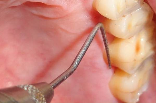
All the defects were classified by a single examiner and for the index of recession by Smith a graduated William’s periodontal probe was used and the measurement was taken to the nearest millimeter. All data collected was subjected to statistical analysis for comparison of readings by both the methods.
Results
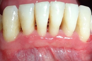
A total of 100 defects were classified out of which 69 defects were Miller’s class I, 3 recession defects fell under Miller’s class II, 25 defects were classified as class III and 3 defects were Miller’s class IV. When Miller’s class I recession defects were studied it was seen that out of the 69 defects there were 38 such defects which showed interproximal bone loss but were ought to be classified as class I as they did not extend beyond the mucogingival junction. (Figure 1)

Figure 1: Millers Class I recession with interproximal bone
loss not extending beyond the mucogingival junction.
When the Miller’s class I was compared with the Smith classification it was seen that 30 out of the 69 defects had a facial-horizontal score of 5 (F5) and 32 out of the 69 defects had a facial-vertical score of 3. In these 69 defects there were 33 defects which also showed palatal recession but couldn’t be classified by the Miller’s system and hence had to be classified only by the Smith index of recession. (Figure 2) Among the 33 defects 16 (48.48%) had a lingual-horizontal score of 5 and 19 defects showed a lingual-vertical score 3 (57.57%). (Table 1)
| Smith IR Score |
Facial-horizontal n (%) |
Facial-vertical n (%) |
Lingual-horizontal n (%) |
Lingual-vertical n (%) |
| 1 | 0 | 4 (5.8) | 0 | 3 (9.09) |
| 2 | 3 (4.3) | 20 (29) | 0 | 8 (24.2) |
| 3 | 18 (26.1) | 32(46.4) | 8(24.2) | 19 (57.57) |
| 4 | 18 (26.1) | 10 (14.5) | 9 (27.27) | 3 (9.09) |
| 5 | 30 (43.5) | 1 (1.4) | 16 (48.48) | 0 |
| 6 | 0 | 2 (2.9) | 0 | 0 |
| Total | 69 (100) | 69 (100) | 33 (100) | 33 (100) |
Table 1: Comparison of Miller’s Class I recession according to Smith index of recession.
When the Miller’s class I with interproximal bone loss was compared with the index of recession 25 out of the 38 defects had a facial-horizontal score of 5 (F-5) and 16 had a facial-vertical score of 3. Out of the 27 defects which were classified only by the Smith classification due to the recession on the lingual side, 14 defects showed a lingual-horizontal score of 5 (F-5) and a lingual-vertical score of 3. (Table 2)
| Smith IR Score |
Facial-horizontal (n) |
Facial-vertical (n) |
Lingual-horizontal (n) |
Lingual-vertical (n) |
| 1 | 3 | |||
| 2 | 1 | 14 | 8 | |
| 3 | 8 | 16 | 6 | 14 |
| 4 | 4 | 6 | 7 | 2 |
| 5 | 25 | 14 | ||
| 6 | 2 | |||
| Total | 38 (100) | 38 (100) | 27 (100) | 27 (100) |
Table 2: Comparison of Miller’s Class I recession with interproximal
bone loss according to Smith index of recession.
Among the 100 defects that were classified 3 defects each were of Miller’s class II and class IV respectively. For Miller’s class II recession
defects, 2(66.7%) had a facial-horizontal and a lingual-horizontal score of 5 and in Miller’s class IV defects all of the defects showed
a facial-horizontal and a lingual-horizontal score of 5. (Table 3)
| Smith IR Score | Miller’s Class II | Miller’s Class IV | ||||||
| Facial-horizontal n (%) | Facial-vertical n (%) | Lingual-horizontal n (%) | Lingual Vertical n (%) |
Facial-horizontal n (%) | Facial-vertical n (%) |
Lingual-horizontal n (%) | Lingualvertical n (%) | |
| 1 | ||||||||
| 2 | 1 (33.3) | 1 (33.3) | ||||||
| 3 | 1 (33.3) | 1 (33.3) | ||||||
| 4 | 1 (33.3) | 1 (33.3) | ||||||
| 5 | 2(66.7) | 1 (33.3) | 2(66.7) | 3(100) | 1 (33.3) | 3(100) | 1 (33.3) | |
| 6 | 1 (33.3) | 1 (33.3) | ||||||
| 7 | 1 (33.3) | 2(66.7) | ||||||
| Total | 3(100) | 3(100) | 3(100) | 3(100) | 3(100) | 3(100) | 3(100) | 3(100) |
Table 3: Comparison of Miller’s Class II & Class IV recession according to Smith index of recession.
The remaining 25 defects were classified as Miller’s class III in which 23 of them fell under the facial-horizontal and lingual-horizontal score of 5. 60% of the defects (15/25) had a facial- vertical score of 4 and 48% (12/25) had a lingual-vertical score of 3. (Table 4)
| Smith IR Score |
Facial-horizontal n (%) |
Facial-vertical n (%) |
Lingual-horizontal n (%) |
Lingual-vertical n (%) |
| 1 | ||||
| 2 | 1 (4) | 1 (4) | ||
| 3 | 5 (20) | 12 (48) | ||
| 4 | 2 (8) | 15 (60) | 2 (8) | 6 (24) |
| 5 | 23 (92) | 3 (12) | 23 (92) | 4 (16) |
| 6 | 1 (4) | 2 (8) | ||
| Total | 25 (100) | 25 (100) | 25 (100) | 25 (100) |
Table 4: Comparison of Miller’s Class III recession according to Smith index of recession.
Discussion
The aim of this study was to establish a correlation of Miller’s classification to Smith’s classification for the evaluation of gingival recession due to various pitfalls of the Miller’s classification. One of the major issue that arises is the classification of cases that have interproximal bone loss and do not extend beyond the mucogingival junction. Such defects cannot fall in any of the classes of Miller’s recession. [11] In our study there were 38 such defects which were accurately classified by the Smith index score for recession but could not be classified by Miller in the true sense. Secondly, recession occurring on the palatal/lingual aspect can definitely change the overall diagnosis and prognosis of a case. This recession could not be classified by Miller’s classification and this is where the need for another classification system arose. In the present study, 33 out of the 69 Miller’s class I defects showed palatal/lingual recession and all of the Miller’s Class II, III, IV defects showed palatal/lingual recession. All such defects could be classified with ease by the Smith’s classification.
By knowing the facial and lingual extent of recession in both the horizontal and vertical directions, a better estimate of the radicular bone loss could be made which further improvised the decision making. In Miller’s classification, the mucogingival junction was used as the reference point while in Smith’s classification CEJ was used as the fixed point. The difficulty in identifying the mucogingival junction creates confusion while classifying the defect as Miller’s class I/class II. [11] In certain clinical situations, a shallow recession may involve the mucogingival junction whereas a deep recession defect may not extend to or beyond the mucogingival junction based on the difference in the width of attached gingiva. [8,12,13] Also, it has been observed that at times the mucogingival junction may be indistinct visually or in some individuals the MGJ may be very near to the gingival margin that accurate assessment of the width of keratinized gingiva may seem difficult. [3,14]
In gingival recession studies identification of the CEJ is obviously facilitated because direct vision is possible and there is no data available till date as to change in CEJ with time. [10] Also, in the current study any conditions obliterating the cement-enamel junction were excluded to avoid any misinterpretations. Maximum of the recession defects classified by the Smith index score fell into the facial-horizontal and a lingual-horizontal score of 5 and a facial-vertical and a lingual- vertical score of 3. These findings are in accordance with the study of Carreno., et al. [15] who also studied the possible precipitating factors for gingival recession. Certain problems which we faced in the Smith classification were that the mid-mesial and mid-distal dimensions were difficult to calculate in cases of an intact papilla. Also, asterisk (*) on the palatal side which has no mucogingival junction could not be placed and lastly the classification is time consuming.
Various systems of classifications have been developed for gingival recession with pros and cons in each of them. Till date no classification has been considered complete and everlasting. Keeping the time constraints and ease of classification in mind, more short term and long term studies need to be conducted to assess the applicability of a new system of classification over Miller’s.
References
- Gorman WJ. “Prevalence and etiology of gingival recession”. Journal of Periodontology 38.41 (1967):316-22.
- American Academy of Periodontology (AAP). Glossary of periodontal terms. 3rded Chicago: American Academy of Periodontology (1992).
- Geiger AM. “Mucogingival problems and the movement of mandibular incisors; a clinical review.” American Journal of Orthodontics 78.5 (1980): 511-527.
- Lost C. “Depth of alveolar bone dehiscence in relation to gingival recessions”. Journal of Clinical Periodontology11.9(1984): 583-589.
- Wennstrom JL., et al. “Mucogingival therapy- Perioplastic surgery. In: Lindhe J, Lang NP, Karring T, editors. Clinical periodontology and implant dentistry. 5th ed. Blackwell Munksgaard; (2008). p. 958-959.
- Olsson M and Lindhe J. “Periodontal characteristics in individuals with varying form of the upper central incisors”. Journal of Clinical Periodontology 18.1 (1991): 78-82.
- Sullivan HC and Atkins JH. “Free autogenous gingival grafts III Utilization of grafts in the treatment of gingival recession”. Periodontics 6.4 (1964): 152-159.
- Mlinek A., et al. “The use of free gingival grafts for the coverage of denuded roots”. Journal of Periodontology 44.4 (1973): 248-254.
- Miller PD Jr. “A classification of marginal tissue recession”. The International Journal of Periodontics and Restorative Dentistry 5.21 (1985): 9-13.
- Smith RG. “Gingival recession. Reappraisal of an enigmatic condition and a new index for monitoring”. Journal of Clinical Periodontology 24.3(1997): 201-205.
- Pini-Prato G. “The Miller classification of gingival recession: Limits and drawbacks”. Journal of Clinical Periodontology 38.3 (2011): 243-245.
- Bowers G. “A study of the width of attached gingiva”. Journal of Periodontology 34.3 (1963): 201-209.
- Bhatia G., et al. “Assessment of the width of attached gingiva using different methods in various age groups: A clinical study”. Journal of Indian Society of Periodontology 19.2 (2015): 199-202.
- Guglielmoni P., et al. “Intra- and inter-examiner reproducibility in keratinized tissue width assessment with 3 methods for mucogingival junction determination”. Journal of Periodontology 72.2 (2001): 134-139.
- Carreno RS., et al. “Precipitating factors in the development of gingival recession”. Odontol Act Venez 40.2 (2002): 1-7.
Citation:
Sameer Saxena., et al. “Application of two Different Classification Systems in Assessing an Enigmatic Condition-Gingival
Recession: A Short Study”. Oral Health and Dentistry 1.3 (2017): 153-159.
Copyright: © 2017 Sameer Saxena., et al. This is an open-access article distributed under the terms of the Creative Commons Attribution License, which permits unrestricted use, distribution, and reproduction in any medium, provided the original author and source are credited.


 Scientia Ricerca is licensed and content of this site is available under a Creative Commons Attribution 4.0 International License.