Research Article
Volume 1 Issue 4 - 2017
Tridimensional Diagnosis: Asimetrys, Mandibular Condyles, Glenoid Cavitie, lower Dental mid line, Patients from Centro de Estudios Superiores de Ortodoncia.
1Andrea Carolina Ramos Cano, Masters degree on Orthodontics and Maxilofacial Orthopedics at the Centro de Estudios Superiores de Ortodoncia (CESO) México City
2Dr. Javier Mendoza-Valdés, Teacher and Clinical coordinator at CESO
3Dr. Beatriz Gurrola Martínez, Teacher at CESO and full time teacher “C” of the pre graduate Dental School at the Facultad de Estudios Superiores Zaragoza UNAM
4Adán Casasa Araujo, Chairman at CESO
2Dr. Javier Mendoza-Valdés, Teacher and Clinical coordinator at CESO
3Dr. Beatriz Gurrola Martínez, Teacher at CESO and full time teacher “C” of the pre graduate Dental School at the Facultad de Estudios Superiores Zaragoza UNAM
4Adán Casasa Araujo, Chairman at CESO
*Corresponding Author: Beatriz Gurrola Martínez, Teacher at CESO and full time teacher “C” of the pre graduate Dental School at the Facultad de Estudios Superiores Zaragoza UNAM.
Received: August 21, 2017; Published: August 30, 2017
Abstract
The human craniofacial complex is not perfectly symmetrical if you compare both sides of an arch, this asymmetry is considered a natural matter if the difference is between a reasonable quantity. The objective of this study is to locate the position and size of the condyles in the Glenoid cavity and the position of the lower dental midline through tridimentional imagenology on adult patients, to establish the mandibular midline discrepancies and condylar asymmetries.
Methodology: This was a descriptive and transversal study. The sample were 40 patients. Measurements were taken from a computerized Tomography Cone Beam, of the lower dental midline, the height of the right and left condyle, and the distance from the condyles to the mandibular midline.
Results: the measurement of the right condyle height was 2.1 mm, and the left one was observed with 2.8 mm. Regarding the distance from the right condyle to the mandibular midline was 106.3 mm, and the left to the mandibular midline (C.I.L.M.M) was 107.6 mm.
Conclusion: all patients were asymmetrical in relation to the heights of the right and left condyles with a difference of 2 mm. In the coronal plane, with the use of Cone-Beam computed tomography, the highest condylar height is not the determinant of the deviation of the mandibular midline.
Keywords: Condyle; Midline; Cone Beam; Levandosky
Introduction
Guilherme 2001, made a study on patients with Class II and ClassI Angles maloclution to determine if there is significative differences regarding asymeties. Demostrated tha the main cause contributing on the deviation of the lower mid line were the first molars on the side of the Class II maloclution. A second cause was the mesial position of the upper first molars on the Class II maloclution side. [1] On other study Lergell and Isberg,1999 noticed tha the displacement of the disc on the temporomandibular joint (TMJ) is a cause to develop a mandibular midline asymmetry. A surgical anterior displacement of the discs on rabbits with growth. The mandible on the sample animals were sonsiderably shorter on the side of the disc displacement, resulting on a lower mid line deviation toward the same side. This concludes that the displacement of the disc on the TMJ appering during growth may cause changes on the mandibular length and midline asymetry on rabbits with growth. [2] On this matter Rilo on 2008, evaluated several oclusal parameters on an adult group with unilateral posterior cross bite without treatment and compared the results of a normal group of adults. The foudings showed that the condylar inclination and the temporal eminence were asymetric on the patients with unilateral crossbite and a higher length on the side of the cross bite, also that the midline was deviated toward the side of the cross bite, showing that the mandible is longer on the side that doesn’t have the cross bite. [3]
Other study by Olate (2013) had a sample of 12 subjects, evaluated through computerized tomography (Cone- Beam) analysis; used a software to capture the images of a model Pax Zenith, brand Vatech (Korea 2011), using 90 kV y 120 mA; measurements were taken from the anterior-posterior, upper-lower, medial-external of the center of the condyle, relating the positiion of the condyle with the mandibular mid line and the facial position between the upper centrals and lower centrals as well as the menton protuberance. The results showed a men deviation of the menton protuberance of 6,5 mm considering an hiperplasic condyle with 2,7 mm longer than the normal size of condyles. The deviation of the lower centrals showed that for each 1 mm there is a 2.2 mm deviation of the menton protuberance. The hiperplasic condyle was biger in size and was almos 2 mm more lateral positioned than the normal condyles. It can be concluded that the hiperplasic condyle showed an influence on the facial asymmetry and it is possible to estimate the relation of the size of the condyle with the degree of facial asymmetry. [4] Akcam (2003) refers an evaluation of 30 panoramic xrays and 30 lateral Xrays on patients with skelletal and dental class II maloclution, with growth to determine the discrepancies between them. The most remarcable data of the descriptive analysis was the overwelming differences between the measurements of the right and left condyles on the panoramic Xrays. Concluded that the panoramic Xray is not enough trustworthy to get an acceptable information compared with lateral Xrays on facial and mandibular asymmetries. [5] Biagi R y Col. (2012). Evaluated 30 panoramic Xrays of children from 7 to 14 years old, 10 linear measurements were taken. There was a domain for the left side than the right side. The data obtained was not statistical significant, with the exception of the mandibular length: the right mandibular length was shorter compared to the left side. This concludes that the Levandoskys Panoramic analysis is a usefull and accurate method to diagnose and measure skelletal and dental asymmetries [6]. On othe study Piedra I (1995) reports facial and dental asymmeties on 41 children from 8 to 12 years old, using Levandosky Panoramic analysis, found a high correlation between the standard facial picture and the linear measurement on the panoramic Xray. 20 patients (48,78%) showed mandibular mid line deviation toward the left side, where the condyle of that side was also companied with a longer coronoid appofisis. [7] Regardint the evaluation of the vertical condylar and ramus asymmetry on teenagers who showed unilateral and bilateral cross bite and a normal control group using computerized tomography, Biagi 201 showed that even thoug condylar asymmetry was higher on the group of unilaterl cross bite, the results wer not statistical significant.
General Objective: Determine the position, size of the condyles inside the Glenoid cavity and the measure to the lower dental midline thoug the Levandosky Panoramic Analysis and tridimentional imagenology on adult patients from Centro de Estudiso Superiores de Ortodoncia (CESO) to establish if the lower mid line deviation is due to the condylar asymetry.
Materials and Methods
Kind of research: Desriptive and tranversal study.
Sample: Convienience sample, n = 40 patients before treatment that full fill the criteria selection.
Materials: 40 tomography’s Cone Beam of patients with lower dental mid line deviation, Cone Beam, Software On- Demans 3D versión APP, computer, Excel spread sheet, Statistical software SSPS numeral 21.
Methods: Registration Technique Técnique: Cone Bam brand (NewTom vg) to take the 40 Cone Beams of the patients.
We used the on demand 3D Software version APP for DVR (Dental volume reformat), figure 1
The measurementes were taken on the “Dental “label. Whit is designed to evaluate all the anathomic structures when you evaluate
and make a visula comparison between the structures. This window offes a sagital, axial and sagital and axial views, figure 2.
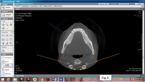
We created our own panoramic image for each patient from the axial view, using the tool Modify: Arch curve, withch allowed us to
cente the basal bone of the patient, figure 3.
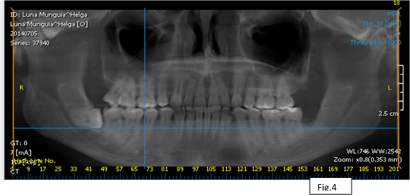
Using the filter 2x, you can improve the image, figure 4.
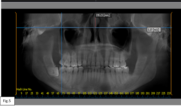
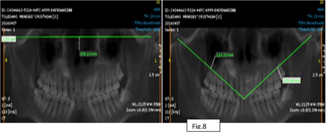
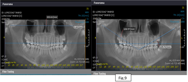
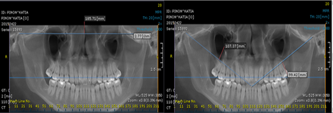
We measure line 2 from Levandoski Analysis to determine the height of the right and left condyle, which is a perpendicular line to the maxilar mid line and tangent to the highest condyle to diagnose the condylar asymmetry, figure 5.
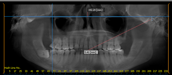
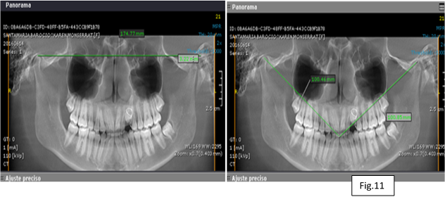
We measure the distance from the left condyle to the lower dental mid line, taking as reference the line 6 of the Levandosky Analysis that goes from the upper border of the head of the left condyle to the lower dental mid line, figure 6.
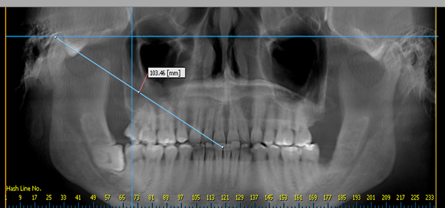
We measure the distance from the right condyle towards the lower dental mid line taking as reference line 6 of the Levandosky Analysis that goes from the upper order of the head of the right condyle to the lower dental mid line, figure 7.
The next are examples of some images of some patients for each measurement and where the grafic regitration were taken, figures
8, 9, 10 11.
Results
Regarding the age the mayority of the patients are in a group between 25 to 28 years old that is 62.5% of the sample, the 15% are between 30 and 31 years old. On regard to the gender 27 patients were female 67.5% and 13 male patients 32.5% The lower dental mid line deviation (LDMLD), 22 patients were deviated to the right side 55% and 18 patients had a deviation to the left side 45%-
Concernign the measurement of the height of the condyles measured though the Cone Beam. On the right side (CHR), we can observe a variation on the asymmetry of the patients; having light differences on 25 patients on the right side 0.mm that represents 62.5% of the total sample, 8 patients had a range of 1-2.8 mm representing 20%. For the measurement of the height of the left condyle (CHL), 20 patients had 0.mm, representing 50% of the total sample, 10 patients had 2–3.7 mm representing 25%. Table 1
| mm | Frecuency | Porcentage % | Valid Percent |
mm | Frecuency | Porcentage | Valido |
| .0 | 25 | 62.5 | 62.5 | .0 | 20 | 50.0 | 50.0 |
| .7 | 1 | 2.5 | 2.5 | .8 | 3 | 7.5 | 7.5 |
| .8 | 1 | 2.5 | 2.5 | 1.1 | 2 | 5.0 | 5.0 |
| 1.0 | 1 | 2.5 | 70.0 | 1.3 | 1 | 2.5 | 2.5 |
| 1.1 | 1 | 2.5 | 72.5 | 2.1 | 1 | 2.5 | 2.5 |
| 1.6 | 1 | 2.5 | 75.0 | 2.4 | 2 | 5.0 | 5.0 |
| 1.8 | 1 | 2.5 | 77.5 | 2.7 | 1 | 2.5 | 2.5 |
| 2.0 | 1 | 2.5 | 80.0 | 2.9 | 1 | 2.5 | 2.5 |
| 2.1 | 2 | 5.0 | 85.0 | 3.2 | 2 | 5.0 | 5.0 |
| 2.8 | 1 | 2.5 | 87.5 | 3.4 | 1 | 2.5 | 2.5 |
| 3.0 | 1 | 2.5 | 90.0 | 3.5 | 1 | 2.5 | 2.5 |
| 3.1 | 1 | 2.5 | 92.5 | 3.7 | 1 | 2.5 | 2.5 |
| 3.5 | 1 | 2.5 | 95.0 | 4.9 | 1 | 2.5 | 2.5 |
| 5.2 | 1 | 2.5 | 97.5 | 5.4 | 2 | 5.0 | 5.0 |
| 8.7 | 1 | 2.5 | 100.0 | 5.5 | 1 | 2.5 | 2.5 |
| Total | 40 | 100.0 | 100.0 | Total | 40 | 100.0 | 100.0 |
Concerning the measure on the Cone Bean for the right condyle to lower dental midline (R.C.M.M.L), 25 patients had a range of 100-110mm being 47.5% and 9 patients on a range 110-120 mm being 25%. On the measurement of the left condyle to the lower dental midline (L.C.M.M.L.) 18 patients had a range of 100- 110 mm being 45% and 8 patients had a range of 110-114 mm being 27.5%
| R.C.L.D.M.L | Frecuency | Porcenta Ge % |
Valid Percent |
L.C.L.D.M.L, | Frecuenciy | % | Valid Percent |
| 95 a 99.5 | 6 | 2.5 | 15 | 95.7 a 99.4 | 7 | 2.5 | 15 |
| 100.4 a 105.3 | 11 | 27.5 | 39.0 | 100.3 a 105.7 | 12 | 48.5 | 63 |
| 106.3 | 2 | 5.0 | 42.0 | 106.2 | 1 | 2.5 | 65.5 |
| 106.8 | 1 | 2.5 | 44.5 | 106.5 | 1 | 2.5 | 68.5 |
| 107.2 | 1 | 2.5 | 46.5 | 106.6 | 1 | 2.5 | 70.0 |
| 107.4 | 1 | 2.5 | 49.0 | 107.4 | 2 | 5.0 | 75.0 |
| 107.8 | 1 | 2.5 | 51.5 | 107.8 | 2 | 5.0 | 81.0 |
| 108.2 | 1 | 2.5 | 53.5 | 107.9 | 1 | 2.5 | 83.5 |
| 108.7 | 1 | 2.5 | 56.0 | - | - | - | - |
| 108.8 | 1 | 2.5 | 58.5 | - | - | - | - |
| 109.1 | 1 | 2.5 | 60.5 | 109.4 | 1 | 2.5 | 85.0 |
| 109.2 a 109.8 | 4 | 10.5 | 70.0 | 110.7 | 1 | 2.5 | 87.5 |
| 110.3 | 1 | 2.5 | 72.5 | 11.0 a 111.5 | 2 | 4.5 | 92.0 |
| 111.4 | 1 | 2.5 | 75.5 | 111.9 | 1 | 2.5 | 94.5 |
| 111.9 | 1 | 2.5 | 77.5 | 112.3 | 1 | 2.5 | 96.5 |
| 112.3 | 1 | 2.5 | 80 | 112.6 | 1 | 2.5 | 98.5 |
| 114.1 | 1 | 2.5 | 82.5 | 113.2 | 1 | 2.5 | 2.5 |
| 114.7 | 1 | 2.5 | 85.5 | 114.1 | 1 | 2.5 | 2.5 |
| 115.6 | 1 | 2.5 | 88.0 | - | - | - | - |
| 117.5 | 1 | 2.5 | 90.5 | - | - | - | - |
| 120.5 | 1 | 2.5 | 92.5 | - | - | - | - |
| Total | 40 | 100.0 | 100.0 | ||||
Discussion
Authors like Guilherme, Moyers, Sağlam, Pinto, De Moraes, Silva, Sezgin, Varoli, Langberg [1,9,10,11,12,13,14,15,16] Mention: human craneofacial complex is not perfetly symmetrical if you comapare right to left side; the asymmetries are considered as a natural phenomena only if it is in a reasonable limits. On the study made at the CESO we found all patients present some asymmetry, presenting some variablity from one side to the other as much as 8 mm but with normal facial charasteristics. On other study Jiménez, [17] evaluated and quantified different causes that may cause mandibular asymmetry or deviation of the lower dental mid line on a patient during or at the end of the growth on 20 children. He usde several points such as Mento, Gonion, lower dental mid line, and articulare, to define a specific mandibular area. On the other hand, the patient may present asymmetry displacement, structural asymmetry or a combination of both, and the last choice could be the most frequent on patients with growth: This was pointe otu by Fuentes, Kambylafkas, Kiki, Kilic [18-21] On this matter we can mention that on this research at CESO, due to the characteristics og the study the points and measurements taken were: condileon, upper dental mid line, its tangent, lower dental mid line, the study was made on the Panoramic image taken by the Cone Beam. Regarding the evaluation throug Computerized Tomography Analysis Cone Beam, Olate & col (2013) report a study on 12 subjects, where they measured the position of the position of the condyles toward mandibular mid line and the position of facial points between upper and lower centrals and menton. The results showed an average of deviation of the menton of 6.5 mm cosiderating an hiperplasic condyle with a size 2.7 mm higher than the normal condyles. They concluded the same as Uysal, Chin, Bermejo, Ueki, Okeson, Kasumi, Westesson, that the hiperplasic codyle represents aa clear influence on the facial asymmetry and it is possible to estimate a relation of the size of the condyle and degree of facial asymmetry, being an important factor to deviate the lower mid line. [4, 22-28] On the study made at CESO all the patients had an asymetry on the condilar heith and lower dental mid line deviation, however, the condyle with the higher measurement was not the one to determine or cause the asymetry, the mayority of the patients, 22, presented a deviation towards the right side , and the right condyle presented a higher heigth on the total average of the sample, and for sure there are several other ethiological factors such as genetics, enviriomental, functional, or developmental that may influence on the presence of facial or dental asymmeties.
Conclusion
Although all the patients presented asymetries on the heith of the condyles right and left, with an average of difference of 2 mm. We can consider that the higher heigth is not determinat of the deviation of the mandibular midline, this was ratified with the coronal plane of the computerized tomography Cone Beam. It is important to use other media such as the Coputerized Tomography Cone Beam that may help to stablish a more accuarate judgement because can provide dental and skelletal accuracy with a lot of advantages: Field of view variable from 5 cm x 5 cm up to 17 cm x 13.5 cm. Integrates on the same sistem the Panoramic View, the Lateral View, and Tridimentional View (3D) with exceptional quality of image: a wide range of clinical applications like focused fiedl, idividual maxilla,double maxilla, individula TMJ or bilateral TMJ. Etc.
References
- Guilherme RP., et al.“Three-dimensional evaluation of skeletal and dental asymmetries in Class II subdivisión malocclusions”. American Journal of Orthodontics and Dentofacial Orthopedics 119.4 (2001): 406-418.
- Legrell PE. And Isberg A. “Mandibular length and midline asymmetry after experimentally induced temporomandibular joint disk displacement in rabbits”. American Journal of Orthodontics and Dentofacial Orthopedics 115.3 (1999): 247-253.
- Rilo Benito., et al. “Midline shift and lateral guidance angle in adults with unilateral posterior crossbite”. American Journal of Orthodontics and Dentofacial Orthopedics 113.6 (2008): 804-808.
- Olate S., et al. “Relación entre el tamaño condilar y la asimetría facial transversal en individuos con hiperplasia condilar”. International Journal of Morphology 31.3 (2013): 937-941.
- Akcam., et al. “Panoramic radiographs: A tol for investigating skeletal pattern.” American Journal of Orthodontics and Dentofacial Orthopedics 123.2 (2003):175-181.
- Biagi R., et al. “Diagnosis of dental and mandibular asymmetries in children according to LevandoskiPanoramic Analysis”. European Journal of Paediatric Dentistry 13.4 (2012):297-300.
- Piedra I. “The Levandoski Panoramic Analysis in the diagnosis of facial and dental asymmetries”. The Journal of Clinical Pediatric Dentistry 20.1 (1995): 15-21.
- Canut Brusola J. Ortodoncia clínica. 1st edición. Barcelona: Editorial: Salvat (2001).
- Moyers Robert E. Manual de Ortodoncia. 4th edición. Buenos Aires: Editorial: Panamericana (1992).
- Sağlam A. “The condylar asymmetry measurements in different skeletal patterns”. Journal of Oral Rehabilitation 30.7 (2003): 738-742.
- Pinto A., et al. “Morphological and positional asymmetries of young children with functional unilateral posterior crossbite”. American Journal of Orthodontics and Dentofacial Orthopedics 120.5 (2001): 513-520.
- De Moraes., et al. “Assimetria da Face associada a mordida Cruzada Medida em Radiografías Panorámicas”. Revista Gaucha de Odontología 52.2 (2004).
- Silva M., et al. “Morfometría del Proceso condilar y Rama de la Mandíbula en individuos adultos de la ciudad de Temuco-Chile”. International Journal of Morphology 22.2 (2004): 169-172.
- Sezgin O., et al. “Mandibular Asymmetry in Different Occlusion Patterns”. The Angle Orthodontist 77.5 (2007).
- Varoli, FP,. et al. “Estudio radiográfico da asimetría condilar por meio de elipsopantomografias de indivíduos descendentes de japoneses”. Revista do Instituto de Ciências da Saúde 23.1 (2005).
- Langberg B., et al. "Transverse skeletal and dental asymmetry in adults with unilateral lingual posterior crossbite." American journal of orthodontics and dentofacial orthopedics 127.1 (2005): 6-15.
- Jiménez V., et al. "Influencia de la mordida cruzada posterior unilateral en el crecimiento mandibular”. Revista Latinoamericana de Ortodoncia y Odontopediatria 127.1 (2006): 1-19.
- Fuentes R., et al. “Morfometría Mandibular en Adultos a través de una Radiografía Panorámica”. International Journal of Morphology 26.2 (2008).
- Kambylafkas., et al. “Validity of Panoramic Radiographs for Measuring Mandibular Asimmetry”. Angle Orthodontist 76.3 (2006): 388-393.
- Kiki A., et al. “Condylar Asymmetry in Bilateral Posterior Crossbite Patients”. Angle orthodontist 133.3 (2008): 382-387.
- Kilic N., et al. “Condylar Asymmetry in Unilateral Posterior Crossbite Patients”. Angle orthodontist 133.3 (2008): 382-387.
- Uysal T., et al. “Condylar and ramal vertical asymmetry in unilateral and bilateral posterior crossbite patients and a normal occlusion simple”. American journal of orthodontics and dentofacial orthopedics 136.1 (2009): 37-43.
- Chin L., et al. “The viscoelastic properties of the human temporomandibular joint disc”. Journal of Oral and Maxillofacial Surgery 54.3 (1996): 315-318.
- Bermejo A., et al. “Morphology of the meniscotemporal part of the temporomandibular joint and its biomechanical implications”. Act Anat 129.3 (1987): 220-226.
- Ueki K., et al. “The relationship between temporomandibular joint disc morphology and stress angulation in skeletal Class III patients”. European Journal of Orthodontics 27.5 (2005): 501-506.
- Okeson J. Signos y síntomas de los trastornos temporomandibulares. In: Okeson J, editor. Oclusión y afecciones temporomandibulares. 3ª edición. Madrid: De Mosby/doyma. (1995): 178- 213.
- Kasumi I and Akira K. “Assessment of optimal condylar position with limited cone-beam computed tomography.” American Journal of Orthodontics and Dentofacial Orthopedics 135.4 (2009): 495-501.
- Westesson P and Paesani D. “MR imaging of the TMJ. Decreased signal from the retrodiskal tissue”. Oral Surgery, Oral Medicine, Oral Pathology 76.5 (1993): 631-635.
- Avila Castro O J. “Estudio retrospectivo sobre los tipos de crecimiento mandibular” Revista Dentista y Paciente 99 (2000).
- Tasaki MM and Westesson PL. “Temporomandibular joint: diagnostic accuracy with sagittal and coronal MR imaging”. Radiology 186.3 (1993): 723-729.
Citation:
Beatriz Gurrola Martínez., et al. “Tridimensional Diagnosis: Asimetrys, Mandibular Condyles, Glenoid Cavitie, lower Dental
mid line, Patients from Centro de Estudios Superiores de Ortodoncia.” Oral Health and Dentistry 1.4 (2017): 187-196.
Copyright: © 2017 Beatriz Gurrola Martínez., et al. This is an open-access article distributed under the terms of the Creative Commons Attribution License, which permits unrestricted use, distribution, and reproduction in any medium, provided the original author and source are credited.


 Scientia Ricerca is licensed and content of this site is available under a Creative Commons Attribution 4.0 International License.