Research Article
Volume 1 Issue 6 - 2017
In Vitro Study of Micro Leakage and Micro Hardness of High Viscosity Glass Ionomer Cement and Resin Modified Glass Ionomer Cement
1Instituto Superior Ciênçia da Saúde Egas Moniz, Monte da Caparica, Alamda Portugal
2Department of Pediatric Dentistry, Instituto Superior Ciênçia da Saúde Egas Moniz, Monte da Caparica, Alamda Portugal
2Department of Pediatric Dentistry, Instituto Superior Ciênçia da Saúde Egas Moniz, Monte da Caparica, Alamda Portugal
*Corresponding Author: Luísa Lopes PhD, Campus Universitário, Quinta da Granja, 2829-511 Monte da Caparica, Almada, Portugal.
Received: October 24, 2017; Published: November 09, 2017
Summary
Aim: The aim of this study was to compare Vickers Micro hardness of three conventional glass ionomer cement (Ketac Molar 3M, Photac Fil 3M and Equia System GC) before and after being submitted to an artificial aging and to evaluate the degree of marginal micro leakage.
Materials and Methods: There were made 45 samples, 15 of each material, from a standard matrix. Each sample was submitted to a first Vickers Micro hardness measurement, and then repeated after artificial aging. Using the same glass ionomer cements, 45 Class I restorations were realized in molars, then immersed in blue methylene 2%. They were then sectioned and the results were read with the use of a magnifying glass with 40 times magnification.
Results: The effect and magnitude of aging are dependent on the type of glass ionomer cement. The Micro hardness differs significantly between the Photac and the other two types of glass ionomer cement (p < 0,001). In Micro leakage, there were no relevant statistical differences between the three different materials (p = 0,053).
Conclusion: Equip System was the one with a smaller difference between the initial and final micro hardness values. All materials presented some degree of micro leakage and Photac Fil was the one with less degree.
Keywords: Glass ionomer cement; Vickers’ Micro hardness; Chewing Simulator; Marginal micro leakage
Introduction
In the last years there has been a great evolution in restorative materials. Glass ionomer cements have appeared due to the low physical and chemical properties of restorative materials and they have shown good chemical bond to enamel and dentin, fluoride releasing, good Biocompatibility and low toxicity. (Seemann, Flury, Pfefferkorn, Lussi, & Noack, 2014)
Wilson and Kent have introduced glass ionomer cements in the 70s, and since then there has been a great evolution of this material. These materials are considered types and subtypes of the original glass ionomer cement. They are divided as restorative materials in conventional glass ionomer cement, resin modified glass ionomer cement, metal modified glass ionomer cement and a subtype of conventional glass ionomer cement, the high viscosity glass ionomer cement. (Vieira, Louro, Atta, Navarro, & Francisconi, 2006)
The resin modified glass ionomer cement emerged around the 80s, to improve the physical and chemical proprieties of conventional glass ionomer cements. The low initial hardness and the water sensibility of glass ionomer cements were disadvantages, which decreased with the adding of resin to their constitution. This adding also gave glass ionomer cement the ability to have micromechanical adhesion to dentin, besides the chemical bond that was given by the acid base reaction of glass ionomer cement. (Wiegand, Buchalla, Attin, 2007).
On the other hand, a subtype of the original conventional glass ionomer emerged with characteristics that favour the mechanical properties of glass ionomer cements. The high viscosity glass ionomer cement has a higher powder liquid ration and smaller glass particles, which allow having a higher resistance to compression and higher hardness. Recently there has been an association of the high viscosity glass ionomer with a resin coating that promotes the sealing of restorations and maintains the integrity of the teeth-restoration interface. (Gurgan., et al. 2015)
ART (A traumatic Restorative Treatment) has been reported as having excellent outcomes. Specifically in terms of restoration retention, and ability to treat large numbers of children in otherwise inaccessible and isolated areas. The ART treatment has been introduced primarily using traditional glass ionomer materials and after specifically formulated glass ionomers have been developed for this ART technique. These are high powder-to-liquid ratio traditional glass ionomer materials, with enhanced physical properties (high viscosity glass ionomer cements). (Gurgan, Kutuk, Ergin, Oztas, Cakir, 2015)
Micro leakage and Micro hardness are ways to evaluate the clinical performance of the materials. In Micro leakage studies, colour dye penetration is the most commonly technique and the use of methylene blue 2% is the simplest and one of the fastest method. Due to the constant appearance of new restorative materials, it is important to have simple and fast in vitro tests to evaluate the performance of the new restorative materials. (Fabianelli., et al. 2007) In Micro hardness, one of the tests that can be used is the Vickers micro hardness test. It is very used once it allows using a small size of the samples in study, obtaining results in short time with a minimal cost. (Wang., et al. 2003)
The abrasion resistance of a material is a factor that must be taken into account when evaluating the performance of the restoration. One of the in vitro tests that can be used to evaluate abrasion resistance is the Chewing Simulator test. The mechanical and thermal cycles done by the Chewing Simulator can reproduce the physiological function and this allows it to be a useful test to study the behaviour of restorative materials in the oral cavity in an in vivo situation. (Guo., et al. 2014)
Materials and Methods
Vickers Micro hardness
Samples of the three different materials were made using a metallic mold of 10 mm diameter and 2 mm thickness (ISO 4049: 2009). To achieve a smooth surface, there were placed a glass plate underneath the metal matrix and between the matrix and the restorative materials an acetate sheet. All materials were prepared according to the manufacturer's instructions. They were made 15 samples of each material. It produced a total of 45 disks divided into 3 groups: 15 Ketac Molar disks, 15 Equia System disks and 15 Photac Fil disks. The disks were stored in distilled water at 37 degrees for 24 hours, in the dark and was subsequently initiated the readings of Vickers Micro hardness of all samples. The Vickers indentations were performed on the polished surface of the samples, each indentation for 5 seconds with 29.42N. 5 indentations were performed on each sample to obtain an average of each disk. There were made 225 measurements Vickers.
Samples of the three different materials were made using a metallic mold of 10 mm diameter and 2 mm thickness (ISO 4049: 2009). To achieve a smooth surface, there were placed a glass plate underneath the metal matrix and between the matrix and the restorative materials an acetate sheet. All materials were prepared according to the manufacturer's instructions. They were made 15 samples of each material. It produced a total of 45 disks divided into 3 groups: 15 Ketac Molar disks, 15 Equia System disks and 15 Photac Fil disks. The disks were stored in distilled water at 37 degrees for 24 hours, in the dark and was subsequently initiated the readings of Vickers Micro hardness of all samples. The Vickers indentations were performed on the polished surface of the samples, each indentation for 5 seconds with 29.42N. 5 indentations were performed on each sample to obtain an average of each disk. There were made 225 measurements Vickers.
After the readings of the initial Vickers Micro hardness of each sample, they were submit to artificial aging, using the Chewing Simulator. In the chambers of Chewing Simulator the force applied to each sample was 50N , with a frequency of 1.5 Hz, lowering speed 40 mm/s and lateral movement defined 0.7 mm to 20 mm/s. 240000 cycles, corresponding to one year in vivo. During artificial aging, it was placed in the chambers distilled water at 37 degrees to simulate the temperature of the oral cavity and to remove particles resulting from the wear of the surface of the samples.
To avoid oscillation movements of the samples during the artificial aging, the discs were wrapped in acrylic resin (Vertex). After the complete cycles, all disks were clean and dry with paper towels. Distilled water was removed from the chambers Chewing Simulator.
After artificial aging, all the samples were again submited to a new Vickers Micro hardness reading. Five indentations were performed on each sample to obtain an average of each disk.
Marginal Micro leakage
Class I cavities were performed with cylindrical diamond drill bits in the 45 teeth with standardized dimensions: depth of 3 mm , mesiodistal width of 6mm and buccolingual width of 2mm . They were restored in accordance with the standards of the manufacturer: 15 teeth with Ketac Molar, 15 with Photac Fil and 15 with Equia System GC . The teeth were fully covered with a lacquer layer except the restoration and 1 mm around the interface restoration/tooth .
Class I cavities were performed with cylindrical diamond drill bits in the 45 teeth with standardized dimensions: depth of 3 mm , mesiodistal width of 6mm and buccolingual width of 2mm . They were restored in accordance with the standards of the manufacturer: 15 teeth with Ketac Molar, 15 with Photac Fil and 15 with Equia System GC . The teeth were fully covered with a lacquer layer except the restoration and 1 mm around the interface restoration/tooth .
The teeth were placed in distilled water at room temperature and then immersed for twenty-four hours in a solution of 2% methylene blue. After that, they were removed and washed with running water for 15 minutes. All the teeth were sectioned longitudinally towards Mesio - Distal direction, using a cutting machine. The observation of micro leakage was held in magnifying glass with magnification of 40 times.
The quantification of micro leakage was determined using the following scale:
Grade 0 - No infiltration ;
Grade 1 - With infiltration that only reaches enamel;
Grade 2 - With infiltration reaching enamel and dentin;
Grade 3 - With infiltration reaching enamel, dentin and pulp.
Grade 0 - No infiltration ;
Grade 1 - With infiltration that only reaches enamel;
Grade 2 - With infiltration reaching enamel and dentin;
Grade 3 - With infiltration reaching enamel, dentin and pulp.
Results and Discussion
Micro leakage
A descriptive analysis was performed between the three materials and it was carried out a statistical analysis taking into account only the material that had suffered micro leakage and without micro leakage (Table 1).
A descriptive analysis was performed between the three materials and it was carried out a statistical analysis taking into account only the material that had suffered micro leakage and without micro leakage (Table 1).
| With Microleakage | Without Microleakage | Total | |
| Ketac Molar | 2 4,4% |
13 28,9% |
15 |
| Photac Fil | 8 17,8% |
7 15,6% |
15 |
| Equia Fil | 7 15,6% |
8 17,8% |
15 |
| Total | 17 37,8% |
28 62,2% |
45 100% |
Table 1: Micro leakage comparison between the three groups of glass ionomer cements.
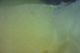
Equia Fil showed no micro leakage in 7 teeth (Figure 1), 6 teeth showed micro leakage in enamel, and 2 teeth dentin micro leakage
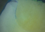
Photac Fil showed no micro leakage in 8 teeth, 5 teeth with micro leakage in enamel (Figure 2) and 2 teeth with dentin micro leakage
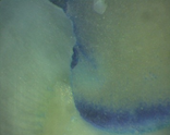
Ketac Molar, showed 2 teeth with no micro leakage, 3 had micro leakage in enamel and 10 showed micro leakage in dentin (Figure 3).
It was performed the chi- square test in order to relate the micro leakage with materials. It was concluded that the micro leakage
existence does not depend on the material ( p = 0.053 ) .
Micro hardness
The values of Micro hardness Vickers, initial and final, were similar in Ketac Molar and Equia Fil (average of 60.29VHN3 and 59.58VHN3 respectively in the initial Micro hardness and 53VHN3 and 53.VHN3 respectively in the final Micro hardness).
The values of Micro hardness Vickers, initial and final, were similar in Ketac Molar and Equia Fil (average of 60.29VHN3 and 59.58VHN3 respectively in the initial Micro hardness and 53VHN3 and 53.VHN3 respectively in the final Micro hardness).
The Micro hardness values for Photac Fil were the lowest, either the initial or the final Micro hardness (an average of 43.48VHN3 and 32.59VHN3 respectively).
IT was applied the ANOVA test with Brown- Forsythe correction so that was possible to check if there were significant differences between the Micro hardness of the three groups.
Proceeding to the test of multiple comparisons (Table 2) and taking into account that there was a significant difference between the groups when the significance is p < 0.005 , it was possible to conclude that there was a significant difference between the values of Photac Fil Micro hardness when compared to Equia and Ketac values (p < 0.0001). In turn, the latter two groups do not have Micro hardness values which differ significantly from each other.
| Depedent variable | Group (I) | Group (j) | Significance |
| Initial Microhardness | Ketac | Equia | 0,985 |
| Photac | 0,000 | ||
| Equia | Ketac | 0,985 | |
| Photac | 0,000 | ||
| Photac | ketac | 0,000 | |
| Equia | 0,000 | ||
| Final Microhardness | Ketac | Equia | 0,979 |
| Photac | 0,000 | ||
| Equia | Ketac | 0,979 | |
| Photac | 0,000 | ||
| Photac | Ketac | 0,000 | |
| Equia | 0,000 |
Table 2: Test of multiple comparisons for initial and final
micro hardness of the GIC Groups.
Following the inferential analysis it was observed that:
- Aging results in a significant decrease in the Micro hardness of the GICs (p = 0.001) observed with a potency of 95.5%.
- The efect and magnitude of aging is dependent on the type of GIC.
- Micro hardness differs significantly between Photac and the other two kinds of IVC (p < 0.001), observed with a potency of 99.9%
- The Tamhane 's multiple comparison analyses test indicates that there were significant differences Photac (p < 0.001) for the other two GIC groups but no significant differences between Ketac and Equia.
Glass Ionomer Cements have advantages like good chemical bond to enamel and dentin, fluoride release and Biocompatibility but they also have disadvantages like low resistance to abrasion and low aesthetics. The conventional glass ionomer bond to tooth structure is achieved by the carboxile group of glass ionomers and the Calcium ions of teeth. This bond is strong, but the bond of resin modified glass ionomer appears to be stronger due to fact that they have de micro-mechanical adhesion to teeth, provided by resin, besides the conventional bond of a glass ionomer.
This is the reason why a great majority of studies showed better results in terms of micro leakage, in resin modified glass ionomers when compared with the conventional glass ionomer. (Tyas, 2006).
The appearance of micro leakage in restorations can be the result of dimensional changes, temperature, mechanical stress, or failure in the adaptation of the restoration, which can result in a flaw of the interface tooth/restoration. Micro leakage can then result in sensibility, secondary caries, marginal discoloration and pulp lesions. (Masih, Thomas, Koshy, & Joshi, 2011)
In the present study it was used methylene blue to evaluate the degree of micro leakage in glass ionomer restorations. Upadhyay and Rao (2011) consider that this is the fastest and simplest method to study micro leakage. Youngson, Jones, Manogue & Smith (1998) affirm that the use of 2% concentration is the most apllied in this type of studies.
In this study Ketac Molar was the material that showed a higher number of teeth with micro leakage. This was also described by Raggio, Rocha & Imparato (2002) and Mufti (2014) when comparing Ketac Molar with resin modified glass ionomers. In the other hand, the Equia system has showed results very similar to resin modified glass ionomer, which was also described by Gurgan., et al. (2015) and Friedl, Hiller & Friedl (2011). This results may be due to the use of a resin coating, Equia coat that grants a good marginal sealing, decreases the fracture risk and prevents an early wear of the restoration.
Micro hardness is one of the most important physical properties of dental materials, with an interplay of various properties such as ductility and malleability, having the ability to pass on important information about the behavior of materials and longevity. The hardness of a GIC, in turn, is influenced by many factors including the particle size and the ratio powder/liquid in their constitution (Aratani, Pereira, Correr-Sobrinho, Sinhoreti, & Consani, 2005). According to Shintome., et al. cited by Perondi., et al. (Perondi., et al. 2014), one CIV with higher ratio powder/liquid has higher Micro hardness values.
In addition of the constitution of the material, also other factors may influence GICs physical and mechanical properties. In attempt to improve the mechanical properties and to make GICs likely to be used in high occlusal stress areas, in addition of adjusting the composition and shape of the glass particles and the polyacrylic acid of these materials , manufacturers have added protective resin (coating ) to this materials. However, as regards the micromechanical characteristics, the use of a resin coating does not appear to change the Micro hardness of CIVCs , although this brings significant advantages in properties such as flexural strength in 90%. ( Zoergiebel & Ilie, 2013b ) ( Pitel, 2010).
In the results obtained in this study, the values of Vickers Micro hardness of Ketac Molar and Equia Fil + Equia Coat did not show very different values, either in the initial measurements or in the final measurements (60.288 VHN3 and 59,575VHN3 as means of initial measurements; 53,008VHN3 and 53.685 VHN3 as averages of the final measurements, respectively) supporting the previously reported.
However, the Micro hardness of Ketac Molar and Equia GC in this investigation was higher when compared to Photac Fil Micro hardness (43.48 VHN3 32,592VHN3 initial and final). The explanation for these values was based on the constitution of Photac Fil whose chemical composition includes methacrylate monomers, which despite the good performance of resin-modified glass ionomer cements in many parameters, regarding to the Micro hardness no advantage or improvement over conventional was considered (Magni., et al. 2010).
Conclusion
All the glass ionomer cements tested showed some level of micro leakage after immersed for 24 hours in methylene blue 2%. However it was not observed micro leakage in enamel, dentin and pulp in any of the materials. The material with the highest number of teeth with micro leakage was Ketac Molar and the material that showed a lower number of teeth with micro leakage was Photac. Equia had very similar results to the Photac Fil.
After artificial aging, all the groups of glass ionomer cements tested showed a decrease of their Vickers Micro hardness. Photac Fil was the one with the lowest values of Micro hardness before and after the artificial aging. The Equia System was the material with better results, showing the best chewing strength results by obtaining a minor difference when comparing the initial and final Micro hardness.
Therefore, the materials in study with best results concerning both micro leakage and Micro hardness was Equia System, a high viscosity glass ionomer cement with a surface protector, Equia Coat.
References
- Aratani M., et al. "Compressive strength of resin-modified glass ionomer restorative material: effect of P/L ratio and storage time". Journal of Applied Oral Science 13.4 (2005): 356-359.
- Fabianelli A., et al. "The Relevance of Micro - Leakage Studies". Modern Dentistry Media 9.3 (2007): 64-74.
- Friedl K., et al. "Clinical performance of a new glass ionomer based restoration system: A retrospective cohort study". Dent Materials 27.10 (2011): 1031-1037.
- Guo J., et al. "Investigation of the time-dependent wear behavior of veneering ceramic in porcelain fused to metal crowns during chewing simulations". Journal of the Mechanical Behavior of Biomedical Materials 40 (2014): 23-32.
- Gurgan S., et al. "Four-year Randomized Clinical Trial to Evaluate the Clinical Performance of a Glass Ionomer Restorative System". Operative Dentistry 40.2 (2015): 134-43.
- Magni E., et al. "Evaluation of the mechanical properties of dental adhesives and glass-ionomer cements". Clinical Oral Investigations 14.1 (2010): 79-87.
- Masih S., et al. "Comparative evaluation of the micro leakage of two modified glass ionomer cements on primary molars. An in vivo study”. Journal of Indian Society of Pedodontics and Preventive Dentistry 29.2 (2011): 135-139.
- Perondi PR., et al. "Ultimate tensile strength and Micro hardness of glass ionomer materials". Brazilian Dental Science 17.1 (2014).
- Pitel ML. "A rapid and aesthetic alternative to a direct posterior composite". Dentistry Today 11.29 (2010): 150-151.
- Raggio DP., et al. "Avaliação da Microinfi ltração de Cinco Cimentos de Ionômero de Vidro Utilizados no Tratamento Restaurador". Jornal brasileiro de odontopediatria & odontologia do bebê 5.7 (2002): 370-377.
- Seemann R., et al. "Restorative dentistry and restorative materials over the next 20 years: A Delphi survey". Dental Materials 30.4 (2014)0: 442-448.
- Tyas MJ. "Clinical evaluation of glass-ionomer cement restorations". Journal of Applied Oral Science 14 (2006): 10-3.
- Upadhyay S and Rao A. “Nanoionomer: evaluation of micro leakage”. Journal of Indian Society of Pedodontics and Preventive Dentistry 29.1 (2011): 20-24.
- Vieira IM., et al. "Cimento de Ionómero de Vidro na Odontologia". RevSaúde.com (2006): 75-84.
- Wang L., et al. Mechanical properties of dental restorative materials: relative contribution of laboratory tests. Journal of Applied Oral Science 11.3 (2003): 162-167.
- Wiegand A., et al. "Review on fluoride-releasing restorative materials-Fluoride release and uptake characteristics, antibacterial activity and influence on caries formation". Dental Materials 23.3 (2007): 343-362.
- Youngson CC., et al. "In vitro dentinal penetration by tracers used in micro leakage studies". International Endodontic Journal 31.2 (1998): 90-99.
- Zoergiebel J and Ilie N. "An in vitro study on the maturation of conventional glass ionomer cements and their interface to dentin". Acta Biomaterialia 9.12 (2013): 9529-9537.
Citation:
Luísa Lopes., et al. “In Vitro Study of Micro Leakage and Micro Hardness of High Viscosity Glass Ionomer Cement and Resin
Modified Glass Ionomer Cement”. Oral Health and Dentistry 1.6 (2017): 283-290.
Copyright: © 2017 Luísa Lopes., et al. This is an open-access article distributed under the terms of the Creative Commons Attribution License, which permits unrestricted use, distribution, and reproduction in any medium, provided the original author and source are credited.


 Scientia Ricerca is licensed and content of this site is available under a Creative Commons Attribution 4.0 International License.