Case Report
Volume 1 Issue 1 - 2016
Transient Coronectomy Procedure to Preserve the Inter-Dental Papilla at the Extraction site: Technique Presentation.
1Department of Oral and Maxillofacial Surgery, The Baruch Padeh Medical Center, Tiberias, Israel
2Private dental clinic Dabburiya, Mount Tavor, Israel
2Private dental clinic Dabburiya, Mount Tavor, Israel
*Corresponding Author: Fares Kablan, Department of Oral and Maxillofacial Surgery, The Baruch Padeh Medical Center, Tiberias, Israel.
Received: September 19, 2016; Published: October 14, 2016
Abstract
The loss of interdental papilla may cause phonetic, functional, and esthetic problems. The reconstruction of the lost interproximal dental papilla has been considered a difficult task with unpredictable outcomes.
The aim of this paper is to present a technique that enhances immediate interdental papilla preservation around natural teeth and implants, which was followed for 4 years with excellent esthetic results.
Keywords: Interdental papilla; Tooth extractions; Coronectomy; Dental esthetics; Transient Decoronation
Introduction
Interdental papilla volume and architecture play a key factor at the final restorative treatment outcome of the teeth, especially in the esthetic zones.
When a tooth is extracted, dimensional alternations at the facial soft and hard tissue take place, therefore the interproximal papilla recedes and collapses, as a result, restoring the lost interdental papilla in the esthetic zone is considered one of the most challenging and least predictable problems [1-5].
Several surgical and non-surgical treatment modalities have been described for papilla preservation and re-construction, around natural teeth and around implants with different levels of risk and predictability [6-16]. Socket preservation procedures have been also considered in the preservation of the ridge contour and to support the soft tissue in the extraction site [17]. The root submergence technique is also used as an alternative to socket preservation, in order to preserve the periodontal tissue at the pontic site of fixed dental prosthesis [18-19].
Coronectomy was performed in the past in order to preserve the dimensions of the alveolar bone ridge by leaving the root submerged in order to improve stability of conventional prosthesis [20]. In addition coronectomy is a process that used as an alternative to extraction of third molars, to minimize the risk of nerve injury, with minimal complications [21-23].
Coronectomy (decoronation) in this paper, is considered transient (Transient Coronectomy), and defined as removal of the clinical crown leaving the root submerged in the bone to save the interdental papilla from collapsing till we finish preparing the temporary prosthesis so working in a clean environment free of blood extract the roots at the end of the appointment it improves the patient compliance during the treatment.
Transient coronectomy is indicated when anterior teeth at the maxilla or at the mandible should be extracted and temporary restoration is needed. The final rehabilitation could be implant or conventional fixed prosthesis.
Technique
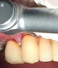
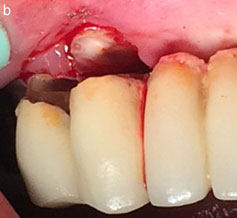
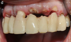
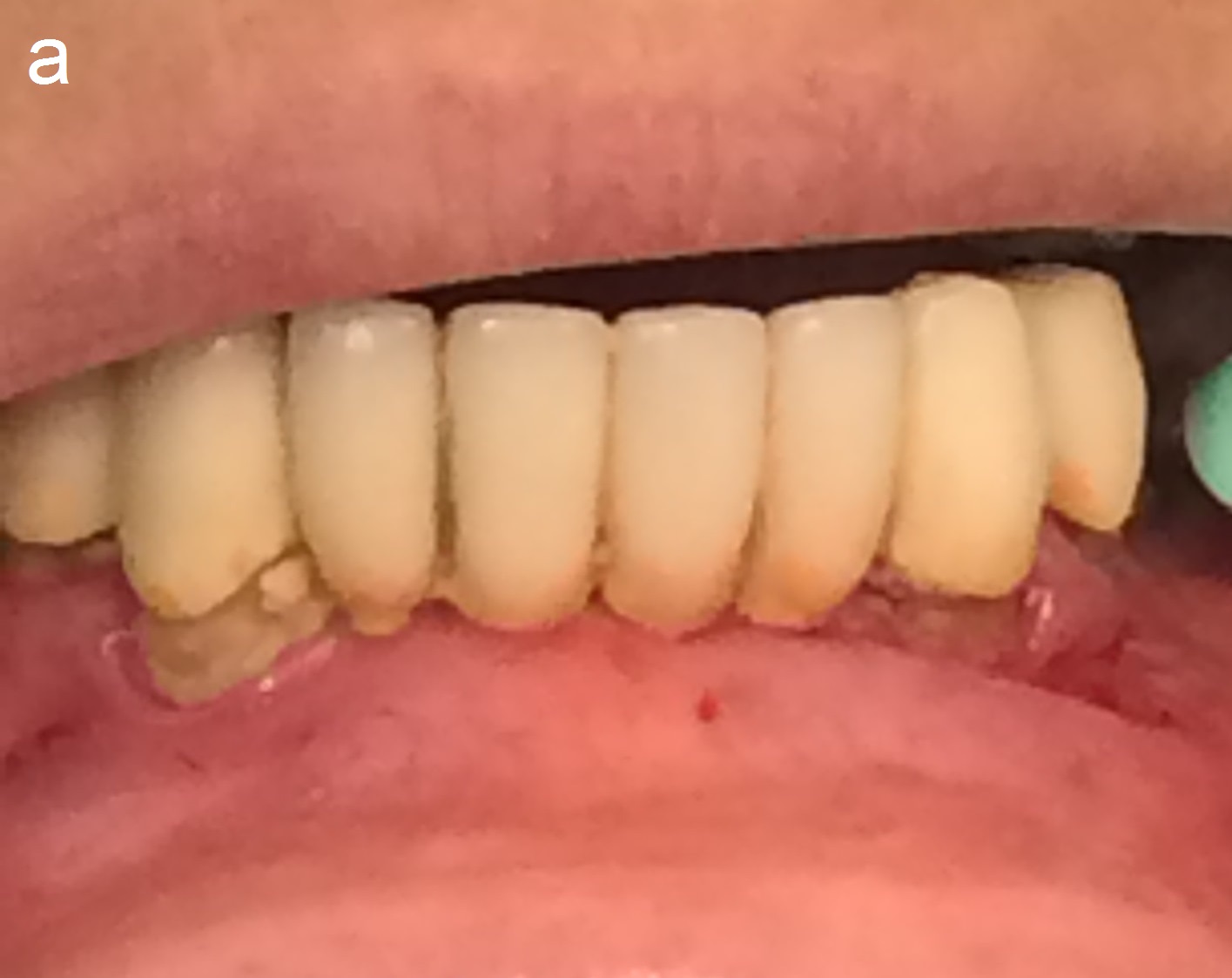
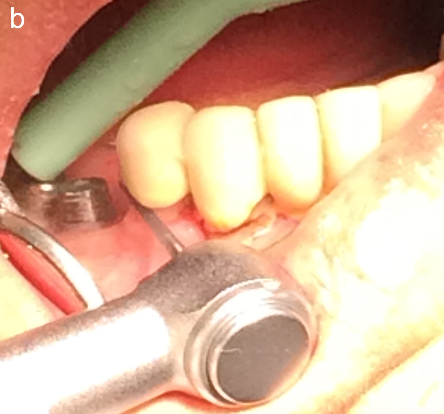
Step 1: The clinical tooth crown (CTC) was cut at the level above the gingiva (decoronation)
In order to prevent injury to the soft tissue (Figure 1a and 1b).
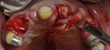
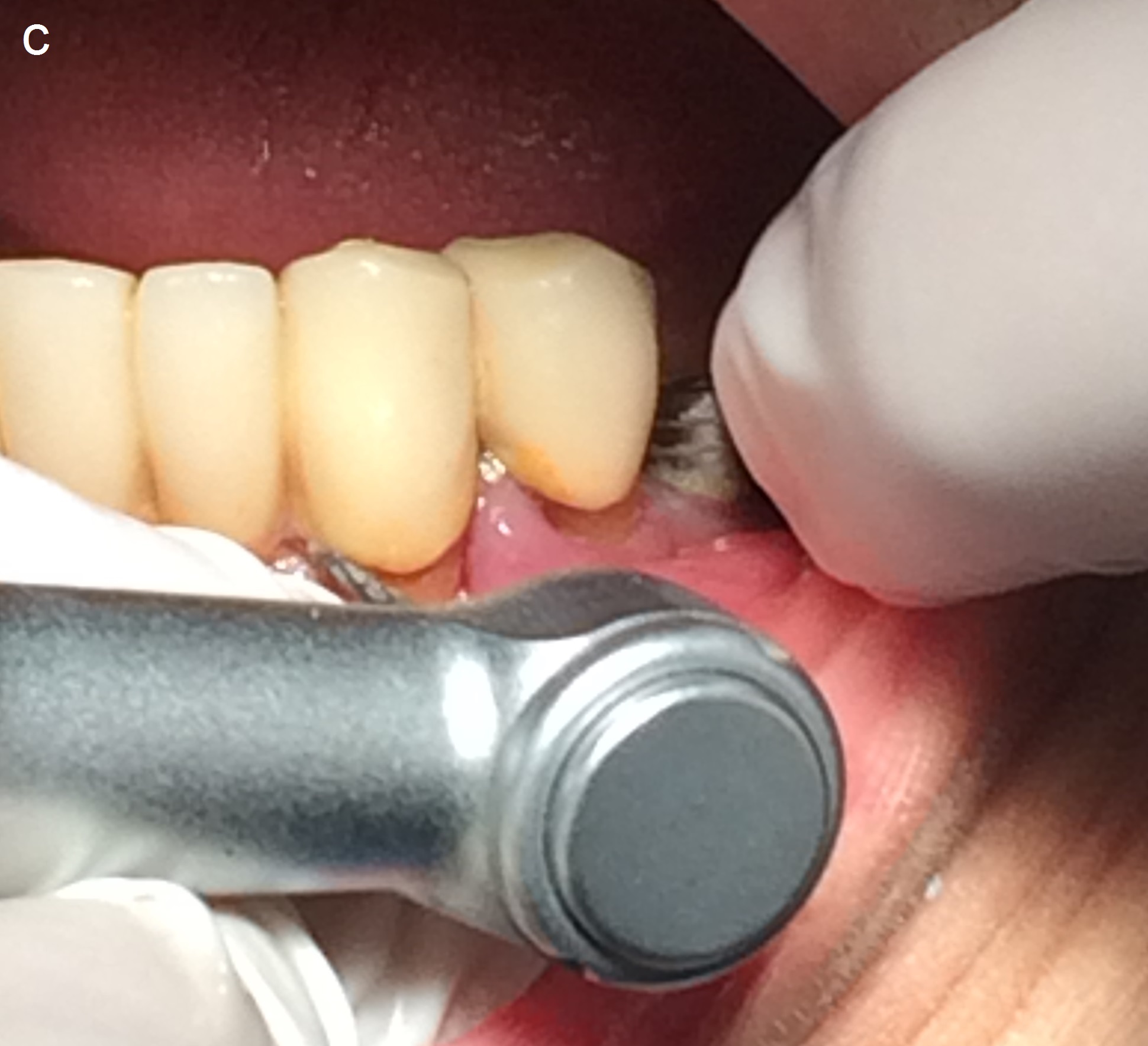
Step 2: Shaving and trimming of the residual roots carefully in concave form leaving the interproximal thin walls that support the interdental papilla. Figure 1c
Step 3: Try in and lining of the temporary bridge sit inside the roots in this way the volume and the shape of the original soft tissue is preserved.
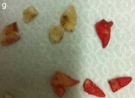
Step 4: Atraumatic extraction of the roots.
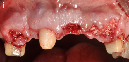
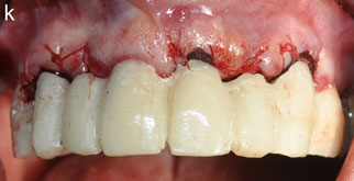
Step 5: Socket preservation and soft tissue suturing.
Step 6: Cementation of the adjusted and provisional temporary bridge that has been prepared earlier in the beginning of the appointment.
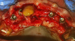
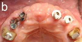
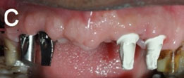
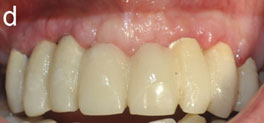
Step 7: The future treatment options, conventional bridge rehabilitation, or rehabilitation over implants. (Figure 2).
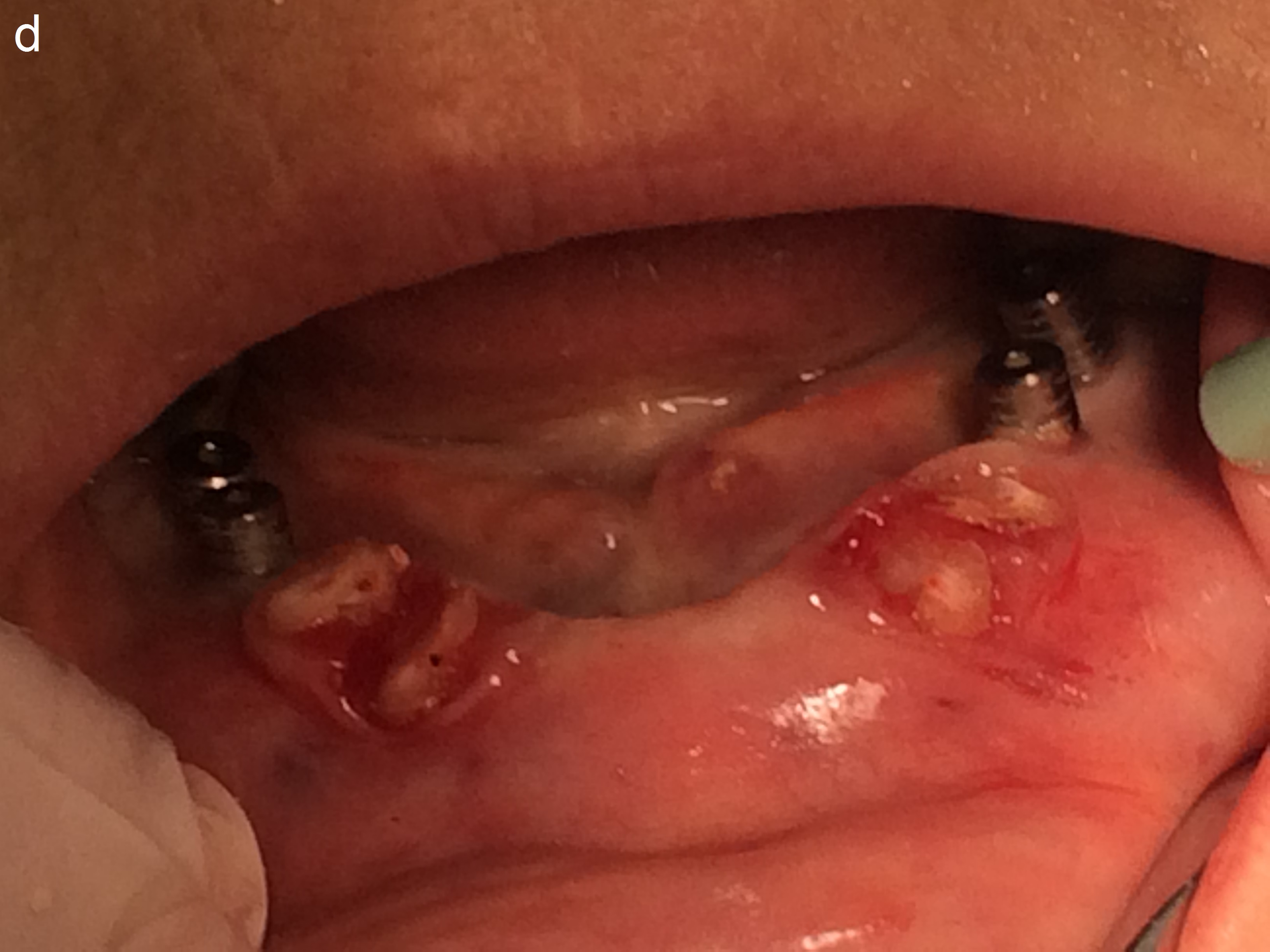
Figure 1: Transient coronectomy:step by step.
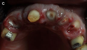
Figure 1C: Grinding of the roots in concave way, and the interproximal root shelf was designed as the papilla shape. Abutment teeth were prepared.
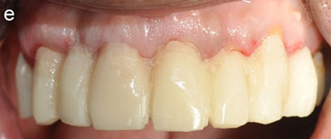
Figure 1E: Passive setting of the temporary bridge inside the submerged roots. With original interdental papilla shape.
4 remaining roots were extracted after adjusting and finishing the temporary bridge
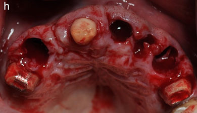
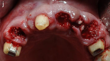
Figure 1H: Sockets after the extraction of the submerged roots with minimal injury to the soft tissue.
 |
 |
| I | J |
Figure 1I and J: Socket preservation, excellent preservation of the interdental papilla and the socket soft tissue.
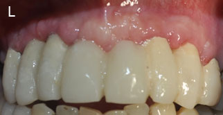
Figure 1L: 3 months follow up, good ridge contour. The interdental papilla was preserved in its original shape and volume.
Figure 2: Implants placement and follow up after the rehabilitation.
Figure 3: Transient Coronectomy in Extraction of teeth with PFM fixed bridge
Discussion
Maintaining the interdental papilla following tooth extraction has been challenge for restorative dentistry. The aim of the present report is to introduce the transient coronectomy as an approach to prevent papilla collapse during the treatment.
Immediately after tooth extraction, there is collapse of the papilla, so to optimally preserve the tissue, surgeons implement to maintain bone architecture and immediate provisionalization to maintain soft tissue .the tissue must be maintained during the surgical procedure.
In 2006 MArgeas reported the use of natural tooth as provisional following implant placement [24].
In 2008 Taleghani., et al. Reported the use of temporary bridge with an ovate pontic at the tissue of extraction to support the proximal papilla, the facial soft tissue, and the healing gingival tissue [25].
The root submergence technique for pontic site development in esthetic implant therapy have been used with excellent esthetic outcomes [18-19]. Leaving of roots under fixed prosthesis has on the other hand several risks, like periapical lesions, external root resorption, late eruption of the submerged root that can lead to late failure of the treatment [26].
The transient coronectomy technique by cutting the root in a way that support the interproximal papilla and matching a temporary restoration (crown, bridge) that fit 100% the original location and shape of the interdental papilla, inhibit immediate papilla collapse. The removal of the roots later during the same appointment and socket preservation eliminates the risk of late failure that can be happened to the submerged roots. Coronectomy technique has several advantages working in a clean environment, less bleeding during the treatment so less acryl contamination and less time consuming while preparing the temporary bridge.
This technique is highly recommended in cases of PFM Bridge on multiple teeth by coronectomy we prevent metal staining to the gingiva and less time consuming trying to remove the bridge.
Conclusion
This report demonstrates a technique with excellent and predictable results, and can be considered as a simple, minimal invasive procedure to preserve the interdental papilla.
References
- Gruder U. “The inlay graft technique to create papilla between implants”. Journal of Esthetic Dentistry 9.4 (1997): 165-8.
- Tarnow DP and Eskow RN. “Considerations for single-unit esthetic implant restorations”. Compendium of Continuing Education in Dentistry 16.8 (1995): 1778-84.
- 3.Chen MC., et al. “Factors influencing the presence of interproximal dental papilla between maxillary anterior teeth”. Journal of Periodontology 81.2 (2010): 318-24.
- Kim TH., et al. “An analysis of the factors responsible for relative position of interproximal papilla in healthy subjects”. Journal of Periodontal & Implant Science 43.4 (2013); 160-7.
- Singh UP., et al. “Black triangle dilemma and it’s management in the esthetic dentistry”. Dental Research Journal 10.3 (2013): 296-301.
- Prato GP., et al. “Interdental papilla management: A review and classification of the therapeutic approaches”. The International Journal of Periodontics and Restorative Dentistry 24.3 (2004): 246-55.
- Takei HH. “The interdental space”. Dental Clinics of North America 24.2 (1980): 169–76.
- Zetu L and Wang HL. “Management of inter-dental/inter-implant papilla”. Journal of Clinical Periodontology 32.7 (2005): 831-9.
- Cortellini P., et al. “The modified papilla preservation technique. A new surgical approach for interproximal regenerative procedures”. Journal of Periodontology 66.4 (1995): 261-6.
- Cortellini P., et al. “The simplified papilla preservation flap. A novel surgical approach for the management of soft tissues in regenerative procedures”. The International Journal of Periodontics and Restorative Dentistry 19.6 (1999): 589-99.
- Azzi R., et al. “Surgical reconstruction of the interdental papilla”. The International Journal of Periodontics and Restorative Dentistry 18.5 (1998): 467-74.
- Azzi R., et al. “Root coverage and papilla reconstruction using autogenous osseous and connective tissue grafts”. The International Journal of Periodontics and Restorative Dentistry 21.2 (2001): 141-7.
- Nordland WP., et al. “Microsurgical technique for augmentation of the interdental papilla: Three case reports”. The International Journal of Periodontics and Restorative Dentistry 28.6 (2008): 543-9.
- Ingber JS. “Forced eruption; A method of treating isolated one and two wall infrabony osseous defects-rationale and case report”. Journal of Periodontology 45.4 (1974): 199-206.
- Blatz MB., et al. “Reconstruction of the lost interproximal papilla: Presentation of surgical and nonsurgical approaches”. The International Journal of Periodontics and Restorative Dentistry 19.4 (1999): 395-406.
- Kokich VG. “Esthetics: The orthodontic-periodontic restorative connection”. Seminars in Orthodontics 2.1 (1996): 21-30.
- Nemcovsky CE and Serfaty V. “Alveolar ridge preservation following extraction of maxillary anterior teeth. Report on 23 consecutive cases”.Journal of Periodontology 67.4 (1996): 390-5.
- Salama M., et al. “Advantages of the root submergence technique for pontic site development in esthetic implant therapy”. The International Journal of Periodontics and Restorative Dentistry 27.6 (2007): 521-7.
- Mohadeb JV., et al. “Effectiveness of decoronation technique in the treatment of ankylosis: A systematic review”. Dental Traumatology 32.4 (2016): 255-63.
- Casey DM and Lauciello FR. “A review of the submerged-root concept”. The Journal of prosthetic dentistry 43.2 (1980): 128-32.
- O'Riordan BC . “Coronectomy (intentional partial odontectomy of lower third molars)”. Oral Surgery, Oral Medicine, Oral Pathology, Oral Radiology, and Endodontology 98.3 (2004): 274-80.
- Kim YB., et al. “Coronectomy of lower third molar in combination with vital pulp therapy”. European Journal of Dentistry 8.3 (2014): 416-8.
- Pogrel MA. “Partial odontectomy”. Oral & Maxillofacial Surgery Clinics of North America 19.1 (2007): 85-91.
- Margeas RC. “Predictable periimplant gingival esthetics: use of the natural tooth as a provisional following implant placement”. Journal of Esthetic and Restorative Dentistry 18.1 (2006): 5-12.
- Taleghani M., et al. “Nonsurgical management of interdental papillae loss following extraction of anterior teeth”. General dentistry 56.4 (2008): 326-31.
- von Wowernn N and Winther S. “Submergence of roots for alveolar ridge preservation. A failure (4-year follow-up study)”. International Journal of Oral Surgery 10.4 (1981): 247-250.
Citation:
Fares Kablan and Abir Abu Subeh. “Transient Coronectomy Procedure to Preserve the Inter-Dental Papilla at the Extraction
site: Technique Presentation.”. Oral Health and Dentistry 1.1 (2016): 21-29.
Copyright: © 2016 Fares Kablan and Abir Abu Subeh. This is an open-access article distributed under the terms of the Creative Commons Attribution License, which permits unrestricted use, distribution, and reproduction in any medium, provided the original author and source are credited.


 Scientia Ricerca is licensed and content of this site is available under a Creative Commons Attribution 4.0 International License.