Case Report
Volume 2 Issue 3 - 2018
An Unusual Case of Large Oral Lipoma in Mental Region: Case Report
Specialist Oral Surgeon, Ministry of Health Saudi Arabia
*Corresponding Author: Shiv Prasad Sharma, Specialist Oral Surgeon, Ministry of Health Saudi Arabia.
Received: December 27, 2017; Published: January 04, 2018
Abstract
Lipomas are very slow growing benign tumor of mature adipose tissues. They tend to involve many sites, even though intra- oral occurrence is very rare. Usual age of predilection according to published literature is in the fourth and fifth decades. In this paper we would like to highlight a case of large vestibular lipoma involving mental region in a 19 year male causing left facial. CT showed a well circumscribed mass of fat attenuation. The lesion in our case was found to be in intimate relation with underlying periosteum and the mental nerve branches but well separable. It is important to rule out other potential pathologies with similar presentations. The diagnostic question in our case whether should we go for Imaging modalities CT or Ultrasonography considering the size of the lesion and demonstration of poor mobility which is quite unusual with lipomas.
Key words: Lipoma; Oral; Mental region
Introduction
Lipomas are the most common benign soft tissue mesenchymal tumors and account for one third of all the benign tumors. Most common sites include trunk, shoulder, axilla, neck, scalp, rarely feet, lower legs, hands. Intraoral lipomas are very rare with incidence from 1-4%. Common intraoral sites of occurrence are buccal mucosa, parotid area, lips, floor of mouth, tongue, hard and soft palate, and vestibule. 50% of oral lipomas have been reported in buccal mucosa. Several varieties of lipoma have been reported which include classic lipoma, fibro lipoma, spindle cell lipoma, intramuscular lipoma, inter muscular lipoma, parosteal lipoma. Classic and fibro-lipomas are the most common histopathological variants reported. Most of the lipomas have a fairly clear clinical diagnosis. In this case we relied on CT scan and USG Doppler study to establish our preoperative diagnosis and to evaluate the exact site, size, relation with adjacent structures and to rule out any internal vascularity.
Another reason for imaging modalities in this case was to decide our surgical approach. The surgical excision was performed Intra orally. In this case the lesion was closely abutting the bony mandible in the region of mental neurovascular bundle causing stretching of the nerve considerably but the patient did not report any paresthesia. Such type of close intimacy to the periosteum is seen in case of a rare subtype ossifying parosteal lipoma (0.3% of all lipomas). Cytogenetic profile studies of lipomas have been studied and reported and may play an important role in avoiding any misdiagnosis and also in predicting the course of the disease. Although it was not done in our case.
Case Report
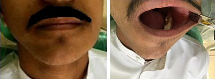
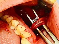
A 19 year old male patient reported with the chief complain of a swelling left side face cheek region of about one year duration. He was asymptomatic. His only concern was esthetic and he was cancerphobic. His medical and dental history was insignificant. On extraoral examination there was a soft to firm non-tender swelling involving left side face along the lateral aspect of mandible. Intraorally, the swelling extended from the region of left canine till the second molar with normal overlying mucosa. Approximate size was 5.5 x 3.5 x 3 cm. The swelling was found to fill the entire vestibule.
The other probable diagnosis which must be kept in mind include fibroma, granular cell tumor, neurofibromas, salivary gland tumors, vascular tumors like hemangioma.
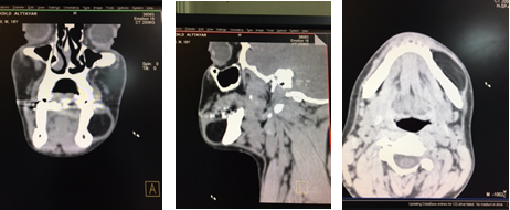
Preop CT scan showed a well-defined lesion with fat attenuation. We also performed USG Doppler to rule out significant vascularity. Both these modalities helped us in preop diagnosis and also to decide our approach to this large lesion.
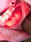
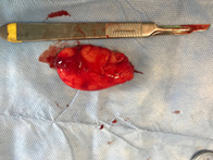
The patient underwent surgical removal of the entire lesion intraorally under local anesthesia. Significant intraop findings included the overlying mental nerve above the capsule of the lesion and the close intimacy of the lesion capsule to the underlying periosteum. The neurovascular bundle appeared stretched even though they were not infiltrated by the lesion. The entire mental neurovascular bundle was preserved and the lesion was removed in total and sent for histopathological examination.
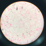
The histopathological report confirmed the diagnosis of lipoma.
Post-operative period was uneventful.
Post-operative period was uneventful.
Discussion
Lipomas are a very rare group of benign tumors of adipose tissue origin, more rare in the oral cavity. Intraoral lipomas account for about 1-4% of all tumors. Common age of occurrence is in the 4th-5th decade of life, even though cases have been reported in all age groups. A rare case of intraoral lipoma has been reported in a 6 year old child.
Our case report is unique due to several reasons. Very few cases have been reported of intraoral lipoma in the mental nerve region. Those reported were usually of smaller size compared to the lesion in our case which was about 5.5 x 3.5 x 3 cm in all dimensions. Another key finding was the close intimacy with the periosteum which has been reported with the rarest of Osteal/Ossifying Parosteal lipomas which are not commonly seen in oral cavities rather found in other locations as thoracic spine. The diagnosis of lipoma is mostly based on clinical appearance alone if it involves any other location in the body but cases like ours where we have a vestibular lesion of a large size, role of imaging cannot be ignored. Imaging helps to rule out other potential diagnosis and gives an exact knowledge about the adjacent structures and also in deciding the approach to surgery. CT can be invaluable in identifying rare parosteal/osteal lipomas which can be more infiltrative and require aggressive management to prevent their recurrence. This case highlights the role of Imaging in surgical management of lipomas.
Several reports have been published regarding lipomas at various sites but very few cases of intraoral lipomas have been reported. An interesting entity to be considered is a rare histologic variant, Intraosseous Lipoma, which accounts for only about 0.1%. It is believed to arise from osseous metaplasia. Cases of intraosseous variety have been reported involving inferior turbinate, maxillary tuberosity, mandibular condyle etc. Mandible is more frequently involved compared to maxilla. An interesting case was reported by Morais AL., et al. where an intraooseous lipoma was found to be associated with the periapical region of the distobuccal root of third molar mimicking apical periodontitis. Intraosseous lipoma should be a part of our differential diagnosis list when evaluating jaw lesions.
Fine Needle Aspiration Biopsy (FNAB) can be useful in cytologic diagnosis of lipoma especially those associated with salivary glands such as intraparotid lipomas. It can rule out other primary salivary gland tumors which can be clinically and radiographically indistinguishable. FNAB could be used in our case in conjunction with imaging modalities to initiate and plan the surgery. However it cannot replace the definitive histopathologic diagnosis.
Our PubMed search showed only three publications with the lipoma occurrence involving the mental region. In the article published by Choi HJ, Byeon JY intraoral lipomas involving mental neurovascular bundle were reported. They highlighted the possibility of sarcomatous changes within the huge lipomas and hence the need for histopathological examination. Kumar LK, Kurian NM reported a case of intraoral lipoma in the mental region in a 77 year old man. Harnisch H., et al. reported a case of intraoral lipoma in close proximity to mental nerve in a female patient. Maximum size of lipoma reported was 2 cm. Existing literature suggests that the occurrence of intraoral lipomas in the mental region is extremely rare. Our case will provide an interesting contribution to the already existing considering the large size of the lesion causing stretching of the mental nerve without undue sensory abnormalities. None of these cases had features similar to our case in terms of size, mental nerve involvement, juxtacortical presentation.
Very few cases have been reported where cytogenetic work up was included in the management of oral lipomas. Cytogenetic studies should be undertaken to learn more about the pathogenesis and tumorigenesis of oral lipomas. More and more case reports will contribute towards clarifying various aspects of this lesion.
Conclusion
Large lipomas in the close proximity of mental nerve are extremely rare and sparsely reported. A thorough clinical assessment is required along with the imaging modalities prior to surgical management. Histopathology confirms the clinical diagnosis and also helps to rule out sarcomatous changes in the lipomas. Immunohistochemistry/cytogenetic profile studies may play a role in establishing the course of the disease. Recurrence after surgical excision is extremely rare.
References
- Choi HJ and Byeon JY. “Symptomatic Intraoral Submuscular Lipoma Located Nearby Mental Foramen”. Journal of Craniofacial Surgery 27.5 (2016): e457-459.
- Kumar LK., et al. “Intraoral lipoma: Case report.” Case Reports in Medicine (2014).
- Harnisch H., et al. “Intraoral lipoma in the region of the mental nerve--report of a case and review of the literature”. Schweiz Monatsschr Zahnmed 117.4 (2007): 372-386.
- Venkateswarlu M., et al. “A rare case of intraoral lipoma in a six year-old child: a case report”. International Journal of Oral Science 3.1 (2011): 43-46.
- Fujimaki H., et al. “A case of an intramuscular lipoma in the mental region”. Plastic and Reconstructive Surgery 2.9 (2014): e209.
- S Kurup., et al. “Intraoral schwannoma–a report of two cases”. BMJ case reports (2012).
- Jaeger F., “Oral Spindle Cell Lipoma in a Rare Location: A Differential Diagnosis”. American Journal of Case Reports 16 (2015): 844-848.
- Sun Z., et al. “Ossifying parosteal lipoma of the mandible: a case report and review of the literature”. Dentomaxillofacial Radiology 42.1 (2013): 57852073.
- Myint ZW., et al. “Ossifying parosteal lipoma of the thoracic spine: a case report and review of literature.” Journal of Community Hospital Internal Medicine Perspectives 3; 5.1 (2015): 26013.
- Naruse T., et al. “Lipomas of the oral cavity: clinicopathological and immunohistochemical study of 24 cases and review of the literature.” Indian Journal of Otolaryngology and Head & Neck Surgery 67 Suppl 1 (2015): 67-73.
- Lee KM., et al. “mDixon-based texture analysis of an intraosseous lipoma: a case report and current review for the dental clinician.” Indian Journal of Otolaryngology and Head & Neck Surgery (2017).
- Cooper TJ., et al. “Intraosseous spindle cell lipoma of the mandible: case report”. British Journal of Oral and Maxillofacial Surgery 55.8 (2017): 839-840.
- Trento GDS., et al. “Extra-Oral Excision of a Buccal Fat Pad Lipoma.” Journal of Craniofacial Surgery 28.3 (2017): e226-e227.
- Egido-Moreno S., et al. “Intraoral lipomas: Review of literature and report of two clinical cases.” Journal of Clinical and Experimental Dentistry 8.5 (2016): e597-e603.
- Brooks JK., et al. “Oral lipoma: report of three cases”. General Dentistry 56.2 (2008): 172-176.
- Tabakovic SZ., “Intraosseous lipoma of the maxillary tuberosity: A case report.” Journal of Stomatology, Oral and Maxillofacial Surgery (2017).
- Nahles G., et al. “An intraosseous lipoma in the frontal bone--a case report”. International Journal of Oral and Maxillofacial Surgery 33.4 (2004): 408-410.
Citation:
Shiv Prasad Sharma. “An Unusual case of Large Oral Lipoma in Mental Region: Case Report”. Oral Health and Dentistry 2.3
(2018): 388-392.
Copyright: © 2018 Shiv Prasad Sharma. This is an open-access article distributed under the terms of the Creative Commons Attribution License, which permits unrestricted use, distribution, and reproduction in any medium, provided the original author and source are credited.


 Scientia Ricerca is licensed and content of this site is available under a Creative Commons Attribution 4.0 International License.