Case Report
Volume 2 Issue 4 - 2018
Guidelines for Restoring Fractured Central Incisors
Department of Fixed Prosthodontics, Research Laboratory of Occlusodontics and Ceramic Prostheses LR16ES15, Faculty of Dental Medicine, University of Monastir, Monastir, Tunisia
*Corresponding Author: Hadyaoui Dalenda, Department of Fixed Prosthodontics, Research Laboratory of Occlusodontics and Ceramic Prostheses LR16ES15, Faculty of Dental Medicine, University of Monastir, Monastir, Tunisia.
Received: January 08, 2018; Published: January 20, 2018
Abstract
Faced with fractured central incisors, many solutions are available and the practitioner has to choose the appropriate one. Rehabilitation of the compromised teeth number 11 and 21 was performed with lithium discilcate veneers. The purpose of this clinical case is to outline the treatment approach and to highlight the different guidelines to establish function and esthetic using ceramic veneers.
Key words: Ceramic veneers; Dental trauma; Central incisors; Function; Esthetic
Introduction
Traumatic dental injury has been confirmed as a current health problem in many recent studies. Nowadays [1,2]. First, trauma of the oral region occurs frequently and makes up 5% of all injuries for which people seek treatment in all dental clinics and hospitals in a country [2].
High-risk age groups for dental and facial trauma were 10-18 years and 19-28 years, which may be attributed to the fact this age-group usually has more intense social interaction and sports activities [3]. The teeth most commonly affected by trauma are the maxillary central incisors [3,4]. There are many causes for these such as falls, sports injiolenceuries, and vehicle accidents; other causes may also exist, depending on a country’s development and local habits [3,4,5]. The most frequent types of permanent teeth fractures are enamel fractures, enamel and dentine fractures, and enamel and dentine fractures with pulp involvement [6].
The conservative dental esthetic reestablishment treatments has been improved and evaluated with the development of adhesive materials. The adhesive dentistry allowed minimally invasive preparation through direct treatments with composite resin and indirect ceramic laminates veneers [7,8]. Despite the contribution of this treatment modality in terms of esthetic outcome, restoration of a fractured tooth in the anterior maxilla remains a challenge for even the most experienced dental practitioner. Several approaches for recovery of the esthetics and the function are available [7,8], Currently, the clinician must consider all diagnostic parameters before making a decision or recommendation to the patient. Direct resin is suitable when compromised structures is minimal allowing a natural look [9], However, Indirect restorations are indicated for greater strength and longevity, but they add a layer of complexity when communication with the laboratory technician is required for an esthetic outcome [10].
A range of dental ceramic materials is presently available on the market for these treatments, though with very different characteristics in terms of the composition, optic properties and manufacturing processes involved. In fact, A.A Font., et al. created a classification based on the objectives of treatment: esthetic and/or functional problems Because of their predictable results and conservation of tooth structure, [11] ceramic veneers are indicated for the esthetic rehabilitation of fractured anterior teeth with anomalous position and appearance. The aim of this paper is to highlight the steps of dental rehabilitation in a 19-year-old patient with fractured central incisors which had been directly restored by composite resin and because of repetitive fracture of the resin. The patient restrained herself from smile due to self-consciousness. Seeking for a permanent restoration, the alternative solution was a fixed restoration using ceramic veneers. Who restrained herself from smile due to self-consciousness, using ceramic veneers.
Case Presentation
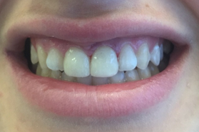
H.F was a 19-year-old female patient reported to the department of prosthetic dentistry, with a chief complaint of unattractive smile because of her fractured tooth number 11 and defective composite restoration in tooth number 21. Complete history of the patient along with preoperative photograph was taken (Figure 1). Medical history was non-contributory. Extra oral examination showed an ovoid face with a convex profile.
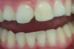
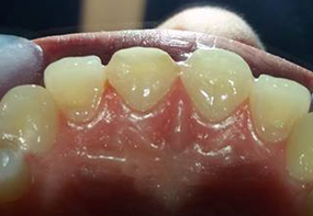
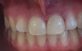
Intraoral examination revealed that the right central incisor was fractured in the middle-third of the crown, involving enamel and dentin without pulp exposure (Figure 2) and without symptoms of concussion or contusion, the left central incisor was restored by composite but she complained from it repetitive loss. Oral prophylaxis was done and dental hygiene maintenance instructions were given. Radiographic examination and tooth vitality tests were positive. Anterior guid¬ance was evaluated.
Several approaches for recovery of the esthetics and the masticatory function, depend on the type and extent of tooth fracture; In this case, the fracture is located in enamel a dent in with a loss of much tooth structure, the use of ceramic veneers is an excellent and suitable alternative.
In order to facilitate the treatment planning, a wax-up and cosmetic mock-up are recommended. The wax-up is a study model that present build- up wax teeth and the mock-up is obtained from silicon matrixfill ED with bis-acrylic resin [4] which provided a real three-dimensional in situ visualization of the final result of the proposed treatment.
Various techniques for accurate tooth reduction have been proposed, including silicone matrices, depth limiting burs and free hand preparation (Cherukara., et al. 2005). It is important that whatever tooth reduction guide method is used, it is based on the definitive wax up and not the original tooth.
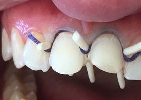
Failure to do this may result in excessive and unnecessary removal of tooth enamel. Tooth should be prepared within the enamel whenever possible. In this case depth limiting burs were used to prepare directly through the bis-acryl mockup, as described by Gurel (2003) (Figure 3). The teeth were prepared with a marginal chamfer labially and interproximal, and a butt fit margin palato-incisally with wrap around onto the palatal aspect as described by Castelnuovo., et al. 2000 (Figure 4). Contact points were not preserved, in order to allow freedom for the technician to change the width and shape in the final restoration. Ensure smooth finish lines and surfaces, using 40 micron diamond abrasives. A smooth surface avoid stress under the veneer and also a more uniform thickness of cement. This also leads to better adhesion.
After adequate gingival retraction (Figure 4), a two-step dual impression was made and sent to the laboratory for fabrication of lithium disilicate (IPS e max cad) veneers.
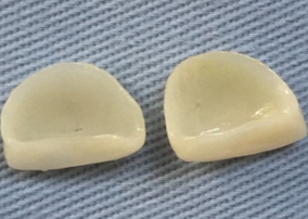
Lithium discilcate veneers were aided by computer (CAD), by coping the contours from the diagnostic wax-up. (Figure 5) Veneers were individually checked intra-orally to control gingival margins adaptation, the complete seating andembrasure opening, and occlusion. Then, shade and esthetics were well checked. To minimize contamination from saliva and blood, the application of rubber dam is strongly recommended; In fact blood can change the completely the color of final restoration and because of esthetic failure. Currently maintaining clean tooth with water and pumice during bonding is very important for the success of this step. Light curing composite resin were used for bonding.
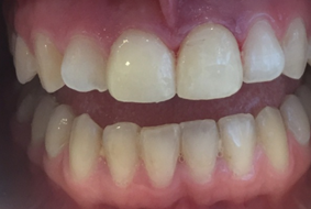
At the end of the treatment, the patient was pleased with the results and no longer hides her smile (Figure 7, Figure 8)
Periodic follow-up was scheduled to evaluate the gingival health and patient comfort and satisfaction. (Figure 9, Figure 10)
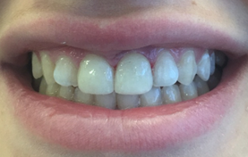
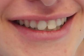
Figure 9, 10: Natural look of teeth restored after two months: Satisfying esthetic
outcome: characterizations reproduction, periodontal integration and function.
Discussion
Unlike dental caries that have been declining over the last decades, Dental fractures are considered an increasing public health problem compromising aesthetic and function.
If this trauma is not treated, personal problems can occur, such as difficulties in eating, laughing, and smiling, as well as emotional problems associated with public contact [12,13]
Aesthetic dentistry has expanded dramatically in the last two decades and re-establishing dental aesthetic appearance is a very important clinical challenge. [14]
Currently, based on the type and the extent of tooth fracture, there are many treatment options and it is possible to restore function and esthetics using very conservative restorative techniques. In our case, the use of composites was well suited for our young patient because it is a very conservative technique for performing repairs without reduction in healthy tooth structure [15], Final restoration using nanoparticles-based composite resin was performed, allows restorations with shades and nuances similar to the adjacent dental structures. However, to achieve good results, this technique requires knowledge of the field of restorative material, dental anatomy, and the skills to reproduce all the characteristics of the tooth [9]. After a short time, there was a repetitive loss of restoration, because the restoration probably doesn’t support the masticatory efforts, mainly because of the insufficient area for bonding, Furthemore, those restorations should be limited for fractures limited in dental enamel or in enamel and dentin without loss of much tooth structure. [8,9]
Currently, the use of ceramic veneers was indicated, it was introduced into dentistry as Hollywood veneers by Pincus [16] with a survival rates ranged from 92% at 5 years to 64 % at 10 years. [16,17]. On another hand, according to a recent systematic review Composite and ceramic veneers were found to have statistically similar survival rates. Indirect composites showed 87% survival rate compared with 100% for ceramic veneers, with all failures occurring within 13 months of placement. No secondary, Caries were seen with either material. Temporary postoperative sensitivity developed with both ceramic (9%) and composite (26%) [18]
According to some clinicians, provisionalization is not necessary because tooth reduction is minimal, but in reality, it’s an important step in the treatment plan as it gives to both patient and clinician the opportunity to access the final planned result [19].
Preparation, cementation, and finishing procedures adopted are considered key factors for the long-term success and aesthetical result of the veneer restorations. In addition with improved mechanical properties of dental ceramics and the optical qualities of these materials have allowed the use of ceramics with esthetic predictability [1].
Conclusion
The success of minimally invasive restoration of fractured teeth dependent on the detailed planning and correct selection of dental materials. Ceramic veneers provided good treatment outcomes and allowed a long-lasting functional and esthetic outcomes.
References
- Eilert-Petersson E., et al. “Traumatic oral vs non-oral injuries”. Swedish Dental Journal 21.1-2 (1997): 55-68.
- Marcenes W., et al. “Epidemiology of traumatic injuries to the permanent incisors of 9–12-year-old schoolchildren in Damascus, Syria”. Endodontics & Dental Traumatology 15.3 (1999): 117-123.
- Andreasen JO and Ravn IJ. “Epidemiology of traumatic dentalinjury to primary and permanent teeth in a Danish population sample”. International Journal of Oral and Maxillofacial Surgery 1.5 (1972): 235-239.
- Lin S., et al. “Dento-alveolar and maxillofacial injuries: a 5-year multi-center study. Part 1: general vs facial and dental trauma”. Dental Traumatology 24.1 (2008): 53-55.
- Altay N and Güngör HC. “A retrospective study of dento-alveolar injuries of children in Ankara, Turkey”. Dental Traumatology 17.5 (2001): 201-204.
- Santos SE., et al. A 9-year retrospective study of dental trauma in Piracicaba and neighboring regions in the State of Sa˜o Paulo. Brazilian Journal of Oral and Maxillofacial Surgery 68.8 (2010): 1826-1832.
- Bora Korkut., et al. “Direct Midline Diastema Closure with Composite Layering Technique: A One-Year Follow-Up”. Case Reports in Dentistry (2016).
- Layton DM and Clarke M. “’A systematic review and meta-analysis of the survival of non-feldspathic porcelain veneers over 5 and 10 years”. International Journal of Prosthodontics 26.2 (2013): 111-124.
- Sakai VT., et al. “Predictable esthetic treatment of fractured anterior teeth: a clinical report”. Dental Traumatology 23.6 (2007): 371-375.
- Venâncio GN., et al. “Conservative esthetic solution with ceramic laminates: literature review”. RSBO Revista Sul-Brasileira de Odontologia 11.2 (2014): 185-191.
- Font AF., et al. “Choice of ceramic for use in treatments with porcelain laminate veneers”. Medicina oral, patologia oral y cirugia bucal 11.3 (2006): E297-302.
- Ivancic Jokic N., et al. “Dental trauma in children and young adults visiting a University Dental Clinic”. Dental Traumatology 25.1 (2009): 84-87.
- Ivancic Jokic N., et al. “Dental trauma in children and young adults visiting a University Dental Clinic”. Dental Traumatology 25.1 (2009): 84-87.
- Kamble VD and Parkhedkar RD. “Esthetic rehabilitation of discolored anterior teeth with porcelain veneers”. Contemporary clinical dentistry 4.1 (2013): 124-126.
- Peumans M., et al. A prospective ten-year clinical trial of porcelain veneers. Journal of Adhesive Dentistry 6.1 (2004): 65-76.
- Pincus CR. “Building mouth personality”. Journal of the California Dental Association 14 (1938): 125-129.
- Decurcio RdA and Cardoso PdC. “Porcelain laminate veneers: A minimally invasive esthetic procedure”. Stomatos 17.33 (2011): 12-19.
- Peumans M., et al. “A prospective ten-year clinical trial of porcelain veneers”. Journal of Adhesive Dentistry 6.1 (2004): 65-76.
- Decurcio RdA and Cardoso PdC. “Porcelain laminate veneers: A minimally invasive esthetic procedure”. Stomatos 17.33 (2011): 12-19.
Citation:
Hadyaoui Dalenda., et al. “Guidelines for Restoring Fractured Central Incisors”. Oral Health and Dentistry 2.4 (2018): 421-
427.
Copyright: © 2018 Hadyaoui Dalenda., et al. This is an open-access article distributed under the terms of the Creative Commons Attribution License, which permits unrestricted use, distribution, and reproduction in any medium, provided the original author and source are credited.


 Scientia Ricerca is licensed and content of this site is available under a Creative Commons Attribution 4.0 International License.