Case Report
Volume 2 Issue 5 - 2018
Concept of Simultaneous Crown-Root Shielding in Endodontics
Department of Restorative Dentistry, School of Dentistry, University of São Paulo, Brazil
*Corresponding Author: Jose Edgar Valdivia Cardenas, School of Dentistry, University of São Paulo, Av. Prof. Lineu Prestes, 2227–Cidade Universitária, 05508-000, São Paulo,SP, Brazil.
Received: February 20, 2018; Published: February 28, 2018
Abstract
The practice of endodontic-restorative treatment requires restorations that both conserve and protect remaining tooth structure. An alternative is the use bulk-fill composites resin restorations and fiberglass posts associated when necessary. In this context, the simultaneous restoration of both root canal and crown represents a restorative possibility. The paper shows two clinical cases of endodontic treatment and tooth restoration using the concept of simultaneous coronal-radicular shielding with resin material in a single treatment visit.
Key words: Restoration; Endodontics; Shielding
Introduction
The restoration of endondontically treated teeth is a a preocupation of Dentistry because these teeth have lower mechanical resistance when compared to teeth with vital pulps [1]. Several scientific and clinical papers have been published and discuss the benefits of the fiberglass posts and adhesive materials. The disclosure of clinical cases that describe techniques that demonstrate longevity of their results is of fundamental importance for the credibility and safety of professionals who will use these systems.
A large loss dentin can be reconstitute using composite resin filling core inserted at the interface between the post and the morphological restoration of the endodontically treated tooth. This reconstruction is important to provide support and retention for the direct or indirect restorative material, and also in the homogenous distribution of stresses [2]. In this context, the contemporary resins such as bulk fill resins and resin sealer are suitable for insertion in a 4 mm bulk placement due to their reduced polymerization stress and high reactivity to light curing [3].
The fiberglass posts and resinous materials have rigidity very similar to dentin, absorbing the tensions generated by masticatory forces and protecting the remaining root, then, they can be used to restoration in root canal using a mechanically homogeneous unit [4]. Moreover, the adequate combination of these materials enables the professional to perform restorations with minimal wear of dental structure and high clinical success.
The coronal portion of the filling core is an important component in the final morphological and aesthetic resconstrution of the tooth. Present composite resins are great options for meeting esthetic requirements and mechanical resistance necessary for direct restorations or as a base for a future prosthetic crown. The evidence supports placement of a crown to encircle the tooth can increase the resistance of posterior teeth to fracture, and a high incidence of failure for posterior endodontically treated teeth without cusp coverage has been reported [5,6]
The concept of simultaneous coronal-radicular shielding consists of the simultaneous root canal preparation and tooth restoration using resinous materials. Thus, this paper shows two clinical cases of endodontic treatment and tooth restoration using the concept of simultaneous coronal-radicular shielding.
Case Presentation
Case 1
A male patient presented with pain in the third lower left molar. The clinical and radiographic examination showed an extensive caries without pulp expusure (Figure 1), and the thermical tests characterize acute irreversible pulpitis.
A male patient presented with pain in the third lower left molar. The clinical and radiographic examination showed an extensive caries without pulp expusure (Figure 1), and the thermical tests characterize acute irreversible pulpitis.

Figure 1: Initial radiograph of the third lower left molar showing
extensive carie in proximity to the pulp chamber (Case 1).
After diagnosis, the operative field was isolated with a rubber dam and the access to the root canal system was performed. After preparation of the entrance to the root canals, the working length was determinated using a foraminal locator (Propex Pixi, Dentsply-Maillefer, Ballaigues, Switzerland). Subsequently, the root canal preparation was performed with Wave One Gold primary and medium files (Dentsply-Maillefer) using 2.5% sodium hypochlorite (Fórmula e Ação, São Paulo, Brazil) as irrigant, and then, final irrigation was performed alternating 2.5% sodium hypochlorite and 17% EDTA (Fórmula e Ação).
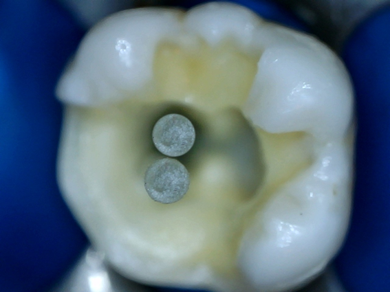
Root canal obturation was performed by single-cone technique using utilizing Wave One Gold medium gutta-percha cones (Dentsply-Maillefer) and AhPlus endodontic sealer (Dentsply-Maillefer). The gutta-percha cutting was performed 2 mm from the entrance of the root canal using heat tip (system B, SybronEndo, Orange, USA) (Figure 2A-B).
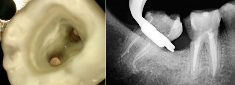
Figure 2: The gutta-percha cutting performed 2 mm from the entrance
of the root canal. Clinical (A) and radiographic visualization (B).
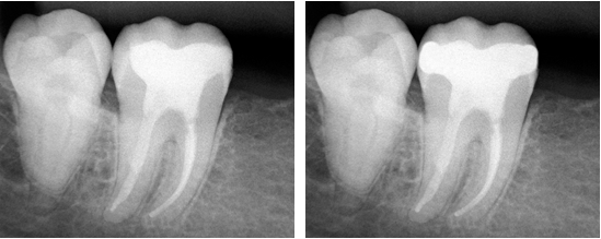
Due to the little loss of dentin structure was chose the direct restoration technique: the pulp chamber was conditioned with the adhesive system (XP Bond, Dentsply-Maillefer). An layer of 4 mm thickness of vertical increment of composite resin bulk-fill flow (Surefil SDR, Dentsply-Maillefer) was inserted, and a light-cured was applied for 40 seconds with a LED light curing unit (Demi Plus, Middleton, USA) with output irradiance of approximately 1100 mW/cm2. (Figure 3A-C). The final restoration was performed with composite resin (Spectra, Dentsply-Maillefer) on occlusal face, and polishing using system Enhance Pogo (Dentsply-Maillefer, Ballaigues, Switzerland). Then, periapical radiograph of the tooth was taken (Figure 4).

Figure 3: (A-C) - An layer of 4 mm thickness of vertical increment of composite resin bulk-fill flow.
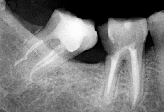
Figure 4: Final periapical radiograph of the case 1 showing complete
endodontic treatment and restoration tooth by direct technique.
Case 2
A male patient presented with discomfort in the second lower left molar. The tooth showed caries but did not respond to vitality tests. No periapical changing was observed radiographically, characterizing the diagnosis of pulp necrosis (Figure 5).
A male patient presented with discomfort in the second lower left molar. The tooth showed caries but did not respond to vitality tests. No periapical changing was observed radiographically, characterizing the diagnosis of pulp necrosis (Figure 5).
Considering the need for endodontic treatment and its restoration regarding to little dental structure, a intraradicular post and a prosthetic crown were planned. Then, Before endodontic treatment, a fiberglass post (Superdont, São Paulo, Brazil) was choosen according to manufacturer recommendation to be used in the distal root canals.
After choice of the post, the endodontic treatment was carried out: the tooth was isolated with a rubber dam and the access to the root canal system was performed. After preparation of the entrance to the root canals, working length was determinated using a foraminal locator (Propex Pixi, Dentsply-Maillefer).
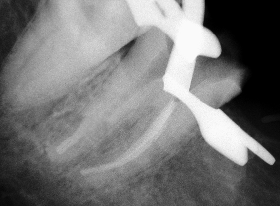
Subsequently, the root canal preparation was carried out as follows according to Valdivia & Machado [7]: In the mesial root canals, the preparation was performed using Wave One Gold primary and medium, and in the distal canals the preparation was carried out using Wave One Gold medium and large to the working length, both using 2.5% sodium hypochlorite as irrigant. During the distal root canals preparation, a ultrasonic tip (POST PREP, Trinks, São Paulo, Brazil) was used to 5 mm from working length (Figure 6A-B) with the purpose of root canal shaping for the fiberglass post. Then, a visual and tactile test was carrried out to verify its adaptation to root canal (Figure 7). Final irrigation was performed alternating 2.5% sodium hypochlorite and 17% EDTA.
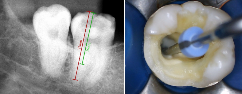
Figure 6: Radiographic planning to ultrasonic tip usage (A).
Ultrasonic tip to 5 mm from working length (B).
The mesial root canal obturation was filled by single-cone technique using utilizing Wave One Gold medium, and the distal root canal using large gutta-percha cones, both with AhPlus endodontic sealer. In the distal root canals, the gutta-percha was cutted 5 mm from working length using ultrasonic tip (POST PREP). Then the endondontic treatment was radiologically verified (Figure 8).
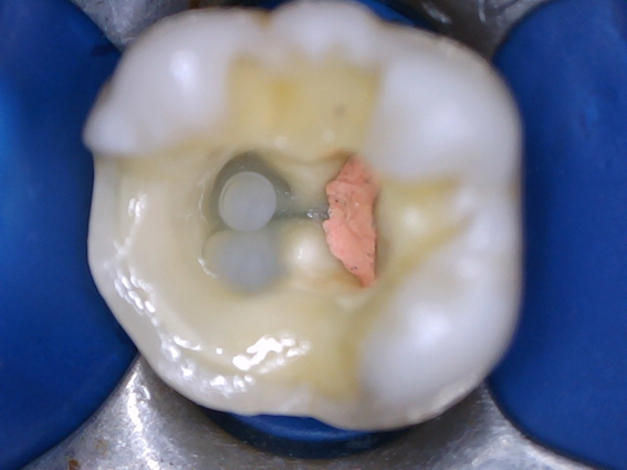
Adhesive system (Dry and Wet bond, Superdont, São Paulo, Brazil) was then applied on root canal wall by means of extra-fine applicators (Endo Tim, Voco GmbH, Cuxhaven, Germany). Then, the fiberglass posts with dual resin sealer (Superpost CoreCement, Superdont, São Paulo, Brazil) were inserted at distal root canals (Figure 9). Next, they were photopolymerized for 40 seconds with a LED light curing unit (Demi Plus).
The coronary build-up was carried out using composite resin (GrandioSO, Voco GmbH, Cuxhaven, Germany) to complete restoration (Figure 10). The final radiograph was take (Figure 11A), and the patient was referred to a prosthodontist.
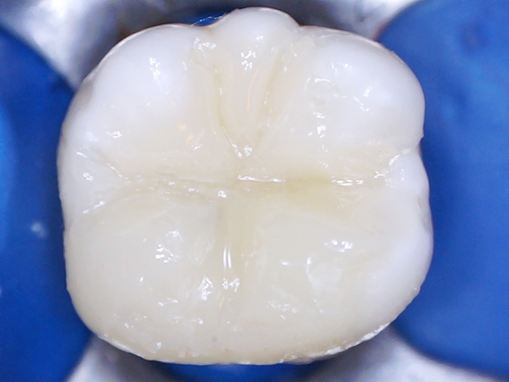
Figure 10: Final restoration with coronary build-up using
bulk-fill flow and additional layer of composite resin.
After 12 months, the patient was asymptomatic and the tooth was rehabilitated with a prosthetic crown (Figura 11B).
Discussion
A pulpless tooth becomes weak due to biomechanical alterations. This is so because the tooth underwent modification to its architecture and morphology, therefore it becomes more fragile due to loss of dental structure by caries, fractures, cavity preparation and furthermore from access and excessive instrumentation of the root canal [8].
The cast metal cores associated with prosthetic crowns are undoubtedly the most traditionally used solution in the process of restoring endodontically treated teeth. However, these cores have the disadvantage of being silver in color. Another factor is that the number of sessions required to make them is larger when compared to the time spent using a prefabricated post. In the cases described, the importance of an applicable technique in the contemporary context is displayed as being a viable option in the immediate rehabilitation of endodontically treated teeth. This technique is executed in a minimally invasive way, associating fiber posts with direct restorations in composite resin. This association represents an alternative, mainly due to the good mechanical and aesthetic properties of fiberglass posts and the current composite resins.
Intra-radicular fiberglass posts are widely used for the restoration of endodontically treated teeth when there is not enough tooth structure to retain a definitive restoration [9]. The advantage of producing satisfactory aesthetic results due to its optical properties have caused an incease in the popularity of fiberglass posts in relation to metallic systems [10]. Fiberglass posts exhibit a modulus of elasticity similar to that of dentin and as such protect the remaining root by absorbing tensions generated by masticatory force by forming a mechanically homogeneous unit (11). It is worth mentioning that the direct use of these pins eliminates laboratory steps. According to Assif., et al. [12], molten metal cores do not meet the needs of the pulpless teeth, since they are made with metals that have a high modulus of elasticity and can therefore induce a high rate of root fracture. It is known that endodontic posts do not increase the resistance of the remaining dental structure of endodontically treated teeth [13]. On the contrary, depending on the post´s design it can weaken the root in relation to the amount of dentin removed during preparation. Tapered posts like that used in the second clininical case have a configuration that is compatible with the tapered preparation of the root canal after instrumentation and in this way provides preservation of the tooth´s root structure, especially in the apical region.
In order to prepare the space for intra-radicular post, prefabricated drills with the same diameter of the fiberglass posts indicated by the manufacturers, or Gates Glidden or Largo drills are commonly used. However, these drills in order to adapt the posts to the root canal can remove an excessive amount of dentin causing a fail adaptation of the post [14,15] and the fact that there are anatomical situations that do not allow a good adaptation.
The lack of adaptation that can be observed with a greater thickness of resin cement and the lack of retention of the pins to the root canal can be explained that despite recent improvements in adhesive dentistry, fiberglass posts cannot mechanically overlap with the walls of the Root canal because these pins have standardized geometry. This paper shows a planned instrumentation technique for the installation of a future intra-radicular post. The use of the POST PREP ultrasonic tip during instrumentation of the root canal aims to prepare the canal where the post will be used. Valdivia and Machado [16] demonstrated that the shapping pcarried out by ultrasonic tip provides better adaptation of the fiberglass post to the root canal preparation, and that it influences the better adhesion and bond strength. It should be noted that in this context, the great advantage of the endodontist performing intra-radicular restorative procedures is that this specialist is familiar with the root canal system and can perform the preparation for posts based on the root anatomy of each case under absolute isolation. This avoids contamination of the root canal by saliva. Additionally, the use of absolute isolation eliminates humidity and fluids of the oral environment that can impair adhesion [17].
At the interface between the fiberglass post and the morphological restoration of the treated tooth, a composite resin filling core will be created, which has the purpose of reconstructing dental structure that had been lost. This reconstruction is important, not only in order to provide support and retention for direct or indirect restorative materials, but also in the distribution of stresses by distributing them more homogeneously throughout the remaining tooth. Several materials have been found to be effective in the construction of fill cores, such as amalgam, composite resin or glass ionomer cement, which have been widely described in literature [18]. With the evolution of the optical and mechanical characteristics of composite resins, there is greater predictability of outcome of restorations of endodontically treated teeth. In the first clincal case, The use of Surefill SDR bulk fill flow is a resinous dentin substitute for insertion in large increments as a basis in dental restorations. It has radiopacity, a high polymerization depth and a component that results in a low contraction voltage generated by the polymerization process, thus allowing the use in large increments. SDR bulk fill flow has a self-leveling feature that allows adaptation to the walls of the prepared cavity. Considering bulk-fill placement technique, it has been demonstrated that Surefil SDR showed better internal adaptation than conventional composites in high C-factor cavities [19]. In the second clinical case, it allowed the creation of a filling core for a future prosthetic crown.
Conflict of Interest: The authors declare that they have no conflict of interest.
References
- Sedgley CM and Messer HH. “Are endodontically treated teeth more brittle?” Journal of Endodontics 18.7 (1992): 332-335.
- Soares CJ., et al. “Longitudinal clinical evaluation of post systems: a literature review”. Brazilian Dental Journal 23.2 (2012): 135-140.
- Ilie N and Hickel R. “Investigations on a methacrylate-based flowable composite based on the SDR technology”. Dent Materials 27.4 (2011): 348-355.
- Santos-Filho PC., et al. “Influence of ferrule, post system, and length on stress distribution of weakened root-filled teeth”. Journal of Endodontics 40.11(2014): 1874-1878.
- Heling I., et al. “Endodontic failure caused by inadequate restorative procedures: review and treatment recommendations”. Journal of Prosthetic Dentistry 87.6 (2002): 674-678.
- Wegner PK., et al. “Survival rate of endodontically treated teeth with posts after prosthetic restoration”. Journal of Endodontics 32.10 (2006): 928-931.
- Valdivia JE and Machado MEL. “Simultaneous crown-root shielding in endodontics: from root preparation to coronary restoration”. Dental Press Endodontics 7.1 (2017): 32-42.
- Figueiredo FE., et al. “Do metal post-retained restorations result in more root fractures than fiber post-retained restorations? A systematic review and meta-analysis”. Journal of Endodontics 41.3 (2015): 309-316.
- Ferrari M., et al. “Clinical evaluation of fiber-reinforced epoxy resin posts and cast post and core”. American Journal of Dentistry 13(Spec No) (2000): 15B-18B.
- Morgano SM. “Restoration of pulpless teeth: application of traditional principles in present and future contexts”. Journal of Prosthetic Dentistry 75.4 (1996): 375-380.
- Asmussen E., et al. “Finite element analysis of stresses in endodontically treated, dowel-restored teeth”. Journal of Prosthetic Dentistry 94.4 (2005): 321-329.
- Assif D., et al. “Photoelastic analysis of stress transfer by endodontically treated teeth to the supporting structure using different restorative techniques”. Journal of Prosthetic Dentistry 61.5 (1989): 535-543.
- Schwartz RS and Robbins JW. “Post placement and restoration of endodontically treated teeth: a literature review”. Journal of Endodontics 30.5 (2004): 289-301.
- Souza EM., et al. “The impact of post preparation on the residual dentin thickness of maxillary molars”. Journal of Prosthetic Dentistry 106.3 (2011): 184-190.
- Pilo R., et al. “Residual dentin thickness in bifurcated maxillary first premolars after root canal and post spacepreparation with parallel-sided drills”. Journal of Prosthetic Dentistry 99.4 (2008):267-73.
- Valdivia JE., et al. “Influencia de la técnica de preparación ultrasónica en la adaptación de pernos de fibra de vidrio cónicos”. Odontologia UCE19.2 (2016): 20-28.
- Goldfein J., et al. “Rubber dam use during post placement influences the success of root canal-treated teeth”. Journal of Endodontics 39.12 (2013): 1481-1484.
- Engelman MJ. “Core Materials”. Journal of the California Dental Association 16 (1988): 41-45.
- Van Ende A., et al. “Bulk-filling of high C-factor posterior cavities: effect on adhesion to cavity-bottom dentin”. Dent Materials 29.3 (2013): 269-277.
Citation:
Jose Edgar Valdivia Cardenas., et al. “Concept of Simultaneous Crown-Root Shielding in Endodontics”. Oral Health and
Dentistry 2.5 (2018): 456-464.
Copyright: © 2018 Jose Edgar Valdivia Cardenas., et al. This is an open-access article distributed under the terms of the Creative Commons Attribution License, which permits unrestricted use, distribution, and reproduction in any medium, provided the original author and source are credited.


 Scientia Ricerca is licensed and content of this site is available under a Creative Commons Attribution 4.0 International License.