Research Article
Volume 3 Issue 1 - 2018
Different 2d Digital Radiographic Methods Versus Cone Beam Computed Tomography for the Diagnosis of Facial asymmetry- In vivo Study.
M.A Rangoonwala Dental College and Research Centre, India
*Corresponding Author: Taabish Contractor, M.A Rangoonwala Dental College and Research Centre, India.
Received: April 16, 2018; Published: May 08 2018
Abstract
Introduction: Facial symmetry refers to a state of balance, where size, form and arrangement of facial tissue and structures on the opposite sides of the median sagital plane correspond to each other. 2D radiographs are an effective tool in evaluating the asymmetrical craniofacial structures in transverse & vertical dimensions. However with the invention of 3D CBCT in the field of dentistry, diagnosing such asymmetries has become more accurate and relatively easier.
Objectives and Methodology: The objective of this study was to measure facial asymmetry diagnosed by different 2D digital radiographic methods & 3D CBCT and to compare the reliability of linear and angular measurements from digital (2-D) radiography method and those derived from CBCT (3-D) radiography for diagnosing facial asymmetry. 20 patients with facial asymmetry were selected. Initial photographs obtained for primary diagnosis of facial asymmetry, 20 CBCTs, 20 OPG’s, 20 PA view radiographs were used. All OPGs were traced and Levandoski analysis was carried out and Grummons analysis was performed on the traced PA cephalograms. The readings obtained from these 2D radiographic analysis was then compared to 3D cbct.
Results: Frontal vertical relation parameters showed significant difference for maxillary ratio and maxilla mandibular ratio, whereas upper and lower facial ratio, total maxillary and total mandibular had comparable accuracy between CBCT and digital techniques. Digital angular cephalometric measurement had comparable accuracy to CBCT technique, whereas linear measurements by Levandoski method had higher values in digital radiographs thus revealing poor accuracy with CBCT.
Conclusion: The results of this study show that 2-D digital radiographic aids have comparable accuracy in most parameters for the diagnosis of facial asymmetry when compared with 3-D CBCT techniques. Hence 2-D radiography serves as a satisfactory tool in the diagnosis of facial asymmetry.
Introduction
Facial symmetry refers to a state of balance, where the size, form, and arrangement of facial tissues and structures on the opposite sides of the median sagittal plane correspond to each other [1]. Thus, the right and left sides in the craniofacial complex, comprising identical structures, must similarly grow and develop to reach symmetry [2]. However asymmetry is a usual finding in human craniofacial bones. The aetiology of the facial asymmetries is more often associated with Class II and Class III malocclusions. Sometimes they are associated secondary to condylar hyperplasia or hypoplasia, ankylosis of the temporomandibular joint, condylar fractures and hemifacialmicrosomia [3].
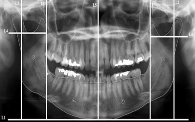
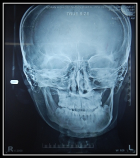
The etiology of mandibular asymmetry includes trauma, infections, developmental abnormalities, myogenic problems, joint pathologies, occlusal interferences and syndromes associated with asymmetries [4]. Lateral and PA cephalograms are widely used for diagnosis and treatment planning of orthodontic problems and to determine growth and treatment effects. PA cephalogram provides beneficial information regarding asymmetry and dimensions of the jaws, and thus can be helpful for treatment planning of surgical cases also. It can be used to compare the right & left structures since they are located at relatively equal distances from the film & X-ray source [5]. Grummons analysis is one of the methods used to calculate the asymmetry in postero-anterior relation [6]. Orthopantomography (OPG) is one of the essential diagnostic aids used in Orthodontics. The Levandoski method on OPG is used for analyzing mandibular asymmetry. (Figure 1) However, PA cephalogram and OPG are both 2D Digital radiographic method and has limitations of image magnification & distortion, improper head position affects the identification of the landmarks etc. [7]. (Figure 2)
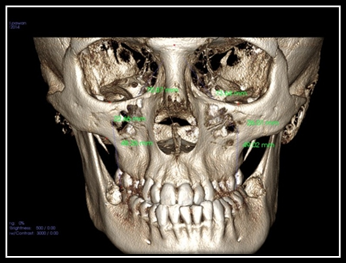
Cone beam computed tomography (CBCT) is considered one of the most accurate diagnostic aid with optimum balance between high diagnostic yield, and low risk (Figure 3). CBCT was introduced in dentistry, with smaller sized machines, high spatial resolution, and rapid scan times and less radiation exposure to patient, however its availability is limited due to the involvement of skilled technicians and technique sensitive procedures. Moreover its high cost and overall management is still a matter of concern
Therefore keeping the concerns of CBCT in mind, this study was designed to evaluate the reliability of 2D digital radiographs by denoting the exact difference between the CBCT and 2D digital radiography readings obtained using frontal vertical proportions, mandibular morphology, and maxillary height from Grummon’s analysis on the PA radiograph and the Levandoski method on the OPG for the diagnosis of facial asymmetry.
Materials & Methods
This study consisted of a sample size of 20 young subjects with facial asymmetry, who were willing to undergo orthodontic treatment. A standardized millimetric ruler, lead acetate paper , and radiographic records of those 20 subjects (i.e20 CBCTs, 20 lateral cephalograms, 20 digital OPGS, 20 extra oral photos view at rest ),digital SLR camera (cannon EOS550D) and a view box. Patients selected from the Department of Orthodontics, interested in orthodontic treatment. Standardized photographic set up was taken into consideration. The frontal view at rest photograph was taken maintaining the natural head position using fluid level device.
Inclusion Criteria
- Facial asymmetry.
- All permenant teeth upto 1st molar erupted.
- Stable occlusion.
- Good periodontal health.
Exclusion Criteria
- Pregnancy.
- Lactating mother.
- Steroid therapy.
- Syndromes.
- Genetic disorder.
- TMJ disorders
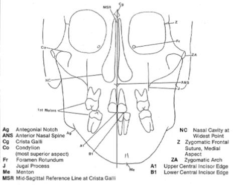
Evaluation of facial deviation, asymmetry, width of the nose, and lower third of face and width of the mouth were assessed with the help of facial photographs. When asymmetry was noted on the facial photograph & confirmed with the facial photographic analysis, patient was advised digital panoramic radiograph. All panoramic radiographs were traced on lead acetate paper for Levandoski panoramic analysis (Figure 1) .After OPG analysis, linear measurements and angles were measured using Grummon’s analysis (Figure 4) Following attaining of the above measurements, the patients were advised a CBCT. The measurements from 2-D digital OPG & P.A cephalogram were compared with the measurements obtained from 3-D CBCT.
The analysis was determined by the construction of series of base reference lines as described by Levandoski which are as follows (Figure 1)
- Line 1 - Ramal lines are drawn along the posterior edge of each ramus.
- Line 2 - Line drawn from condylon to the contact point of the upper central incisors mesially
- Line 3 -Line drawn from condylon to the contact point of the lower incisorsmesially.
- Line 4- Line drawn from condylon to gonion. [Condylar length].
- Line 5 -Line drawn from coronoid to gonion. [Coronoid length].
The parameters considered for diagnosis of facial asymmetry on P.A Cephalogram using Grummons Analysis:-
- Frontal Vertical Portions
- Upper facial ratio - Cg-ANS/ Cg-Me
- Lower facial ratio - ANS-Me/ Cg-Me
- Maxillary ratio - ANS-A1/ ANS-Me
- Total maxillary ratio - ANS-A1/ Cg-Me
- Mandibular ratio - B1-Me/ ANS-Me
- Total mandibular ratio - B1-Me/ Cg-Me
- Maxillo-mandibular ratio - ANS-A1/ B1-Me
B. Mandibular Morphology
- Distance from - Co to Ag
- Distance from - Ag to Me
- Distance from - Co-Me
- Angle of the mandible - <Co-Ag-Me
C. Maxillary Height: -
- Or to J
- Maxillary dental height - Or-U6
- Maxillary height by - N-J
Once the above measurements were obtained, the patient was asked to take a CBCT- view. The measurements from 2D digital OPG & P.A cephalogram were now compared with the measurements obtained from 3D-cone beam computed tomography for the difference in between 2D radiography & 3D-cone beam computed tomography.
Statistical Analysis: Descriptive statistical analysis was carried out in the present study to explore the distributions of several characteristics of the cases studied across two study groups (two radiographic techniques of measurements). Data on continuous variables were presented with Mean Standard Deviation across two study groups.
The statistical significance of difference of various parameters studied (such as frontal vertebral relation parameters, Mandibular morphology parameters etc) across two study groups (Inter-group comparisons) was carried out using independent sample t test, after confirming the underlying normality assumption. The intra-observer comparison for each parameters (pair-wise differences) was tested using Paired t test, after confirming the underlying normality assumption of difference of Reading 1 and Reading 2.
The p-values less than 0.05 were considered to be statistically significant. All the hypotheses were formulated using two tailed alternatives against each null hypothesis (hypothesis of no difference).
Results
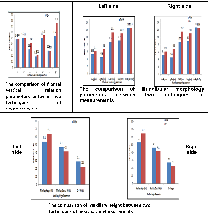
The average upper facial ratio and lower facial ratio did not differ significantly between Digital and CBCT techniques. (Figure 5a). The average mandibular ratio and total mandibular ratio did not differ significantly between Digital and CBCT techniques. (Figure 5b). The average maxillary ratio was significantly higher in CBCT compared to Digital technique. The average total maxillary ratio did not differ significantly between Digital and CBCT techniques. (Figure 5c).
| Parameters | Digital (n = 20) | CBCT (n = 20) | P-value (Digital v/s CBCT) |
| Upper facial ratio | 0.48 ± 0.047 | 0.49 ± 0.042 | 0.330 (NS) |
| Lower facial ratio | 0.51 ± 0.043 | 0.50 ± 0.039 | 0.311 (NS) |
| Maxillary ratio | 0.31 ± 0.059 | 0.42 ± 0.040 | 0.001 (S) |
| Total maxillary ratio | 0.19 ± 0.068 | 0.21 ± 0.033 | 0.222 (NS) |
| Mandibular ratio | 0.51 ± 0.18 | 0.55 ± 0.046 | 0.418 (NS) |
| Total mandibular ratio | 0.29 ± 0.029 | 0.28 ± 0.035 | 0.063 (NS) |
| Maxillomandibular ratio | 0.54 ± 0.11 | 0.76 ± 0.11 | 0.001 (S) |
The average maxilla mandibular ratio was significantly higher in CBCT than Digital technique. The average Co-Ag-Me did not differ significantly between Digital and CBCT techniques on left side of the face. (Figure 5b). The average Co-Ag, Ag-Me, Co-Me and N-Ag is significantly higher in CBCT compared to Digital technique on the left side. (Figure 5b).
| Parameters | Digital (n = 20) | CBCT (n=20) | P-value (Digital v/s CBCT) |
| Linear | |||
| Co-Ag (mm) | 53.8 ± 6.4 | 61.1 ± 5.9 | 0.001 (S) |
| Ag-Me (mm) | 44.9 ± 4.7 | 67.3 ± 6.6 | 0.001 (S) |
| Co-Me (mm) | 87.5 ± 6.2 | 113.5 ± 8.2 | 0.001 (S) |
| N-Ag (mm) | 91.2 ± 14.4 | 110.2 ± 6.4 | 0.001 (S) |
| ANGULAR | |||
| Co-Ag-Me (Deg) | 125.5 ± 8.5 | 125.9 ± 7.8 | 0.847 (NS) |
The average Co-Ag-Me did not differ significantly between Digital and CBCT techniques on the right side of the face. (figure 5b). The average Co-Ag, Ag-Me, Co-Me and N-Ag was significantly higher in CBCT compared to Digital technique on the right side. (Figure 5b).
| Parameters | Digital (n = 20) | CBCT (n = 20) | P-value (Digital v/s CBCT) |
| LINEAR | |||
| Co-Ag (mm) | 53.8 ± 6.4 | 61.1 ± 5.9 | 0.001 (S) |
| Ag-Me (mm) | 44.9 ± 4.7 | 67.3 ± 6.6 | 0.001 (S) |
| Co-Me (mm) | 87.5 ± 6.2 | 113.5 ± 8.2 | 0.001 (S) |
| N-Ag (mm) | 91.2 ± 14.4 | 110.2 ± 6.4 | 0.001 (S) |
| Angular | |||
| Co-Ag-Me (Deg) | 125.5 ± 8.5 | 125.9 ± 7.8 | 0.847 (NS) |
The average Maxillary height was significantly higher in CBCT compared to Digital technique on the left side. (figure 5c).The average Maxillary dental height and Or-J length was significantly higher in Digital compared to CBCT technique on the left side.(Figure 5c)
| Parameters (mm) | Digital (n = 20) | CBCT (n = 20) | P-value (Digital v/s CBCT) |
| Maxillary Height (N-J) | 54.1 ± 9.6 | 64.1 ± 10.9 | 0.004 (S) |
| Maxillary Dental Height | 47.1 ± 4.7 | 41.8 ± 5.7 | 0.003 (S) |
| Or-J Height | 29.1 ± 5.1 | 22.5 ± 4.9 | 0.001 (S) |
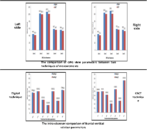
The average Line-1, Line-2, Line-4 and Line-5 readings did not differ significantly between Digital and CBCT techniques on the left or right side. (Figure 6a). The average Line-3 reading was significantly higher in Digital compared to CBCT technique on the right side. (figure 6a). The average Line-1 and Line-3 reading was significantly higher in Digital compared to CBCT technique on right side of the face. (Figure 6a).
| Parameters (mm) | Digital (n = 20) | CBCT (n = 20) | P-value (Digital v/s CBCT) |
| Line-1 | 44.6 ± 4.9 | 41.9 ± 3.5 | 0.060 (NS) |
| Line-2 | 102.5 ± 8.1 | 100.3 ± 5.8 | 0.318 (NS) |
| Line-3 | 107.3 ± 8.2 | 101.5 ± 6.2 | 0.015 (S) |
| Line-4 | 58.7 ± 6.2 | 56.7 ± 7.1 | 0.349 (NS) |
| Line-5 | 56.4 ± 6.6 | 53.7 ± 5.8 | 0.178 (NS) |
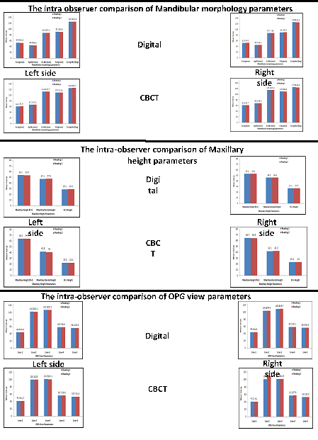
The average reading 1 and reading 2 of frontal vertebral relation parameters did not differ significantly. The average reading 1 and reading 2 Co-Ag, Ag-Me, Co-Me and N-Ag did not differ significantly. (Figure 7a). The average reading 1 and reading 2 Co-Ag-Me did not differ significantly. (Figure 7a)
| Parameters | Reading 1 (n = 20) | Reading 2 (n = 20) | P-value (Paired t test) |
| Linear | |||
| Co-Ag (mm) | 53.8 ± 6.4 | 53.2 ± 5.6 | 0.244 (NS) |
| Ag-Me (mm) | 44.9 ± 4.7 | 44.5 ± 4.2 | 0.483 (NS) |
| Co-Me (mm) | 87.5 ± 6.2 | 87.1 ± 6.5 | 0.516 (NS) |
| N-Ag (mm) | 91.2 ± 14.4 | 90.5 ± 12.7 | 0.530 (NS) |
| Angular | |||
| Co-Ag-Me (Deg) | 125.5 ± 8.5 | 125.6 ± 8.8 | 0.728 (NS) |
The average reading 1 and reading 2 Co-Ag, Ag-Me, Co-Me and N-Ag did not differ significantly the intra observer comparison of Mandibular morphology parameters (Digital or CBCT left sides). (Figure 7a). The average reading 1 and reading 2 Co-Ag-Me did not differ significantly (P-value>0.05) the intra observer comparison of Mandibular morphology parameters (Digital or CBCT Right sides). (Figure 7a).
| Parameters | Reading 1 (n = 20) | Reading 2 (n = 20) | P-value (Paired t test) |
| Linear | |||
| Co-Ag (mm) | 52.9 ± 5.8 | 53.2 ± 5.8 | 0.647 (NS) |
| Ag-Me (mm) | 46.1 ± 5.0 | 45.7 ± 5.1 | 0.491 (NS) |
| Co-Me (mm) | 87.7 ± 6.9 | 88.0 ± 7.8 | 0.617 (NS) |
| N-Ag (mm) | 90.0 ± 13.1 | 90.4 ± 13.2 | 0.297 (NS) |
| Angular | |||
| Co-Ag-Me (Deg) | 126.4 ± 8.5 | 125.5 ± 8.6 | 0.181 (NS) |
The average reading 1 and reading 2 Maxillary heights did not differ significantly intra-observer comparison of Maxillary height parameters (Digital Right or Left sides). (Figure 7b). The average reading 1 and reading 2 Maxillary heights did not differ significantly intra-observer comparison of Maxillary height parameters (CBCT Left or Right sides) (Figure 7b)
| Parameters | Reading 1 (n = 20) | Reading 2 (n = 20) | P-value (Paired t test) |
| Upper facial ratio | 0.48 ± 0.047 | 0.47 ± 0.039 | 0.170 (NS) |
| Lower facial ratio | 0.51 ± 0.043 | 0.51 ± 0.039 | 0.677 (NS) |
| Maxillary ratio | 0.31 ± 0.059 | 0.31 ± 0.049 | 0.368 (NS) |
| Total maxillary ratio | 0.19 ± 0.068 | 0.19 ± 0.067 | 0.519 (NS) |
| Mandibular ratio | 0.51 ± 0.18 | 0.51 ± 0.15 | 0.977 (NS) |
| Total mandibular ratio | 0.29 ± 0.029 | 0.29 ± 0.038 | 0.999 (NS) |
| Maxillomandibular ratio | 0.54 ± 0.11 | 0.53 ± 0.11 | 0.245 (NS) |
The average reading 1 and reading 2 OPG view parameters did not differ significantly intra-observer comparison of OPG view parameters (Digital Left and Right sides as well as CBCT Left and Right sides) (Figure 7c)
Discussion
Craniofacial symmetry and balance is referred to as the ‘state of equilibrium’, where there is a correspondence in size, form and arrangement of the various structures on the opposite sides of the median sagittal plane [8]. Cephalometry is a valuable tool for diagnosing skeletal imbalance and for assessing growth, response to treatment, and long-term stability after orthodontic treatment. Cephalometric evaluation of patients with orthodontic needs has traditionally been performed by lateral and frontal cephalograms [9]. Unlike conventional cephalograms, computed tomography has no inherent distortion or superimposition of anatomic structures. The introduction of CBCT has made 3-D imaging more readily available for dental applications. In recent studies, the reproducibility of measurements obtained from cone beam computed tomography scans was found to be greater than the reproducibility of those obtained from conventional frontal 2D radiographs [13].
The accuracy of CBCT imaging in determining the characteristics of asymmetry is not only important for diagnosis and evaluating treatment outcomes but it may also enable more precise planning of surgical treatment. The PA cephalograms were 2-D replica of the 3-D images which were less accurate, when compared with the CBCT images used for the detection of the characteristics of the mandibular asymmetry [14]. This is important because differences in the mandibular ramus and body length are important factors in detection of chin deviation. In PA cephalometry, landmarks have their own magnification error since the structures are located at a distance from the film. However, due to the positioning of the head in the cephalostat, the magnification error of bilateral landmarks should be the same since the bilateral structures are located at relatively the same distances from the X-ray source and the film [16]. This suggests that the comparison of left and right side structures is possible with PA cephalograms. Hence, in the present study comparison of the right and left side was also conducted.
Levandoski., et al, used their method to detect the asymmetry using OPG radiograph [17]. The results showed that the information obtained using this method supports its use in diagnosing facial and dental asymmetries and its relationships, when compared to digital radiographic techniques. It is not advisable to undergo CBCT as a routine diagnostic record for all the patients for the same reason. In the cases of facial asymmetry, we, routinely advise frontal cephalogram and OPG radiograph as diagnostic record. This study served to find out the accuracy of PA cephalograms and OPG in the diagnosis of facial asymmetries when compared to CBCT images taken for the same patients. When a routine digital radiographic technique shows no significant differences when compared with CBCT images, CBCTs can be avoided whenever possible. If there is significant difference in the measurements, then CBCT techniques should be routine in facial asymmetry diagnosis.
Statistically significant difference was found in frontal vertical proportion parameters in the maxillary ratio showed an increase in the values of the CBCT when compared with the digital radiographic technique. Hence, when the lower anterior facial height region information is required, CBCT is best. When upper facial ratios are required, digital radiography will suffice. When comparing the mandibular morphology, the linear measurements showed higher values in the CBCT. Denoting when any surgical treatment was suggested in the mandibular region, a CBCT should be made. When comparing the mandibular morphology for angular measurements, there was no statistical difference found in the angular measurements between the two radiographic techniques. Hence, when the surgery is required in the mandibular angular region, digital radiographic techniques gives the best required information.
Statistical difference was found when maxillary height showed higher values in the CBCT technique when compared with digital technique. The identification of the Nasion (N) in this region is difficult; due to superimposition of various bones of the anterior cranial region on the digital radiographs thus CBCT is considered to be more accurate and is the choice of diagnostic aid. There was also an increased difference noted in the maxillary dental height and only maxillary height in the digital technique when it was compared with the CBCT technique.
There was a statistical significant difference found in the Levandoski OPG analysis which showed the increased difference in the Line-3 readings on the left side in the digital radiographic technique when compared with the CBCT technique. The same increased difference was noted on the right side Line -1 and Line -3 in the digital radiographic techniques when compared with the CBCT technique. There was no statistical difference for the Line-2, Line-4, Line-5 found for the right side. Studies suggest that laterality in the normal asymmetry of the face, which is consistently found in humans, is likely to be a hereditary rather than an acquired trait. This study supports the readings for the right side dental asymmetry i.e. Line-1 and Line-3 which has increased dimensions and values [18]. Other studies suggest that the asymmetries show right side dominance over left side. [19] our study therefore supports the significant statistical readings for the right side i.e. Line-1 and Line-3. CBCT can better evaluate craniofacial morphology when compared with digital 2D images.
The study was conducted to compare the accuracy of PA cephalogram and OPG measurements for diagnosing asymmetry with the 3-D CBCT image measurements. The readings for the frontal vertical relation parameters from Grummon’s analysis showed significant differences for the maxillary ratio and maxillomandibular ratios. Whereas, upper and lower facial ratio, total maxillary ratio, mandibular ratio, total mandibular ratio had comparable accuracy between CBCT and digital techniques. Maxillary height (N-J), maxillary height (Or-J), maxillary dental height (Or- U6) according to Grummon’s analysis had significantly higher values when comparison was done between digital radiography and CBCT techniques. Mandibular morphology, according to Grummon’s analysis showed significant difference in the linear measurements, which signifies digital cephalometric measurements do not have comparable accuracy to the CBCT measurements. On the other hand, digital cephalometric angular measurements under the same parameters have comparable accuracy to the CBCT technique. Linear measurements, according to Levandoski method, showed significantly higher values in the digital radiography which helps to conclude that it does not have comparable accuracy with the CBCT technique.
Therefore, this study took the radiation exposure into consideration and selected samples who were interested in orthodontic treatment by their own will, in our department. Basic diagnostic tools such as OPG, PA, lateral cephalogram, are usually advised, patients were advised for additional CBCTs, to prove the reliability of the CBCT technique and the digital radiography for the future human benefit.
Conclusion
Though 3D CBCT stands as the most precise radio diagnostic aid in dentistry, this study enlightens the fact that 2D digital radiographs still manages to be reasonably accurate for the diagnosis of facial asymmetry in certain parameters when compared to that of 3d CBCT and can be a reliable source for diagnosing dental and skeletal abnormalities especially when the resources and availability for 3d CBCT are deficient.
References
- German O., et al. “Prevalence of mandibular asymmetries in growing patients.” European Journal of Orthodontics August 33.3-1 (2010): 1-7.
- Shah SM and Joshi MR. “An assessment of asymmetry in the normal craniofacial complex”. Angle Orthodontist 48.2 (1978): 141-148.
- You-Wei Cheong and Lun-Jou Lo. “Facial Asymmetry: Etiology, Evaluation and Management”. Chang Gung Medical Journal 34 (2011): 341-51.
- Dana C Van Elslande., et al. “Mandibular asymmetry diagnosis with panoramic imaging”. American Journal of Orthodontics and Dentofacial Orthopedics 134.2 (2008): 183-192.
- Damstra J., et al. “Evaluation & comparison of postero-anterior cephalogram& cone-beam computed tomography images for the detection of mandibular asymmetry”. European Journal of Orthodontics 35.1 (2011).
- Robert M Rickets. Duane Grummons Frontal Cephalometrics: Practical Application, Part I. World Journal Orthodontics 4 (2003): 297-316.
- Matthew M., et al. “Asymmetry assessment using cone beam CT. A Class I and II patient comparison”. Angle Orthodontist 82.3 (2012). 410-417.
- Brent E Larson. “Cone-beam computed tomography is the imaging technique of choice for comprehensive orthodontic assessment”. American Journal of Orthodontics and Dentofacial Orthopedics 141.4 (2012): 402-411.
- Kumar V., et al. “In vivo comparison of Conventional & Cone Beam CT Synthesized Cephalograms”. Angle orthodontist 78.5 (2008): 873-879.
- Ludlow JB., et al. “Dosimetry of two extraoral direct digital imaging devices: NewTom cone beam CT and Orthophos plus DS panoramic unit.” Dentomaxillofacial Radiology32.4(2003): 229–234.
- Sukovic P. “Cone beam computed tomography in craniofacial imaging”. Orthodontics & Craniofacial Research2003; 6(suppl 1): 31–36; discussion 179-182.
- Farman AG and Scarfe WC. “Development of imaging selection criteria and procedures should precede cephalometric assessment with cone-beam computed tomography”. American Journal of Orthodontics and DentofacialOrthopedics130.2 (2006); 130:257–265.
- Cho PS., et al. “Cone-beam CT for radiotherapy applications”. Physics in Medicine & Biology 40.11 (1995): 1863–1883.
- Chidiac JJ., et al. “Comparison of CT scanograms and cephalometric radiographs in craniofacial imaging”. Orthodontics & Craniofacial Research 5.2 (2002): 104-113.
- Hwang HS., et al. “Maxillofacial 3-dimensional image analysis for the diagnosis of facial asymmetry”. American Journal of Orthodontics and Dentofacial Orthopedics 130.6 (2006): 779-785.
- Peck S., et al. “Skeletal asymmetry in esthetically pleasing faces”. Angle Orthodontist 61.1 (1991): 43-46.
- Bishara SE., et al. “Dental and facial asymmetries: a review”. Angle Orthodontist 64.2 (1994): 89-98.
- Issabella Piedra. “The Levandoski Panoramic Analysis in the diagnosis of facial & dental asymmetries”. Journal of Clinical Pediatric Dentistry 20.1 (1995): 15-21.
- Seiji Haraguchi., et al. “Asymmetry of the Face in Orthodontic Patients”. Angle Orthodontist 78.3 (2008): 421-426.
- Mari Eli Leonelli de Moraes., et al. “Evaluating craniofacial asymmetry with digital cephalometric images and cone beam computed tomography”. American Journal of Orthodontics and Dentofacial Orthopedics 139.6 (2011): e523-e531.
Citation:
Taabish Contractor., et al. “Different 2d Digital Radiographic Methods Versus Cone Beam Computed Tomography for the
Diagnosis of Facial asymmetry- In vivo Study.”. Oral Health and Dentistry 3.1 (2018): 542-553.
Copyright: © 2018 Taabish Contractor., et al. This is an open-access article distributed under the terms of the Creative Commons Attribution License, which permits unrestricted use, distribution, and reproduction in any medium, provided the original author and source are credited.


 Scientia Ricerca is licensed and content of this site is available under a Creative Commons Attribution 4.0 International License.