Research Article
Volume 3 Issue 2 - 2018
Ricketts Cephalometric Analysis for the 9 to 14 Year Old Population of Central Mexico
1Center for Research and Advanced Studies in Dentistry, Autonomous University of the State of Mexico (UAEM)
2Faculty of Dentistry, Autonomous University of the State of Mexico (UAEM)
2Faculty of Dentistry, Autonomous University of the State of Mexico (UAEM)
*Corresponding Author: Norma Margarita Montiel Bastida, School of Dentistry, Autonomous University of the State of Mexico, Jesús Carranza of Paseo Tollocan. C.P.50130. Toluca, Mexico.
Received: May 08, 2018; Published: May 21, 2018
Abstract
Background: Ricketts cephalometric analysis is one of the most widely used, however, it is structured from an Anglo-Saxon population. Most orthodontic treatments in other populations may be inaccurate.
Objective: To establish a cephalometric analysis for the population of central Mexico aged 9-11 years and 12-14 years.
Materials and Methods: Descriptive, comparative, cross-sectional study. A sample of 135 Mexican individuals was taken. The craniofacial analysis of Ricketts was performed by tracing the lateral cephalogram of the cranium. Data by age and sex were analyzed and cephalometric polygons were created.
Results: Significant differences in craniofacial growth were observed between the female and male sex, as well as in various measurement parameters.
Conclusions: The values obtained allow us to establish a cephalometric standard for the central population of Mexico from 9 to 11 years old and 12 to 14 years old.
Key words: Cephalometric analysis; Mexican population; Craniofacial growth
Introduction
According to the World Health Organization (WHO), cranio-facial-dental anomalies and malocclusions are of medium prevalence worldwide. In Mexico, approximately 96% of the population has some type of malocclusion, whether dental, skeletal, functional or mixed, indicating a public health problem [1]. These anomalies cause alterations in essential functions such as phonation, chewing and swallowing, which have repercussions on the quality of life. In addition, the bone complex being the supporting framework of the soft tissues, the disharmony between them impacts on the individual in the aesthetic aspect.
Most of these discrepancies are the result of a discordance between the size of the teeth and their maxillary support, the result of the development and growth of each individual, which is genetically determined and closely related to age and sex. According to Woodside, between the ages of 3 and 15, different periods of significant growth can be observed. Within these ages, orthodontics and orthopedics are essential for the treatment and prevention of malocclusions [2].
To establish a correct diagnosis, the orthodontic specialist must perform a clinical, radiographic and model study analysis of each patient, which allows him/her to determine the appropriate treatment plan. Among the elements necessary for orthodontic diagnosis is cephalometry, which facilitates the observation of the relationship between the skeletal, dental and soft tissue structures of each individual. Likewise, applied in the various stages of growth, it allows the craniomaxillofacial development process to be studied [3].
There are several cephalometric analyses, based on a pattern of normality that is specific to the population analysed. One of the most commonly used is the one described by Robert Murray Ricketts. Considered the father of computerized cephalometrics, he created the cephalometric analysis that bears his name in the 1950s, thus suggesting the possibility of predicting growth and treatment plan, expressing this idea in a term called "visual treatment objective" [4].
Over time, the analysis has undergone several modifications, it is currently composed of 33 factors grouped into six fields that analyze from aesthetics to internal structure, so it is considered the most complete5. However, since the parameters from which this examination emerged were obtained from an Anglo-Saxon population, they cannot be applied to the Mexican population [6,7].
Therefore, the objective of the present work was to establish a cephalometric standard for the population of central Mexico from 9 to 14 and 12-14 years of age from selected x-rays, using the craniofacial analysis of Ricketts. With the results obtained, an instrument is provided to facilitate better diagnoses and treatment plans with which to obtain functional and aesthetic results suitable for this population group.
Materials and Methods
A descriptive, comparative and transversal study was carried out. From a universe of 3463 children and adolescents belonging to four primary schools, a sample was taken of 135 Mexican patients from Mexican parents and grandparents, with facial symmetry, apparently normal growth and development according to their age group, with mixed or permanent teeth, according to their age, free of cavities, with molar relation of Angle class I and skeletal class I, without previous orthodontic or orthopedic treatment and without a maxillofacial surgical history.
The sample was selected by clinical dental review with prior informed consent. Each subject had a lateral cranium x-ray taken within the facilities of the Center for Research and Advanced Studies in Dentistry of the School of Dentistry of the Autonomous University of the State of Mexico. To take the x-rays, the patient was placed seated with the Frankfurt plane parallel to the floor, a cephalostat was used to support the head with the help of stems in the ears, aligned in the central axis of the X-ray tube radiation.
The x-rays were obtained with a Panoura 10 C orthopantomographer (Yoshida Dental MFG. Co., LTD Tokyo, Japan), using Kodak X-Omat blue-sensitive (8 x 10" CAT 6031876) x-rays, on a Kyokko PS-II, Kasei Optonix, LTD chassis of the same size, with a three-second exposure time and 88kvp at a standard target-to-film distance of 1.65m. The development of the X-rays was automatic by means of a yoshida Dental MFG device. Co LTD (Tokyo, Japan).
Measuring technology
The lateral cephalogram of the cranium was performed by a single person to standardize the technique. It was included the soft tissue and bone profile, jaw profile, posterior profile of the cerebral case, odontoid process, anterior edge of the foramen magnum, full quadrilateral of the sphenoid, profile of the Turkish saddle, ceiling of the orbit, cribriform plate, lateral and inferior edges of the orbit, profile of the pterygomaxillary fissure, floor of the nose, roof of the palate and body of the hyoid. The first permanent molars and the most prominent incisors were also traced. The measurements were made in duplicate, repeating them when there was a difference of ±1mm between the two.
The lateral cephalogram of the cranium was performed by a single person to standardize the technique. It was included the soft tissue and bone profile, jaw profile, posterior profile of the cerebral case, odontoid process, anterior edge of the foramen magnum, full quadrilateral of the sphenoid, profile of the Turkish saddle, ceiling of the orbit, cribriform plate, lateral and inferior edges of the orbit, profile of the pterygomaxillary fissure, floor of the nose, roof of the palate and body of the hyoid. The first permanent molars and the most prominent incisors were also traced. The measurements were made in duplicate, repeating them when there was a difference of ±1mm between the two.
Data were tabulated by age group and sex. A descriptive analysis was performed based on frequencies, mean and standard deviation. Student t was applied to establish statistical differences. Data were analyzed using SPSS version 19.
Results
The entire sample (135 patients) aged 9-14 years were analyzed. The frequency of male patients was 47.4% and female patients 52.6%. We analyzed the data by age and sex.
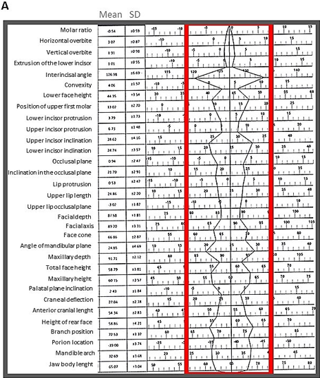
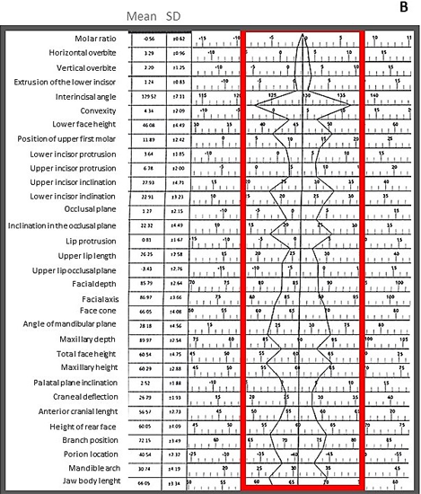
For the 9 to 11 year old group the frequency of male patients was 53.7% and for the female group it was 46.3%. Data on the measures found in this age group for both sexes are summarized in Table I.
| Type of analysis | Measurements | Female | Male | p* | ||
| Mean | DS | Mean | DS | |||
| I: Dental analysis | Molar ratio A6-B6 | -0.54 | 0.59 | -0.56 | 0.62 | 0.43 |
| Horizontal overbite B1-A1 | 3.07 | 0.87 | 3.29 | 0.96 | 0.16 | |
| Vertical overbite B1-A1 | 1.91 | 0.98 | 2.20 | 1.25 | 0.14 | |
| Extrusion of the lower incisor B1-PI.OCI. | 1.01 | 0.55 | 1.24 | 0.83 | 0.09 | |
| Interincisal angle AI-A2/B1-B2 | 126.98 | 6.69 | 129.52 | 7.11 | 0.06 | |
| II: Skeletal analysis (maxillo-mandibular ratio) | Convexity A/N/P/g | 4.06 | 1.57 | 4.34 | 2.09 | 0.27 |
| Lower face height ENA-XI-PM | 44.35 | 3.54 | 46.08 | 4.49 | 0.04 | |
| III: Dentoeskeletal analysis | Position of upper first molar | 13.02 | 2.70 | 11.89 | 2.42 | 0.03 |
| Lower incisor protrusion B1/A-Pg | 3.79 | 1.73 | 3.64 | 1.85 | 0.36 | |
| Upper incisor protrusion A1/A-Pg | 6.73 | 1.48 | 6.78 | 2.00 | 0.46 | |
| Upper incisor inclination A1-A2/A-Pg | 28.62 | 4.16 | 27.93 | 4.71 | 0.26 | |
| Lower incisor inclination B1-B2/A-Pg | 24.74 | 3.57 | 22.91 | 3.23 | 0.01 | |
| Occlusal plane -XI | 0.94 | 2.47 | 1.27 | 2.15 | 0.27 | |
| Inclination in the occlusal plane Xi-pn | 21.70 | 2.91 | 22.32 | 4.49 | 0.25 | |
| IV aesthetic analysis | Lip protrusion Li/Pn-D T | 0.53 | 2.47 | 0.81 | 1.67 | 0.29 |
| Upper lip length Ena-En | 24.86 | 2.20 | 26.25 | 2.58 | 0.01 | |
| Upper lip occlusal plane Em-TI. OcI. | -3.02 | 1.82 | -3.43 | 2.76 | 0.24 | |
| V: craniofacial ratio | Facial depth Po-Or/N-PG | 87.58 | 1.81 | 85.79 | 2.64 | 0.00 |
| Facial axis Ba-N/Pt-GnI | 89.20 | 3.31 | 86.97 | 3.66 | 0.00 | |
| Face cone | 66.86 | 2.87 | 66.05 | 4.08 | 0.17 | |
| Angle of mandibular plane Go-Me/Po-Or | 24.85 | 4.69 | 28.18 | 4.56 | 0.00 | |
| Maxillary depth Po-Or/NA | 91.71 | 2.12 | 89.97 | 2.54 | 0.00 | |
| Total face height | 58.79 | 3.81 | 60.54 | 4.75 | 0.05 | |
| Maxillary height N-Cf-A | 60.15 | 3.57 | 60.29 | 2.88 | 0.42 | |
| Palatal plane inclination Po-Or/Ena-Enp | 2.43 | 1.84 | 2.52 | 1.88 | 0.42 | |
| VI: Internal structure | Cranial deflection Ba-Na/Po-Or | 27.04 | 2.38 | 26.79 | 1.93 | 0.31 |
| Anterior cranial length Cc-Na | 54.34 | 2.83 | 56.57 | 2.73 | 0.00 | |
| Height of rear face Go I-Cf | 58.86 | 4.21 | 60.05 | 4.09 | 0.12 | |
| branch position Po-Or/Cf-XI | 72.53 | 3.32 | 72.15 | 3.49 | 0.32 | |
| Porion location Po/PtV | -39.00 | 3.74 | -40.54 | 2.32 | 0.02 | |
| Mandible arch Dc-XI/XI-Pn | 32.69 | 3.68 | 30.74 | 4.19 | 0.02 | |
| Jaw body lenght XI-Pm | 65.07 | 3.04 | 66.05 | 3.34 | 0.10 | |
| * t student test, p≤0.05 | ||||||
Table 1: Comparison of Mean (SD) values (Load) of both the groups.
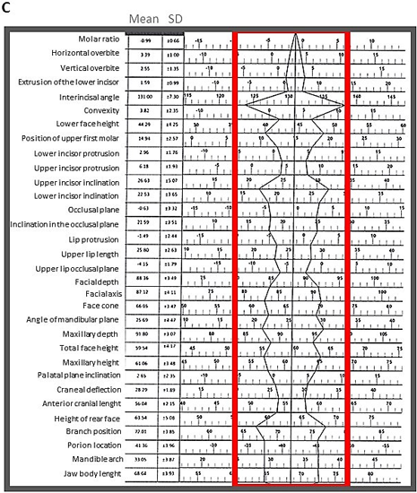
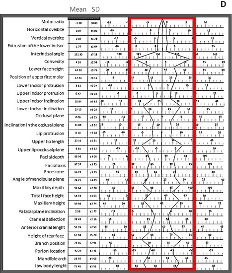
Since the sample is representative of patients from the central region of Mexico, cephalometric polygons were developed for the female and male population aged 9 to 11 and 12 to 14 years, based on the mean and standard deviations of the measurements that included the 6 fields analyzed. (Figures 1 and 2).
For the 12 to 14 year old group, the frequency of male patients was 51.5% and for the female group it was 48.5%. Data on the measures found in this age group for both sexes are reported in Table II.
| Type of analysis | Measurements | Female | Male | p* | ||
| Mean | DS | Mean | DS | |||
| I: Dental analysis | Molar ratio A6-B6 | -0.99 | 0.66 | -1.18 | 0.83 | 0.14 |
| Horizontal overbite B1-A1 | 3.39 | 1.00 | 3.07 | 1.02 | 0.09 | |
| Vertical overbite B1-A1 | 2.55 | 1.35 | 2.02 | 1.28 | 0.05 | |
| Extrusion of the lower incisor B1-PI.OCI. | 1.59 | 0.99 | 1.77 | 1.04 | 0.24 | |
| Interincisal angle AI-A2/B1-B2 | 131.00 | 7.30 | 131.10 | 7.18 | 0.46 | |
| II: Skeletal analysis (maxillo-mandibular ratio) | Convexity A/N/P/g | 3.82 | 2.35 | 4.26 | 2.98 | 0.24 |
| Lower face height ENA-XI-PM | 44.29 | 4.25 | 44.30 | 3.75 | 0.49 | |
| III: Dentoeskeletal analysis | Position of upper first molar | 14.04 | 2.57 | 17.73 | 3.15 | 0.00 |
| Lower incisor protrusion B1/A-Pg | 2.96 | 1.76 | 3.53 | 2.27 | 0.13 | |
| Upper incisor protrusion A1/A-Pg | 6.18 | 1.93 | 6.42 | 2.22 | 0.31 | |
| Upper incisor inclination A1-A2/A-Pg | 26.63 | 5.07 | 25.84 | 4.83 | 0.25 | |
| Lower incisor inclination B1-B2/A-Pg | 22.53 | 3.65 | 23.19 | 3.28 | 0.21 | |
| Occlusal plane -XI | -0.63 | 3.32 | 0.06 | 2.25 | 0.15 | |
| Inclination in the occlusal plane Xi-pn | 21.59 | 3.51 | 21.08 | 2.52 | 0.24 | |
| -1.49 | 2.44 | 0.10 | 2.28 | 0.00 | ||
| IV aesthetic analysis | Lip protrusion Li/Pn-D T | 25.80 | 2.63 | 27.15 | 5.31 | 0.09 |
| Longitud del labio superior Ena-En | -4.15 | 1.79 | -3.65 | 3.63 | 0.24 | |
| Upper lip occlusal plane Em-TI. OcI. | ||||||
| 88.36 | 3.49 | 88.93 | 3.08 | 0.23 | ||
| V: craniofacial ratio | Facial depth Po-Or/N-PG | 87.12 | 4.11 | 87.57 | 3.75 | 0.31 |
| Facial axis Ba-N/Pt-GnI | 66.05 | 3.47 | 66.73 | 3.39 | 0.20 | |
| Face cone | 25.69 | 4.47 | 24.25 | 4.89 | 0.10 | |
| Angle of mandibular plane Go-Me/Po-Or | 91.80 | 3.07 | 92.64 | 2.70 | 0.11 | |
| Maxillary depth Po-Or/NA | 59.54 | 4.17 | 58.93 | 4.63 | 0.28 | |
| Total face height | 61.06 | 3.48 | 59.98 | 2.74 | 0.07 | |
| Maxillary height N-Cf-A | 2.65 | 2.35 | 2.59 | 1.77 | 0.44 | |
| Palatal plane inclination Po-Or/Ena-Enp | 28.29 | 1.89 | 28.49 | 2.16 | 0.34 | |
| VI: Internal structure | Cranial deflection Ba-Na/Po-Or | 56.04 | 2.15 | 59.39 | 2.99 | 0.00 |
| Anterior cranial length Cc-Na | 63.54 | 5.08 | 67.58 | 5.20 | 0.00 | |
| Height of rear face Go I-Cf | 72.01 | 3.85 | 73.36 | 2.91 | 0.05 | |
| branch position Po-Or/Cf-XI | -41.36 | 3.96 | -42.24 | 3.41 | 0.16 | |
| Porion location Po/PtV | 33.05 | 3.87 | 33.40 | 4.63 | 0.36 | |
| Mandible arch Dc-XI/XI-Pn | 68.64 | 3.93 | 71.80 | 3.93 | 0.00 | |
| Jaw body lenght XI-Pm | -0.99 | 0.66 | -1.18 | 0.83 | 0.14 | |
| * t student test, p ≤ 0.05 | ||||||
Table II: Measures found in the 12-14 year old female and male age group with normal occlusion.
Discussion
According to Downs [8], Sassouni [9] and Ricketts [10] there are significant differences in cephalometric parameters between races and ethnic groups. In the present study, it was found that in field I: dental analysis, the 9 to 11 year old age group had similar average values to the Anglo-Saxon population studied by Ricketts [5]. No statistically significant differences were found in this area with respect to sex, coinciding with what was reported previously by Lara., et al. [11].
In this same field, but for the 12 to 14 year old group, a statistically significant difference in vertical overbite between men and women was found, which could be related to the fact that the size of the dental arch of men is wider than in women and also to the fact that in this age group the dental eruption is being completed to second molars. In general, this group presents similar average values to the Anglo-Saxon population studied by Ricketts [5] but also coincides with the Peruvian and Korean populations previously reported [12,13].
As for field II: skeletal analysis, for both the 9 to 11 year old group and the 12 to 14 year old group, no statistically significant differences were found between men and women, however, the average values are different from those reported by Ricketts [5], with the measurement of convexity being higher in the Mexican population and lower in the facial height than in the Anglo-Saxon population, which may result in a more convex profile and with greater horizontal growth. Previous studies conducted by Chavez., et al. [12] in the Peruvian population and by Podadera and Col. [13] in Cuban subjects reported similar values to those of this study.
The results of field III: Dentoeskeletal analysis, showed statistically significant differences in the position of the upper first molar and the inclination of the lower incisor which was greater in the girls. These differences may be related to the condition that the width of the dental arches is narrower in women than in men. This result is typical of the 9 to 11 age group. The 12 to 14 year-old group had statistically significant differences only in the position of the upper first molar. The characteristics that were different from those established by Ricketts [5] were: protrusion of the lower and upper incisor and inclination of the lower incisor, so that the dental protrusion of this population is greater than in the Anglo-Saxon population.
Field IV: aesthetic analysis. In this aspect, for the 9-11 year-old group, significant differences were found between both sexes in the length of the upper lip, which may be due to the fact that girls have finer aesthetic characteristics than boys. In the 12-14 age group there were significant differences in lip protrusion. When comparing the aesthetic analysis in the study population with the results obtained by Ricketts [5], similarities were observed only in the upper lip occlusal plane measurement. The length of the upper lip and the protrusion are greater in the Mexican population which gives it a more convex profile. Background studies in other populations reflect similar results [14].
On the other hand, for the 9 to 11 year old group in the measurements obtained in field V: craniofacial relationship, most of the measurements show statistically significant differences between men and women, which could be related to the disparity in terms of growth times described in this age group. This can be corroborated by the results of the 12 to 14 year old group where no significant differences were found in any of the eight measurements. Most of the measurements present values like those of the Anglo-Saxon population except for the slope of the palatal plane and the maxillary height, being higher in the Mexican population, which may be a factor that influences the conformation of a convex profile in our population, coinciding with different reports [15-17].
Finally, the analysis of field VI: internal structure, for the younger age group, showed results where it is observed, significant differences between men and women that could be related to the strength of the sex-differentiated musculature and that influences jaw development and condyle position. In the 12 to 14 year old group, this was the field where the greatest differences were observed between boys and girls, which may be due to the fact that in this age range there are differential peaks of pubertal growth between men and women. According to this study, only the average value of porion location is similar between the Mexican and Anglo-Saxon populations studied by Ricketts [5].
Conclusions
- The cephalometric values obtained in this study allow us to establish a cephalometric standard for the population of central Mexico from 9 to 11 and from 12 to 14 year old based on the measurements of the craniofacial analysis of Ricketts.
- The average measurements that constitute the present standard will facilitate the diagnosis and treatment plan of craniofacial alterations in patients with similar characteristics to the sample studied.
References
- Murrieta Pruneda JF., et al. “prevalencia de maloclusiones dentales en eun grupo de adolescentes mexicanos y su relación con la edad y género”. Acta odontológica Venezolana 45.1 (2007): 1-7.
- Woodside D. Some effects of treatment on the growth rate of the mandibule: trnsactions of the third international orthodontic congress. Londres Crosby Lockwood Staples. (1973).
- Proffit WR. Ortodoncia: teoría y practicva. 2a ed. España. Mosby-Doyma (1994).
- Montes de Oca ZEC. Compendio de cefalometría, Análisis clínico y práctico. Colombi Amolca (2004): 1-6.
- Ricketts RM., et al. Bio-Progressive Therapy. Denver: Rocky Mountain Orthodontics; (1979).
- Montt J., et al. “Características Cefalométricas en Jóvenes con Oclusión Normal y Perfil Armónico en Población Chilena”. International Journal of Morphology 33.1 (2015): 237-244.
- Conde-Suárez HF., et al. “Confidence interval for the standards of the Ricketts´summarized cephalogram in Cuban children”. Revista Médica Electrónica 40.1 (2018): 35-47.
- Downs WB. “Analysis of the dentofacial profile”. The Angle Orthodontist 26.4 (1956): 191-212.
- Sassouni V. The face in five dimensions. Philadelphia: Growth Center publication; (1960).
- Ricketts RM. “A four-step method to distinguish orthodontic changes from natural growth”. Journal of Clinical Orthodontics (1975): 208-215.
- Lara CE. “Establecimiento de un estándar cefalométrico según Harvold por grupo de edad y sexo en la población de Toluca, México”. Universidad Autónoma del Estado de México. (2005).
- Chávez ME. Valores cefalométricos de una población de escolares peruanos, con oclusión normal, según el análisis lateral de Ricketts. Universidad Nacional Mayor de San Marcos, Perú, (2004).
- Podadera VZ y col. “Cefalometría lateral de Ricketts en adolescentes de 12 a 14 años con oclusión normal, 2001-2003”. Revista Cubana de Estomatología 40.3 (2003): 1-10.
- Merina J., et al. “Sagittal lip positions in different skeletal malocclusions: a cephalometric analysis.” Progress in Orthodontics (2015).
- Eun-Ju-Bae y col. “Changes in longitudinal craneofacial growth in subjets with normal oclusión using the Ricketts analysis”. The Korean Journal of Orthodontics 44.2 (2014): 77-87.
- Balut GM., et al. “Establishing cephalometric norms for a Mexican population using Ricketts, Steiner, Tweed and Arnett analysesear. APOS 3.6 (2013): 171-177.
- Lourenço BRH., et al. “Correlation between transverse and vertical measurements in Brazilian growing patients, evaluated by Ricketts-Faltin frontal analysis”. Dental Press Journal of Orthodontics 18.1 (2013).
Citation:
Norma Margarita Montiel Bastida., et al. “Ricketts Cephalometric Analysis for the 9 to 14 Year Old Population of Central
Mexico”. Oral Health and Dentistry 3.2 (2018): 574-582.
Copyright: © 2018 Norma Margarita Montiel Bastida., et al. This is an open-access article distributed under the terms of the Creative Commons Attribution License, which permits unrestricted use, distribution, and reproduction in any medium, provided the original author and source are credited.


 Scientia Ricerca is licensed and content of this site is available under a Creative Commons Attribution 4.0 International License.