Case Report
Volume 3 Issue 6 - 2019
Cone Beam Computed Tomography - Assisted Endodontic Management of an Atypical Four Canalled Mandibular Second Premolar - A Rare Case Report
1Ex. Assistant Professor, Department of Conservative Dentistry and Endodontics, Government Dental College and Hospital, Mumbai,St. Georges Hospital Compound, P.D'Mello Road, Fort, Mumbai -400001.
2MDS, MA, LLB, Associate Professor, Department of Conservative Dentistry and Endodontics, Government Dental College and Hospital, Mumbai, St. Georges Hospital Compound, P.D'Mello Road, Fort, Mumbai -400001
3BDS, Private Dental Practitioner, Pinky’s Dental Care, Borivali (West), Mumbai-400092
2MDS, MA, LLB, Associate Professor, Department of Conservative Dentistry and Endodontics, Government Dental College and Hospital, Mumbai, St. Georges Hospital Compound, P.D'Mello Road, Fort, Mumbai -400001
3BDS, Private Dental Practitioner, Pinky’s Dental Care, Borivali (West), Mumbai-400092
*Corresponding Author: Manoj Mahadeo Ramugade, MDS, MA, LLB, Associate Professor, Department of Conservative Dentistry and Endodontics, Government Dental College and Hospital, Mumbai, St. Georges Hospital Compound, P.D'Mello Road, Fort, Mumbai -400001.
Received: July 15, 2019; Published: July 25, 2019
Abstract
Successful endodontic treatment warrants proper access opening, effective cleaning-shaping and three-dimensional obturation of the root canal system. Mandibular second premolars have been acknowledged as 'Enigma to Endodontists' due to their aberrant root anatomy and canal configuration which leads to complexity in the endodontic procedure. Ineptitude to diagnose such diversities may cause missed canals with incomplete cleaning and shaping of the root canal space; which may result in harbouring of the microorganisms in the canal space causing persistent pain or recurrent apical periodontitis. This article reports the successful endodontic management of an unusual case of mandibular second premolar exhibiting two roots with four canals, using cone beam computed tomography (CBCT) imaging as a guiding tool.
Keywords: Aberrant canal anatomy; Cone beam computed tomography; Four canals; Mandibular second premolar
Introduction
Successful endodontic treatment mandates proper access preparation, thorough cleaning and shaping, copious irrigation, three-dimensional obturation and adequate coronal seal. At the same time, detail knowledge of the root morphology and their variations along with the possible racial predilection is essential for predictable treatment outcome. In human dentition, the major anatomical variations have reported in the third molars followed by mandibular first and second premolars. [1,2] Like mandibular first premolars, second premolars have also been recognised with complex root canal anatomy by having an extra root, additional canal or C- shaped canal configuration etc. [3-5] Such aberrations in the root canal system must be identified and managed strategically in order to avoid future procedural accidents and to prevent non-healing of the peri-radicular lesion.
It is difficult to evaluate the root canal configuration correctly by using only an intraoral peri-apical (IOPA) radiograph; as it is a 2-dimensional (2D) image of a 3-dimensional (3D) structure. It possesses the limitation of providing a clear image of root canal anatomy due to super-imposition. To identify an extra canal, multi-angulated IOPA radiographs are often required. When an IOPA radiograph shows a sudden disappearance of a radiolucent canal, it gives a suspicion of possible bifurcation or trifurcation of the canal; widely known as the Fast-break principle. [1] With the newer advances in the imaging techniques such as cone beam computed tomography (CBCT) and Spiral CT, the identification of root canal anatomy becomes easier due to its accuracy and virtual 3D reconstruction. [6,7]
With the dawn of magnification with loupes and dental operating microscope (DOM) in Endodontics, treating such complex anomalies have become convenient due to a clear and magnified view of the pulp chamber as well as the canal orifices. This also results in a fewer missed canal, reduced procedural accidents and increased success rate of endodontic treatment. This case report describes the successful endodontic management of the mandibular second premolar, showing 2 roots and 4 canals with the aid of CBCT and magnification.
Case Report
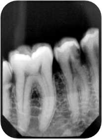
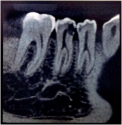
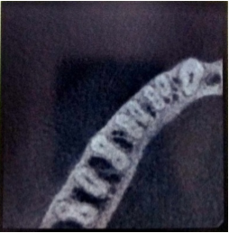
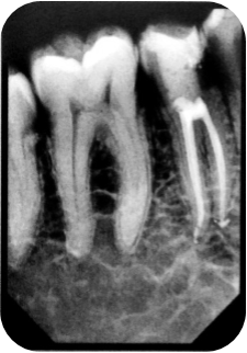
A 29-years-old healthy male patient reported with spontaneous pain in tooth #45. Clinical examination showed mesial caries in tooth #45. Electric pulp test showed an exaggerated response when compared to the adjacent and contra-lateral tooth, suggestive of symptomatic irreversible pulpitis (SIP) in tooth #45. IOPA radiograph showed caries involving the pulp and bifurcated roots with both teeth #44 and #45 (Figure 1). Due to the suspicion of complex root canal configuration; a CBCT scan was advised after obtaining the patient’s informed consent. The CBCT scan revealed four root canals, two in the mesial and two in the distal root starting at the level of the middle third of the root. The mesial canals were observed to be merging further apically (Figure 2a, 2b). Patient’s informed consent was taken and root canal treatment was initiated in tooth #45.
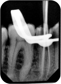
The tooth #45 was isolated under the rubber dam and access preparation was done using 3.5x magnifying loupes. Access cavity was modified to gain access to all the four canals. After access opening, the pulp chamber was flooded with 3% sodium hypochlorite solution for a few seconds. Careful observation of the floor of the pulp chamber and exploring it with a DG 16 endodontic explorer (Dentsply, UK) revealed four root canals. The canals were then negotiated with #10 K files (MANI Inc., Japan). Working length (W.L.) was measured with an electronic apex locator (Root ZX, J Morita, Tokyo, Japan) and confirmed using W.L. IOPA radiograph (Figure 3).
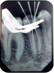
The canal orifices were enlarged with Protaper Gold Sx rotary file (Dentsply, Maillefer). The root canal preparation was carried out with hand K- files (MANI Inc. Japan) followed by NiTi rotary 4% taper files. The final preparation was done with 25/04 Flexicon X5 files (Flexicon, Johnson City, Tennesse, USA). The root canals were copiously irrigated with 3% sodium hypochlorite and 17% ethylene-diamine-tetra-acetic acid (EDTA) solution and normal saline sequentially. After completion of root canal preparation, the canals were flooded with 3% sodium hypochlorite solution and agitated with an Endo-activator tip (Dentsply Tulsa, Dental Specialities). An IOPA radiograph of tooth#45 was taken after placing all the master-cone gutta-percha points in the prepared canals (Figure 4). The root canals were dried with 4% paper points (DiaDent, Dia-ProTTM, Korea). The canals were then coated with AH Plus sealer (Dentsply International Inc.) and the obturation was carried out with warm vertical compaction of the gutta-percha. The access cavity was restored immediately with temporary restoration (Cavit, 3M ESPE, Germany) and a post-operative radiograph was taken (Figure 5).
Discussion
Mandibular premolars are one of the most difficult teeth to treat endodontically and have earned the recognition as ‘Enigma to the Endodontist’, due to their unpredictable and complex root canal configuration. Slowy [8] Zillich and Dowson [9] have reported the prevalence of three root canals in the mandibular premolars as 0.4% in the studied population. Although it is rare, very few authors have reported the cases of mandibular premolars with four and five root canals using dye penetration method and CBCT scan (Table 1).
| Investigator | Number of teeth studied | % teeth showing single canal | % teeth showing two canals | % teeth showing three or more canals and C- shaped canal configuration | Method of investigation |
| Vertucci., et al. (1978) [4] | 400 | 97.5% | 2.5% | 0 | Dye penetration and clearing |
| Rahimi., et al. (2007) [10] | 103 | 80.5% | 17.5% | 2% | Dye penetration and clearing |
| Hajihassani., et al. (2017) [11] | 145 | 78% | 22% | 0 | CBCT scans |
| Burklein., et al. (2017) [12] | 871 | 96% | 3.6% | 0.4% | CBCT scans |
Table 1: Studies indicating prevalence of variations of canal configuration in mandibuar premolars.
Various classifications have been put forth in the literature to categorize the root canal configurations of teeth. The earliest classifications were given by Weine, Pineda and Kuttler, and Vertucci. Among these, Vertucci’s classification described the canal configuration into Type I to Type VIII. [13] Considering Vertucci’s classification as a reference, Sert and Byrili have recently reported 14 additional root canal types (Type IX to Type XXIII). [14] The presented case showed two roots, mesial and distal, with Vertucci’s Type IV (2) canal configuration in the distal root, and Type II (2-1) configuration in the mesial root.
In any tooth, the number of root and canals is determined by the extension of the Hertwig’s epithelial root sheath (HERS) at the cervical area of the root. The number of roots and root canal configuration may also exhibit variations based on the age, gender, ethnic background and the genetics etc.
The correct identification of such aberrant root canal anatomy may be difficult using an IOPA radiograph alone since it provides a 2D image of the 3D tooth. Radiographs with two different angulations may give an idea about multiple canals, by applying Clark’s same-lingual-opposite-buccal (SLOB) rule. However, accurate diagnosis of the canal configuration could be revealed better using a CBCT scan. In CBCT, the clinician may study the root canal anatomy in the axial, coronal and sagittal sections which offer identification of the possible divisions, confluences as well as the ramifications. It also allows the operator to calibrate the level of bifurcation of the canal, which can avoid further procedural errors.
It has been well established that a single root having a single canal with one apical exit is an exception. In reality, the root canal may show divisions, transverse anastomoses, lateral canals, and confluences frequently. Similarly, one of the common reasons contributing to endodontic failure is a missed canal. The game-changing advancement in the field of endodontics is the magnification which with the help of dental loupes and the operating microscope has improved precision because of improved visibility and co-axial illumination. Apart from magnification, other methods to identify the location of canal orifices include; the “champagne bubble’ test, staining the pulp chamber floor with 1% methylene blue dye or troughing of the internal grooves with ultrasonic tips etc.
In the presented case report, a CBCT scan of the tooth #45 showed four root canal orifices at the level of the middle third of the root. Two canals in the mesial root showed fusion to form a single canal in the apical third while two distal canals were separate. The access cavity was modified from a conventional oval shape to a large ovoid outline, to uncover all four root canal orifices. Since the canals were very narrow as per the root size; to preserve root dentin, the canals were prepared with 4% NiTi rotary files. A Crown-down technique of instrumentation was adopted due to its greater tactile control, facilitating more volume of irrigating solution to the canal and lesser possibility of the iatrogenic errors. Agitation of the irrigant with the Endo-activator agitation tip allows the removal of the pulp tissue remnants from both primary and the lateral canals.
Warm vertical compaction technique of obturation was adopted which ensured the 3-D flow of the gutta-percha into most of the anatomical intricacies; like isthmi and accessory canals. In cases of bifurcated canals as in our case, for ease of visibility and convenience one canal was obturated at a time. The master cone GP was seared off at 4-5 mm from the apex and vertically condensed with a heated plugger. The rest of the canal portion was backfilled with thermo-plasticised GP method to achieve 3 D obturation.
Conclusion
Knowledge regarding tooth morphology and anatomical variations is mandatory to predict the successful treatment outcome. Magnification using loupes or dental operating microscope helps in identification of the additional canals. Advance imaging technique like CBCT plays an indispensable role in diagnosing the anatomical divergences. Thus, management of such atypical cases requires magnification, advanced imaging, copious irrigation and thermo-plasticised obturation technique for the predictable endodontic success.
References
- Frank J and Vertucci. “Root canal morphology and its relationship to endodontic procedures”. Endodontic Topics 10.1 (2005): 3–29.
- Bansal R., et al. “Classification of root canal configurations: A review and a new proposal of nomenclature system for Root canal configuration”. Journal of Clinical and Diagnostic 12.5 (2018): ZE01-ZE05.
- Izaz S., et al. “Unusual root canal morphology of mandibular first premolar and its management: A rare case report”. Journal of Conservative Dentistry 21.3 (2018): 344-347.
- Vertucci FJ. “Root canal morphology of mandibular premolars”. Journal of the American Dental Association 97.1 (1978): 47-50.
- Daneshvar F., et al. “Mandibular first premolars with one root and three canals: A case series”. Journal of Contemporary Dental Practice 16.6 (2015): 519-522.
- Scarfe WC., et al. “Use of cone beam computed tomography in endodontics”. International Journal of Dentistry (2009).
- Kamble AP., et al. “Cone-beam computed tomography as advanced diagnostic aid in endodontic treatment of molars with multiple canals: Two case reports”. Journal of Conservative Dentistry 20.4 (2017): 273-277.
- Slowey RR. “Root canal anatomy. Road map to successful endodontics”. Dental Clinics of North America 23.4 (1979): 555–573.
- Zillich R and Dowson J. “Root canal morphology of mandibular first and second premolars”. Oral Surgery, Oral Medicine, Oral Pathology 36.5 (1973): 738-744.
- Rahimi S., et al. “Root canal configuration of mandibular first and second premolars in an Iranian population”. Journal of Dental Research, Dental Clinics, Dental Prospects 1.2 (2007): 59–64.
- Hajihassani N., et al. “Evaluation of root canal morphology of mandibular first and second premolars using cone beam computed tomography in a defined group of dental patients in Iran”. Scientifica (Cairo) (2017).
- Bürklein S., et al. “Evaluation of the root canal anatomy of maxillary and mandibular premolars in a selected German population using cone-beam computed tomographic data”. Journal of Endodontics 43.9 (2017): 1448-1452.
- Vertucci FJ. “Root canal anatomy of the human permanent teeth”. Oral Surgery, Oral Medicine, Oral Pathology 58.5 (1984): 589-599.
- Sert S and Bayirli GS. “Evaluation of the root canal configurations of the mandibular and maxillary permanent teeth by gender in the Turkish population”. Journal of Endodontics 30.6 (2004): 391-398.
Citation:
Apurva Sagale., et al. “Cone Beam Computed Tomography - Assisted Endodontic Management of an Atypical Four Canalled Mandibular Second Premolar - A Rare Case Report”. Oral Health and Dentistry 3.6 (2019): 789-795.
Copyright: © 2019 Apurva Sagale., et al. This is an open-access article distributed under the terms of the Creative Commons Attribution License, which permits unrestricted use, distribution, and reproduction in any medium, provided the original author and source are credited.


 Scientia Ricerca is licensed and content of this site is available under a Creative Commons Attribution 4.0 International License.