Case Report
Volume 4 Issue 3
Case Report of Impacted Bilateral Mandibular Fourth Molar
1
BDS, AGD, Restorative, Consultant Restorative, Prince Sultan Military Medical City
2BDS – Ms Dsc, Restorative & Cosmetic Consultant. Prince Sultan Military Medical City
3ARD Consultant, Prince Sultan Military Medical City
4Ibn Sina National College
5Riyadh Elm University, College of Dentistry
6Dental Center, Prince Sultan Military Medical City
2BDS – Ms Dsc, Restorative & Cosmetic Consultant. Prince Sultan Military Medical City
3ARD Consultant, Prince Sultan Military Medical City
4Ibn Sina National College
5Riyadh Elm University, College of Dentistry
6Dental Center, Prince Sultan Military Medical City
*Corresponding Author: Al Matrafi Badria, B.D.S., AGD, Restorative, Consultant Restorative, Prince Sultan Military Medical City, Saudi Arabia.
Abstract
Supernumerary teeth is rare dental anomaly in maxilla and mandible can be classified by shape and by the position in the jaw. It might cause complications such as caries, periodontal disease and delay or impaction of permanent teeth, malposition or displacement of adjacent teeth.
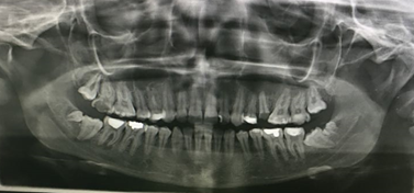
This article presents 21 years old female patient medically fit come to the dental clinic complain from pain in the lower right quadrant. Upon clinical examination and orthopantomogram it showed a rare case of bilateral impacted of mandibular 3rd and 4th molars (distomolars) without any associated syndrome and discuss the management of such a case and explication to the patient.
Introduction
Supernumerary teeth is rare dental anomaly in maxilla and mandible can be classified by shape and by the position in the jaw. It might cause complication such caries, dental disease and delay or impaction of permanent teeth.
Supernumerary tooth or hyperdontia, is not as common as hypodontia: the prevalence in primary dentition 0.2% to 0.8% and in the permanent dentition, 0.5% to 5.3%, with geographic variation (1).
The fourth molar is a kind of supernumerary tooth they have been classified as a type of para molar or distomolar tooth (2). They 8-10 times more frequently in the maxilla than in the mandible (3-5) they appear more frequently in men than in woman with a ratio 2:1 respectedly. There are theories suggest that with “dental lamina hyperactivity theory” to be the most accepted theory fourth molars or distomolars usually are impacted and in this way cause impaction of third molar. They can be associated with complications or stay asymptomatic. (6)
Case Presentation
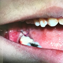
21 year old female patient medically fit come to the dental clinic complaining from pain in the lower right quadrant upon clinical examination and orthopantomogram. It showed bilateral impacted of mandibular 4th and 3rd molars multiple caries teeth, remaining root and incomplete root canal treatment. According to her mother said her sister has also impaction fourth molars. Caries control, root canal treatment, extraction of remaining root was done.
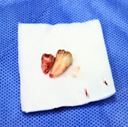
Regarding the supernumerary teeth inferior alveolar block local anesthesia was given , modified triangular flap was done , surgical extraction of 4th and 3rd molar, suturing with 4-O vicryl suture, antibiotic and analgesic prescribed.
Diagram: OPG

The OPG shows a rare case of bilateral impacted mandibular 4th molar also shows impacted 3rd molar, incomplete root canal treatment, remaining roots and multiple caries teeth.
Discussion
Supernumerary teeth (ST) or hyperdontia can be defined as any teeth in excess of the usual configuration of the normal number of deciduous or permanent teeth (7). Its more in permanent dentition compared to primary dentition their prevalence range between 0.1% to 5.3% in permanent dentition and 0.2 to 0.8% in primary dentition (1,8,9,10,11,12,14,15). Supernumerary teeth occur more often in males than females and the reported ratio is almost 2-3:1 respectively (4,10,13,14,15,16). Could be due to the association of ST with the autosomal recessive gene, which has greater penetration in males (17). They 8-10 times more frequently in the maxilla than in the mandible (18,19,20,21), in the maxillary anterior and molar regions (22).
Supernumerary teeth may occur singly, multiply, unilaterally or bilaterally and in one or both jaws. (23)
Supernumerary teeth are classified into two types: supplemental normal size and shape, and rudimentary teeth are of smaller size and abnormal shape, including conical, tuberculate, molariform and odontomas (7,16,24).
Supernumerary teeth in molar area are classified as paramolars or distomolars based on location. They occur frequently in maxilla, but only rarely in the mandible (25,26,27).
A distomolar (dens distomolar) is a supernumerary tooth that is located distally to a wisdom tooth (27,28,29). Paramolar is a supernumerary molar situated buccally or palatally/linguaiiy to one of the molars (3,12,28,30,31). Stafne (3) reported that approximately 90% of all supernumerary teeth in his study occurred in the maxilla and that half of these were found in the anterior region (incisives). Those in the molar region accounted for 38.9% of supernumerary teeth, but the mandibular supernumerary molars were rare.
A mesiodens, is the most frequent supernumerary tooth present in the maxillary central incisor region (32). The most frequent locations are the maxillary incisor (mesiodens) and the third molar areas (19).
The etiology of supernumerary teeth has not been yet completely clarified (1) and various theories have been suggested relating this anomaly (5, 6,33) such as atavism (8), tooth germ dichotomy (5,34,38), hyperactivity of the dental lamina (13,36,37) and heredity-genetic and environmental factors (5,9). The majority of supernumerary teeth are considered to develop as a result of horizontal proliferation or a hyperactivity of the permanent or deciduous dental lamina (35). The local factors such as inflammatory processes, scarring, pressure and injuries may be the causes of dental lamina hyperfunction (39-43).
Heredity is an important factor in the occurrence of supernumerary teeth. Supernumerary teeth occasionally occur within the same family (44,45). Like in our case to our knowledge, the occurrence of multiple supernumerary teeth without any other associated disease or syndrome is rare in individuals (46).
Multiple supernumerary teeth, which are generally impacted (47,48), are characteristically seen in cleidocranial dysplasia, cleft lip and palate, Down syndrome and Familial adenomatous polyposis, including Gardner syndrome.
Ehlers-Danlos syndrome, Nance-Horan syndrome, Fabry disease, Ellis-Van Creveld syndrome, richo-Rhino-Phalangeal syndrome & Robinow syndrome. (1,48-55)
Complications associated with the presence of fourth molars (6): Delayed eruption or non-eruption of third molars (14), Displacement of supernumerary tooth itself with lingual, palatal or parietal direction. Moreover they can migrate into the sinus or stay close to sinus floor (56), Occlusal problems, Displacement of permanent teeth (crowding, rotation), Subacute pericoronitis, periodontal abscess, fistulas and odontomas (57), Resorption of third molars’ root(58), Cystic formation with prevalence that ranges from 2 to 9%.
The treatment may be performed in two ways: surgical extraction or maintenance of the tooth and periodic monitoring at least once a year (59). The prognosis will be better if we diagnosed and treat the supernumerary teeth early. (60).
Conclusion
Early diagnosis and appropriate treatment of supernumerary teeth will give good prognosis ether surgical or periodic follow up at least once a year. In our case we used surgical approach for removal of supernumerary 4th molar.
References
- Wang XP and Fan J. “Molecular Genetics of Supernumerary Tooth Formation”. Genesis. 49.4 (2011): 261-277.
- Christopher S.D., et al. “Fourth Molars-Bilateral Impaction-A Case Report”. Journal of Dental Research Updates 1.1 (2014): 79-81.
- Stafne EC. ‘Supernumerary teeth”. Dent Cosmos 74 (1932): 653-659.
- Rajab LD and Hamdam MAM. “Supernumerary Teeth: Review of the Literature and a Survey of 152 Cases”. International Journal of Paediatric Dentistry 12 (2002): 244-254.
- Grimanis GA., et al. “A Survey on Supernumerary Molars”. Quintessence International 22 (1991): 989-995.
- Lambros Ζouloumis., et al. “Supernumerary Molars: Fourth or Distomolars.Clinical Study and Review of the Literature”. Balkan Journal of Stomatology 13 (2009): 167-171.
- Garvey MT., et al. “Supernumerary Teeth -An Overview of Classification, Diagnosis and Management”. Journal of the Canadian Dental Association 65 (1999): 612‑616.
- Stafne EC. ‘Supernumerary teeth”. Dent Cosmos 74 (1932): 653-659.
- Rajab LD and Hamdam MAM. “Supernumerary Teeth: Review of the Literature and a Survey of 152 Cases”. International Journal of Paediatric Dentistry 12.4 (2002): 244-254.
- Acikoz A., et al. “Characteristics and Prevalence of Non-Syndrome Multiple Supernumerary Teeth: A Retrospective Study”. Dentomaxillofacial Radiology 35.3 (2006): 185-190.
- Qaradaghi IF. “Supernumerary tooth: Report of a rare case of a fourth mandibular molar”. Rev Clín Pesq Odontol Curitiba 5 (2009): 157-160.
- Nayak G., et al. “Paramolar - A Supernumerary Molar: A Case Report and an Overview”. Dental Research Journal 9.6 (2012): 797-803
- Liu JF. “Characteristics of Premaxillary Supernumerary Teeth: A Survey of 112 Cases”. ASDC journal of dentistry for children 62.4 (1995): 262-265.
- Fernández-Montenegro P., et al. Retrospective Study of 145 Supernumerary Teeth. Medicina Oral, Patología Oral y Cirugía Bucal 11.4 (2006): E339-E344.
- Hyun HK., et al. “Clinical Characteristics and Complications Associated With Mesiodentes”. Journal of Oral and Maxillofacial Surgery 67.12 (2009): 2639-2643.
- Kokten G. “Supernumerary Fourth and Fifth Molars:A Report of Two Cases”. The journal of contemporary dental practice 4.4 (2003): 67-76.
- Ozan F., et al. “Impacted Mandibular Permanent Incisors Associated With a Supernumerary Tooth: A Case Report”. European Journal of Dentistry 3.4 (2009): 324‑328.
- Woelfel JB. Dental Anatomy 3rd ed. Philadelphia: Lea & Fabiger (1984): 226-228.
- A Piattelli and S Tetè. “Bilateral Maxillary and Mandibular Fourth Molars. Report of a Case”. Acta stomatologica Belgica 89.1 (1992): 57-60
- Menardia -Pejuan V., et al. “Supernumerary Molars. A Review of 53 Cases”. Bull Group Int Rech Sci Stomatol Odontol 42.2 (2000): 101-105.
- Yashiro M., et al. “Radiographical Study of Supernumerary Teeth With Special Reference to the Upper Molar Region”. Shigaku 75.5 (1987): 1013-1021.
- Pindborg JJ. “Pathology of the dental hard tissues”. Copenaghen (1970).
- Sasaki H., et al. “Multiple Supernumerary Teeth in the Maxillary Canine and Mandibular Premolar Regions: A Case in the Postpermanent Dentition”. International Journal of Paediatric Dentistry 17.4 (2007): 304-308.
- Batra P., et al. “Non-syndromic Multiple Supernumerary Teeth Transmitted as an Autosomal Dominant Trait”. Journal of Oral Pathology & Medicine 34.10 (2005): 621-5.
- Dhull KS., et al. “Bilateral Mandibular Paramolars”. International Journal of Clinical Pediatric Dentistry 7.1 (2014): 40-42.
- Kato H and Kamio T. “Diagnosis and Endodontic Management of Fused Mandibular Second Molar and Paramolar With Concrescent Supernumerary Tooth Using Cone-beam CT and 3-D Printing Technology: A Case Report”. The Bulletin of Tokyo Dental College 56.3 (2015): 177-184.
- Mansur Rahnama., et al. “A rare case of retained fourth molar teeth in maxilla and mandible. Case report”. Current Issues in Pharmacy and Medical Sciences 27.2 (2014): 118-120.
- Goaz PW and White WE. “Oral Radiology”. 2nd ed. St. Louis: Mosby (1987).
- Janas A. “Całkowicie zatrzymany czwarty ząb trzonowy u 21-letniej pacjentki”. Dental Forum 37.2 (2009): 89-91.
- Shah A., et al. “Diagnosis and Management of Supernumerary Teeth”. Dental Update 35.8 (2008): 510-512.
- Ozkan A., et al. “Bilateral Double Maxillary Paramolars: A Rare Case Report”. Journal of Clinical and Diagnostic Research 11.8 (2017): ZD04-ZD05.
- Limbu S., et al. “Mesiodens: A Hospital Based Study”. Journal of Nepal Health Research Council 15.2 (2017): 164-168.
- Yagüe-García J., et al. “Multiple Supernumerary Teeth Not Associated With Complex Syndromes: A Retrospective Study”. Medicina Oral Patologia Oral y Cirugia Bucal 14.7 (2009): E331-E336.
- Primosh RE. “Anterior supernumerary teeth - assessment and surgical intervention in children”. Pediatric Dentistry 3.2 (1981): 204-215.
- Regezi & Sciubb. “Oral Pathology”. W.B. Saunders Co.2nd edition (1993).
- Spauge JD. “Oral Pathology”. St. Louis: Mosby Co (1973).
- Juuri E and Balic A. “The Biology Underlying Abnormalities of Tooth Number in Humans”. Journal of Dental Research 96.11 (2017): 1248-1256.
- Gallas MM and Garcia A. “Retention of Permanent Incisors by Mesiodens: A Family Affair”. British Dental Journal 188.2 (2000): 63-64.
- Hattab FN., et al. “Supernumerary Teeth: Report of Three Cases and Review of the Literature”. Journal of dentistry for children 61.5-6 (1994): 382-393.
- Białkowska-Głowacka J. et al. “Rzadkie przypadki występowania zębów trzonowych czwartych w szczęce”. Mag. Stomatol 12.3 (2002): 34-35.
- Lewandowski B., Lech M. “Zatrzonowcowe zęby zatrzymane przyczyną zębopochodnych okołowierzchołkowych stanów zapalnych”. Stomatol. Współcz 13.2 (2006): 26-30.
- Rosak P. et al. “Dziewiątki – atawizm zębowy?” Twój Prz Stomatol 3 (2008): 60-63.
- Zadurska M. et al. “Nadliczbowość zębów – na podstawie piśmiennictwa”. Czas. Stomat 58.4 (2005): 265-272.
- Zappa J., Cieślik T. “Zatrzymane czwarte zęby trzonowe w żuchwie i szczęce – opis przypadku”. Dental Forum 34.1 (2006): 91-94.
- Marya CM and Kumar BR. Familial occurrence of mesiodens with unusual findings: case repots. Quintessence International 29.1 (1998): 49-51.
- Brook AH. “A Unifying Aetiological Explanation for Anomalies of Human Tooth Number and Size”. Archives of Oral Biology 29.5 (1984): 373-378.
- Gündüz K., and Mu lali M. “Non-syndrome Multiple Supernumerary Teeth: A Case Report”. The Journal of Contemporary Dental Practice 8.4 (2007): 81-87.
- Mangalekar SB., et al. “Molariform Mesiodens in Primary Dentition”. Case Reports in Dentistry (2013): 750107.
- Sapp JP., et al. “Contemporary oral and maxillofacial pathology”. 2nd. Edition (2004): p.4-7.
- Kumar DK and Gopal KS. “An Epidemiological Study on Supernumerary Teeth: A Survey on 5,000 People”. Journal of Clinical and Diagnostic Research 7.7 (2013): 1504-1507.
- Gorlin RJ., et al. “Syndromes of the head and neck”. 4th edition. Oxford University Press (2001): pp.306-309.
- Gorlin RJ., et al. “Syndromes of the head and neck”. 4th edition. Oxford University Press (2001): pp.437-441.
- Regattiery LR and Parker JL. “Supernumerary Teeth Associated With Fabry-Anderson's Syndrome”. Oral Surgery, Oral Medicine, Oral Pathology, and Oral Radiology 35 (1973): 432-433.
- Melamed Y., et al. “Multiple supernumerary teeth (MSNT) and Ehlers-Danlos syndrome (EDS): A case report”. Journal of Oral Pathology & Medicine 23 (1994): 88-91.
- Gorlin RJ., et al. “Syndromes of the head and neck”. 4th edition. Oxford University Press (2001: pp.850-864.
- Mangalekar SB., et al. “Molariform Mesiodens in Primary Dentition”. Case Reports in Dentistry (2013): 750107.
- Pracy JPM., et al. “Nasal Teeth”. The Journal of Laryngology & Otology 106 (1992): 366-367.
- Grimanis GA., et al. “A Survey on Supernumerary Molars”. Quintessence International 22 (1991): 989-995.
- Messer JG. “Supernumerary molar teeth. A case report”. British Dental Journal 133 (1972): 261-262.
- Koo S., et al. “Bilateral Maxillary Fourth Molars and a Supernumerary Tooth in Maxillary Canine Region--A Case Report”. South African Dental Journal 57.10 (2002): 404-406.
- Shashikiran ND., et al. “Molariform supernumerary tooth--a case report”. J Indian Soc Pedod Prev Dent 18.1 (2000): 18-20.
Citation:
Al Matrafi Badria., et al. “Case Report of Impacted Bilateral Mandibular Fourth Molar”. Oral Health and Dentistry 4.3 (2020): 64-68.
Copyright: © 2020 Al Matrafi Badria., et al. This is an open-access article distributed under the terms of the Creative Commons Attribution License, which permits unrestricted use, distribution, and reproduction in any medium, provided the original author and source are credited.


 Scientia Ricerca is licensed and content of this site is available under a Creative Commons Attribution 4.0 International License.