Research Article
Volume 4 Issue 3
Accuracy of Different Guide Plane Transferring Techniques in
Removable Partial Denture
Removable Partial Denture
Department of Dental Material and Prosthodontics, Univ. Estadual Paulista – UNESP – Institute of Science and Technology, São José dos Campos, Brazil
*Corresponding Author: Penteado MM, Department of Dental Material and Prosthodontics, Univ. Estadual Paulista – UNESP – Institute of Science and Technology, São José dos Campos, SP, Brazil.
Abstract
The evaluation of preliminar models, in a surveyor, for constructing removable partial dentures (RPD) is neglected by dentists and is directly related to the impairment of the prosthesis and the remaining teeth. The purpose of this study was to evaluate the angular differences obtained by four guide plane transfer techniques to the mouth, since excessive contour modification or lack thereof leads to the absence of ideal parallelism in the supporting teeth. Chosen techniques were: guided by pin, cast guide, resin guide and the Magalhães technique. Plaster models were built simulating a lower jaw, Kennedy Class III (n = 10). In each model, 3 contour modification guides were performed: in distal face of canine, mesial and lingual faces of molar, totaling 120 prepared guide plane surfaces. For statistical analysis ANOVA and Tukey tests were used for the interpretation of the results. Each surface and each technique were compared. Regarding the techniques, statistical differences were observed (p <0.05) and when submitted to Tukey test, it was possible to conclude that the Magalhães technique achieved the best results, leaving the nearest surface to be parallel with respect to insertion path, whereas the technique guided by pin presented the worst results. Regarding the prepared surfaces, molar lingual showed the worst results among all techniques, probably due to its difficulty of execution.
Keywords: Removable partial denture; guide plane; angular differences
Introduction
The success or failure of the rehabilitation treatment through Removable Partial Denture (RPD) is directly related to the planning and pre-preparation of the remaining supporting teeth.[1] However, the determination of the insertion path during the design of the study models, as well as the preparation of guide planes, correction of the occlusal plane, and creation of niches are often neglected.[2-5] Guide planes are two or more vertical surfaces of the supporting teeth, parallel among themselves, with the function of directing the insertion and removal of the RPD, defining an axis. With the effect of frictional retention, potential displacements during the function are limited [6], stabilizing the prosthesis and favoring treatment prognosis.[7]
Several techniques are available in the literature to perform guide plane contour modification and avoid exaggeration or lack thereof, since both lead to the absence of ideal parallelism between the supporting teeth, becoming expulsive or retentive with each other and not achieving the performance expected by RPD.[6] According to Krikos [5], there is great difficulty in obtaining perfect parallelism between the supporting teeth when the preparations are performed inside the patient's mouth. This difficulty increases as it is directed to the posterior region, where the presence of tongue, jugal musculature and interocclusal distance limit access to the site. [8]
In this sense, several authors have described techniques for transferring guide plans, previously planned in a study model, to the mouth. These techniques have as main objective to facilitate the accomplishment of these preparations and to promote them in a more precise way, so that the insertion trajectory selected for the clinical case is really followed during the insertion and removal of the prostheses, which will favor the preservation of the biological structures of support.[5]
Within the most widespread techniques, we have the free hand technique”, where, without reference, just observing the wear that was made in the study model, the guides planes are prepared. This technique has a lot of precision limitation because it is extremely impirical. Other techniques, called guided techniques, have much greater precision and predictability, these can be guided by pins or caps.[9,10]
Pin-guided techniques use fixed pins as a reference, parallel to the insertion path, in devices, which are fitted in the mouth. This pin will then assist the professional in relation to the inclination of the cylindrical diamond bur, during the guide plane wear. The techniques guided by cap, use lab-made caps, which present information on the inclination and the amount of wear necessary to create the parallelism. Within these techniques we have the fused metallic caps, those of acrylic resin, and these of resin can be separated or joined on an acrylic base as recommended by Magalhães.[11]
Considering this scenario, the purpose of this study was to evaluate the effect of four techniques in the preparation of parallel surfaces in supporting teeth. The null hypothesis is that all techniques provide adequate parallelism.
Materials and Methods
Preparation of Samples
A harmonious dental arch (MOM) dummy was used as the basis for obtaining type IV gypsum models, through duplication with alginate. Ten models were prepared for each group, and in one of them the transfer guide was performed. The path of insertion was determined by the Roach Method (3 points) in surveyor (B2 Bio-Art Equipamentos Odontológicos Ltda) and a transfer plate from model to model was prepared to standardize all samples in the same path of insertion (Fig. 1).
A harmonious dental arch (MOM) dummy was used as the basis for obtaining type IV gypsum models, through duplication with alginate. Ten models were prepared for each group, and in one of them the transfer guide was performed. The path of insertion was determined by the Roach Method (3 points) in surveyor (B2 Bio-Art Equipamentos Odontológicos Ltda) and a transfer plate from model to model was prepared to standardize all samples in the same path of insertion (Fig. 1).
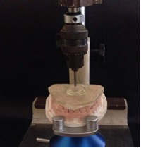
For guide preparation, an acrylic proof base was built and a metal rod was fixed in the region close to the surfaces, in the path of insertion, with the aid of a milling machine (Fresadora 1000N Bio-Art Equipamentos Odontológicos Ltda).
Guide Plane Preparation
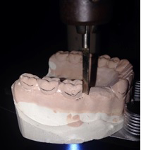
The employed techniques were: G1 - use of guide pins; G2 - metallic cap; G3 - resin cap and G4 – the Magalhães method, obtaining 4 groups (n = 10). Preparations were executed in a direct fashion, simulating the absence of teeth 44 and 45. Prepared surfaces were distal of tooth 43, mesial and lingual of tooth 46. One hundred and twenty preparations of guide planes were prepared, with a vertical knife tip coupled to the surveyor (Fig 2). A transfer guide was used according to the predetermined path of insertion and the parallelism obtained in each group was compared regarding the path of insertion.
The employed techniques were: G1 - use of guide pins; G2 - metallic cap; G3 - resin cap and G4 – the Magalhães method, obtaining 4 groups (n = 10). Preparations were executed in a direct fashion, simulating the absence of teeth 44 and 45. Prepared surfaces were distal of tooth 43, mesial and lingual of tooth 46. One hundred and twenty preparations of guide planes were prepared, with a vertical knife tip coupled to the surveyor (Fig 2). A transfer guide was used according to the predetermined path of insertion and the parallelism obtained in each group was compared regarding the path of insertion.
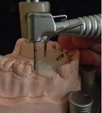
For G1, sample contour modifications were performed with a flat-top cylindrical diamond tip (4102 KG Sorensen) mounted in a micro motor, positioned parallel to the reference pin in the guide (Fig. 3). For G2, a wax cap for incrustation (Kota Imp) was built, with a height of approximately 5mm and sharp angles. They were included in a silicone ring with phosphate coating (CALIBRA EXPRESS VIPI Produtos Odontológicos) and cast in a CuAl alloy (GOLDENT). They were then cleaned, finished and adapted in the corresponding models, with the aid of a cylindrical sanding bit mounted onto a milling machine (Milling Machine 1000N Bio-Art Equipamentos Odontológicos Ltda.) (Fig. 4a-4b). For G3, caps were prepared using the same standards, but with red acrylic resin.
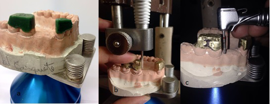
Figure 4: a. Wax caps; b. Contour modification of excess metal from guide-plane regions;
c. Simulation of mouth contour modification in plaster model with metal cap (G2).
c. Simulation of mouth contour modification in plaster model with metal cap (G2).
For G2 and G3 the subsequent procedure was the same. The models with caps were locked on the platen and, in path of insertion, excess metal (G2) or acrylic resin (G3) were removed from the cap, according to the guide plane. Caps were transferred to working models, positioned correctly, and contour modification was performed on the 3 surfaces of each model, with a flat-top cylindrical diamond tip (4102 KG Sorensen), mounted onto a micro motor. (Fig. 4c and 5). For G4, the same steps of G3 were used, however, the caps were fused with an acrylic resin base. The subsequent sequence was maintained (Fig. 6).
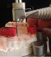
Figure 5: Simulation of mouth contour modification in the plaster model through the acrylic resin cap (G3)
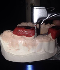
Figure 6:Simulation of mouth contour modification in the plaster model through caps attached to a test base, functioning as a model transfer device for the mouth (Magalhães – G4)
Evaluation of Angulations
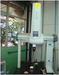
Samples were submitted to coordinate-measuring machine Global Classic (Hexagon - Fig. 7), composed of a body that moved in the three axes, X, Y and Z and had a spherical measuring tip of 1mm in diameter. Four surfaces were evaluated in each group to compare techniques (Dim 1: lingual molar, Dim 2: mesial molar, Dim 3: distal canine). Obtained data and mean values between techniques were submitted to ANOVA (p <0.05) and Tukey (5%), in cases of evidence of equality rejection.
Samples were submitted to coordinate-measuring machine Global Classic (Hexagon - Fig. 7), composed of a body that moved in the three axes, X, Y and Z and had a spherical measuring tip of 1mm in diameter. Four surfaces were evaluated in each group to compare techniques (Dim 1: lingual molar, Dim 2: mesial molar, Dim 3: distal canine). Obtained data and mean values between techniques were submitted to ANOVA (p <0.05) and Tukey (5%), in cases of evidence of equality rejection.
Results
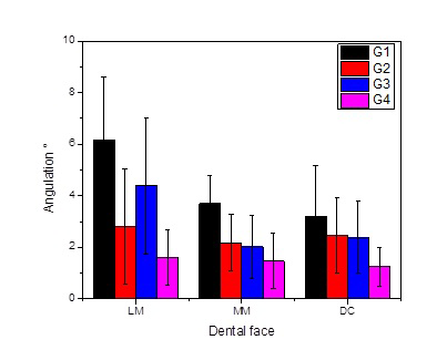
Results are shown in tables 1 and 2. Analysis of the means between the techniques showed that there is a statistically significant difference between G1 and the other groups, as well as similarity between G3 and G4 (Fig. 8).
| Groups | n | Media | Standard Deviations | Minimum | Median | Maximum |
| G1 | 10 | 4,3° | 2,2 | 0,6 | 4,4 | 10,4 |
| G2 | 10 | 2,5º | 1,6 | 0,3 | 2,3 | 6,4 |
| G3 | 10 | 2,9º | 2,0 | 0,2 | 2,9 | 7,3 |
| G4 | 10 | 1,4º | 0,9 | 0,1 | 1,3 | 4,3 |
Table 1: Absolute values of the 2 surfaces of each sample and each of the techniques performed, with 0º being the guide plane surface parallel to the path of insertion.
| Technical | Degrees |
| G1 | 4,3a |
| G2 | 2,5b |
| G3 | 2,9b |
| G4 | 1,4c |
Table 1: Absolute values of the 2 surfaces of each sample and each of the techniques performed, with 0º being the guide plane surface parallel to the path of insertion.
Regarding the lingual surface of the molar (Dim 1 p=0.000270), worse angulations were found in G1. There was no difference between G2 and G3, but there were better results with G2 and G4.
The guide plane surface in the mesial molar (Dim 2 p=0.000476) presented a statistically significant difference only in relation to G1 and to the other groups. G2, G3 and G4 were similar to each other. As for the distal surface of the canine (Dim 3 p=0.036727), G4 presented a statistically significant difference in relation to the other groups, therefore G1, G2 and G3 were similar.
Discussion
The obtained results demonstrated differences among evaluated techniques, thereby rejecting the null hypothesis.
In the literature, a number of transfer techniques of model guide planes to the mouth are described, each with its advantages and disadvantages related to the operator's ability and clinical time to execute [9-12]. Even with its importance in RPD, only a few experiments demonstrate a viable technique for clinical application [13-16].
The methodology used in this research using coordinate-measuring machine Global Classic (Hexagon) was programmed to touch 4 points of each contour modification surface. A plane was formed, and the angle between the surface and the spherical measuring tip was obtained. This measurement provided great reading precision for the points and consequently reliable data.
The lingual molar will probably be the surface that presents the greatest difficulty to be prepared, regardless of the employed technique. This is due to the limited space in the posterior regions, in addition to the presence of the tongue in inferior arches. Nevertheless, G1 presented the worst performance in this matter, which may be explained by the difficulty in positioning the drill correctly parallel to the reference pin, even in the model. It is believed that when performing contour modification on the mouth, obstacles increase because it is a technique that needs good visibility of the surface to be worn.
The lower mean values appeared in the mesial molar and in the distal canine, resembling the study by Yamamoto et al.[17], in which the best results were in the anterior and upper arch. These findings also corroborate with Fornaziero[18], where the preparation of guide planes, guided by acrylic resin caps, adapted to the tooth, allows good control and visualization of the surface to be prepared by the professional, and in addition the union with an acrylic base, favors the device stability, bringing to a better precision. The author also mentions that its manufacturing has low associated costs [17] and eliminates the cap casting step, consequently avoiding casting errors.
The limitations of this study were the fact that the performed was an “in vitro” test, however, although the preparations were made in a plaster model, without the inherent difficulties of the oral cavity, the results show the tendency of the difficulties and limitations of each technique. In the oral cavity, these difficulties and limitations tend to be even greater. This allows us to assess the real need, in our patients, to use the most accurate technique in carrying out this stage of rehabilitation treatment.
Conclusion
Despite the limitations of this research, it can be conclude that:
- The Magalhães technique presented the best results.
- The technique guided by pin is the most critical to obtain guide plane, mainly in posterior region.
- The surfaces of guide planes in the molar lingual presented the worst results across all techniques.
References
- Smith CS., et al. “A predictable all-digital workflow to retrofit a crown to an existing removable prosthesis”. Journal of Prosthetic Dentistry 121.6 (2019): 876-878.
- Davenport JC., et al. “Communication between the dentist and the dental technician”. British Dental Journal 189.9 (2000): 471-474.
- Smith GP. “The responsibility of the dentist toward laboratory procedures fixed and removable dentures prosthesis”. Journal of Prosthetic Dentistry 13.2 (1963): 95-103.
- Mac Entee MI., et al. “Attitudes of dentists in British Columbia to dental technicians, dental mechanics and removable prosthodontics”. Journal of the Canadian Dental Association 46.12 (1980): 768-771.
- Krikos AA. “Preparing guide planes for removable partial dentures”. Journal of Prosthetic Dentistry 34.2 (1975): 152-155.
- Campbell SD., et al. “Removable partial dentures: The clinical need for innovation”. Journal of Prosthetic Dentistry 118.3 (2017): 273-280.
- Haeberle CB., et al. “A Technique to Facilitate Tooth Modification for Removable Partial Denture Prosthesis Guide Planes”. Journal of Prosthodontics 25.5 (2016): 414-417.
- Rudd RW and Rudd KD. “A review of 243 errors possible during the fabrication of a removable partial denture: part I”. Journal of Prosthetic Dentistry 86.3 (2001): 251-261.
- Kaiser DA and Jones JD. “Proximal contour modifications for fixed partial dentures: a clinical report”. Journal of Prosthetic Dentistry 89.4 (2003): 344-345.
- Bezzon OL., et al. “Device for recording the path of insertion for removable partial dentures”. Journal of Prosthetic Dentistry 84.2 (2000): 136-138.
- Magalhães Filho O., et al. “Removable partial dentures: A method of parallel guiding planes transfer, by study model through surveyor to mouth of patient”. Revista da Associação Paulista de Cirurgiões Dentistas 38 (1984): 394- 406.
- Rudd RW and Rudd KD. “A review of 243 errors possible during the fabrication of a removable partial denture: Part II”. Journal of Prosthetic Dentistry 86.3 (2001): 22-76.
- Rudd RW and Rudd KD. “A review of 243 errors possible during the fabrication of a removable partial denture: part III”. Journal of Prosthetic Dentistry 86.3 (2001): 277-288.
- Davenport JC., et al. “Tooth preparation”. British Dental Journal 190 (2001): 288-294.
- Lee JH. “Fabricating a reduction guide for parallel guiding planes with computer-aided design and computer-aided manufacturing technology”. Journal of Prosthetic Dentistry 121.5 (2019): 749-753.
- Jorge JH., et al. “Preparing abutment teeth for removable partial denture”. Revista de Odontologia da UNESP 35.3 (2006): 215-222.
- Yamamoto ETC., et al. “Evaluation of degree of parallelism at prepar support tooth by three techniques”. Revista Gaúcha de Odontologia 54.4 (2006): 328-333.
- Fornaziero CC., et al. “Resolution of Removable partial dentures case using the method of guide for parallel planes transfer”. Semina 18 (1997): 78 -82.
Citation:
Penteado MM., et al. “Accuracy of Different Guide Plane Transferring Techniques in Removable Partial Denture”. Oral Health and Dentistry 4.3 (2020): 83-89.
Copyright: © 2020 Penteado MM., et al. This is an open-access article distributed under the terms of the Creative Commons Attribution License, which permits unrestricted use, distribution, and reproduction in any medium, provided the original author and source are credited.


 Scientia Ricerca is licensed and content of this site is available under a Creative Commons Attribution 4.0 International License.