Case Report
Volume 1 Issue 1 - 2017
A Case of Keratoconjunctivitis Secondary to Aquarium Zoanthid Coral Exposure
Department of Ophthalmology, Mater Dei Hospital, Tal-Qroqq, Malta
*Corresponding Author: Dr James Vassallo MD MRCSEd, Ophthalmology Department, Mater Dei Hospital, Tal-Qroqq, Malta.
Received: April 06, 2017; Published: April 13, 2017
Abstract
This is a case of a 32-year-old male who presented with bilateral eye redness and burning sensation after aquarium coral had squirted
fluid into his eyes. There was a large number of small-to-moderate size, fine, dendritiform epithelial lesions scattered all over
the cornea and conjunctiva; there were no other abnormal findings. He was immediately started on fluorometholone drops and the
symptoms resolved after 2 days. The recovery was uneventful with complete resolution of the epithelial changes by day 17. This
could represent a case of a relatively mild palytoxin reaction which was successfully treated with topical mild steroid and lubricant.
Keywords: Aquarium; Coral; Keratoconjunctivitis; Palytoxin; Zoanthid
Introduction
The aim of this report is to provide a brief account of a case of keratoconjunctivitis secondary to likely aquarium Zoanthid coral (also
known as zoas [1]) toxin exposure that was successfully managed with topical steroid. Zoanthids are common aquarium toxin-producing
corals [2]. Palytoxin is found in some species of zoanthids and may be fatal as it affects the Na+/K+ ATPase pump. Ophthalmologists and
health care providers in general should be aware of this possible presentation and its associations.
Case Report
A 32-year-old man presented to ophthalmic casualty one day after doing aquarium maintenance at home. His past medical history
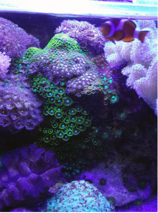
was unremarkable and had not worn contact lenses for at least 3 days. He reported that a fluid squirt from a coral (Figure 1) directly
struck his eyes from close range after cutting through the coral with a scalpel. He washed his face and eyes immediately. Initially he was
asymptomatic but the following day he developed worsening redness and burning sensation in both eyes. There was no blurring of vision.
He denied systemic symptoms and also denied having inhaled or ingested the fluid or sustaining skin cuts.

Figure 1: The Zoanthid coral present in the patient’s aquarium (image provided and used with permission by the patient).
On examination at presentation, the day following the incident, the tear fluid pH was ~7, estimated using a urine dipstick with the
pH pad pressed directly on the tear lake. Spectacle corrected Snellen visual acuity was 6/6-1 in both eyes. Slit lamp assessment did not
reveal any apparent corneal stromal changes. However, the conjunctival and corneal epithelium revealed numerous very small to moderate
size, fine, dendritiform lesions with fluorescein uptake; there were a few larger lesions on the bulbar conjunctiva, and there were
some lesions on the subtarsal conjunctiva without a significant papillary reaction. No superficial foreign bodies were found. Intra-ocular
pressure (IOP) by Goldmann applanation tonometer was 16 mmHg in the right eye and 13 mmHg in the left. The anterior chambers were
quiet and the fundi normal. A detailed diagram of the lesions and their distribution was drawn. He was started on topical fluorometholone
0.1% drops and lubricating drops, both four times/day. The patient was told not to wear contact lenses. He was initially reviewed daily.
Follow-up
On day 1 the patient felt somewhat better; there was foreign body sensation in his right eye only. The eyes were not injected and the conjunctival lesions were mostly resolved. The appearance of the corneas was the same. He was continued on the same treatment regimen and by the following day he was asymptomatic. There was no corneal fluorescein uptake by day 6 with very small, scattered, dendritiform epithelial opacities persisting. On day 8 the steroid was gradually tapered by a drop every 3 days as the epithelial opacities gradually faded and the patient remained asymptomatic. At the end of the steroid taper the corneal epithelial opacities had resolved completely and the IOP was unchanged.
On day 1 the patient felt somewhat better; there was foreign body sensation in his right eye only. The eyes were not injected and the conjunctival lesions were mostly resolved. The appearance of the corneas was the same. He was continued on the same treatment regimen and by the following day he was asymptomatic. There was no corneal fluorescein uptake by day 6 with very small, scattered, dendritiform epithelial opacities persisting. On day 8 the steroid was gradually tapered by a drop every 3 days as the epithelial opacities gradually faded and the patient remained asymptomatic. At the end of the steroid taper the corneal epithelial opacities had resolved completely and the IOP was unchanged.
Discussion
Signs of coral toxin injury can be mild to potentially sight-threatening. A metallic taste is a symptom of possible toxin exposure, but
this was absent in this case. Possibly, if the epithelial barrier is intact the prognosis is expected to be good. If the epithelium is compromised,
as in contact lens use, corneal infiltration is more likely and a higher dose of steroids would be indicated. This case suggests
that a non-intensive regimen of a mild topical steroid seems to be effective in managing the toxic effects of accidental superficial ocular
exposure to Zoanthid coral without stromal keratitis. The frequency, potency, and duration of steroid therapy should be tailored to the
presenting features. A search on PubMed revealed 15 cases of ocular exposure to aquarium and marine coral reported in the literature in
English but no cases of direct fluid jets from coral hitting the eyes [2-4].
The toxicity can be severe, including corneal melting/perforation and scarring, and therefore these patients should be followed up
closely; a systemic enquiry should be undertaken on initial presentation given the potential lethal consequences of palytoxin exposure. It
seems that withholding steroids results in worsening of symptoms [2]. While there is a paucity of scientific literature, there are numerous
online forums in which aquarium enthusiasts and divers share their experience. It is recommended that information about the species
of coral is sought from the supplier and known palytoxin-producing types should be avoided. Goggles and gloves should be worn when
handling these corals. As in other chemical injuries, immediate copious irrigation is advisable.
Conclusion
Coral-related ocular injuries are an uncommon presentation. There should be awareness of the possible serious systemic effects and
topical steroids should be initiated early on to deal with the ocular features.
References
- http://www.reefaquarium.com/2013/Zoanthid-Corals/(last modified on 12th April 2013; last accessed on 6th April 2017).
- M Moshirfar., et al. “Aquarium coral keratoconjunctivitis”. Archives of Ophthalmology128.10 (2010): 1360-1362.
- NL Chaudhry J., et al. “Unique case of palytoxin-related keratitis”. Clinical & Experimental Ophthalmology44.9 (2016): 853-854.
- Farooq AV., et al. “Corneal toxicity associated with aquarium coral palytoxin”. American Journal of Ophthalmology 174 (2017): 119-125.
Citation:
James Vassallo. “A Case of Keratoconjunctivitis Secondary to Aquarium Zoanthid Coral Exposure”. Ophthalmology and Vision
Science 1.1 (2017): 33-35.
Copyright: © 2017 James Vassallo. This is an open-access article distributed under the terms of the Creative Commons Attribution License, which permits unrestricted use, distribution, and reproduction in any medium, provided the original author and source are credited.


 Scientia Ricerca is licensed and content of this site is available under a Creative Commons Attribution 4.0 International License.