Research Article
Volume 1 Issue 4 - 2017
The Influences of High Refractive Error on Macular Thickness, Ganglion Cells Layers and Retinal Detachment Incidence
1Faculty of vision and science, Al-Neelain University, KHartoum, Sudan
2Department of Physics (Optometry) - School of Science University of Minho, 4710-057 Gualtar - Braga (Portugal)
2Department of Physics (Optometry) - School of Science University of Minho, 4710-057 Gualtar - Braga (Portugal)
*Corresponding Author: Ahmed Alqassass, Faculty of vision and science, Al-Neelain University, KHartoum, Sudan.
Received: September 27, 2017; Published: October 17, 2017
Abstract
Objective: To investigate the influence of high refractive errors on the incidence of retinal detachment.
Design: A cross sectional study.
Material and Methods: One Hundred subjects were recruited in this study. A total of 191 eyes were examined. Vision was tested using Snellen ‘s test type (E-test), refraction was done objectively by Streak Retinoscope (Heine Beta 200), the axial length was measured with A/B Scan index US 400 Jap ultrasound and the optical coherence tomography (OCT) was used to measure the macula and ganglion cells thickness and to detect the presence of retinal detachment.
Results: Myopia was the most common refractive errors in this study. 183 myopic eyes ( 95.81%) and 8 hypermetropic eyes (4.19%) were diagnosed in this study. The results showed that the maximum degree of myopic subjects were located between 5 to 6 diopters (42.08%), while the lowest degree were registered between 13 to 24 diopters (3.82%). The mean age and standard deviation (SD) for myopic degree were -8.22 (5.04), while hypermetropic degree had 4.32 (1.49).
Conclusions: High refractive errors for instance myopia decrease the thickness of macula and ganglion cells.
Introduction
In the past few decades noted that people who were diagnosed with high myopic refractive errors are more liable to retinal detachment (RD) complications. In this regards, our study aimed to investigate how high refractive errors particularly high myopia influences on the incidence of retinal detachments.
Retinal detachment (RD) refers to separation of the neurosensory retina (NSR) from the retinal pigment epithelium (RPE). This results in the accumulation of subretinal fluid (SRF) in the potential space between the NSR and RPE (Kanski, 2016). RD its can occur at any age, but it is more common in people over age 40. It affects men more than women and Whites more than African Americans. (Loewenstein, 2002).
(Bowling, 2016) has reported that symptoms of RD may include a sudden or gradual increase in either the number of floaters, which are little “cobwebs” or specks that float about in the field of vision and/or light flashes in the eye. Another symptom is the appearance of a curtain over the field of vision and reduction of vision. Retinal detachment is a medical emergency.
(Ai-Ping Song., et al. 2014) have been showed the total average macular thickness, inner/outer macular thickness, and macular volume decreased with increasing myopia/axial length. Meanwhile, Average foveal thickness had negative correlations with refractive error, and positive correlations with axial length. Furthermore, Nancy., et al. 2015; have showed the foveal and macular thickness in myopia is influenced by the axial length.
To our knowledge, there was not any study regarding detecting the influence of high refractive errors on the incidence of retinal detachment in Sudan. This study has provided normative data of high refractive errors on retinal detachment influences in Sudan as for references in Sudan population. Our hope is to impact on the current practice especially in evaluation and treatment of retinal detachments among populations and consequently on knowledge.
Materials and Methods
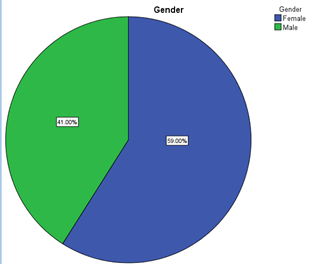
This was a cross-sectional study involving one hundred of Sudanese young people aged 16 to 28 years old; 41 males and 59 females. This study was conducted in the refraction clinic of Makkah Eye Complex, Sudan from May 2016, until November 2016, according to the Declaration of Helsinki. The study protocol was approved by the Research and Ethics Committee, School of optometry science, University of Anileen and conformed to the provisions of the Declaration of Helsinki. Written informed consent was obtained from all parents in both groups before participation of their child.
A special data form was designed for data collection including gender, age, visual acuity, refraction, A-Scanning and OCT Scanning. Subjects between 16 to 28 years and had myopic, Hypermetropic, and astigmatism ≤ 0.50 were included in this study. On the other hand, subjects who had traumatic RD or systemic diseases and astigmatism > 0.5D were excluded from this study. If the subjects had ≤ 0.50 Astigmatism, the degrees were directly added to the spherical power.
Projected Snellen’s visual acuity test chart (E) was used to examine the patient visual acuity, A/B Scan was used to measure the axial length, and Optical coherence tomography (OCT) was used to measure the retinal macula and ganglion cells thickness.
Results
Figure 1 shows the subject’s gender. Majority of them were female and represented 59%. Meanwhile, Table 1 shows the age of subjects’ distributions, the majority of them were between 21 to 24 and represented (47%) of the total subjects.
| Age | Frequency | Percentage |
| 16-20 | 15 | 15% |
| 21-24 | 47 | 47% |
| 25-28 | 38 | 38% |
| Total | 100 | 100% |
Table 1: Distributions of subject’s age.
Table 1 shows the refractive errors. 183 myopic eyes and 8 hypermetropic eyes were recruited in this study. Concurrently, Table 2 shows the common refractive errors in this study. Myopic subjects had registered the maximum percentage (95.81%), while Hypermetropic subjects registered (4.19%) of total subjects.
| Refractive error | Frequency | Percentage |
| Myopia | 183 | 95.81% |
| Hypermetropia | 8 | 4.19% |
| Total | 191 | 100% |
Table 2: Type of refractive error (191 eyes).
Table 3 reports the degree of myopia in this study. The results showed that the maximum degree of myopic subjects were located between 5-6 diopters (42.08%), while the lowest degree were registered between 13 to 24 (3.82%). The mean age and standard deviation (SD) for myopic degree were -8.22 (5.04), while Table 4 reports hypermetropic degree had 4.32 (1.49).
| Myopia | Frequency | Percentage |
| 1-4 | 70 | 38.25% |
| 5-8 | 77 | 42.08% |
| 9-12 | 29 | 15.85% |
| 13-24 | 7 | 3.82% |
| Total | 183 | 100% |
| Maximum | -0.75 | |
| Minimum | -24.00 | |
| Mean | -8.22 | |
| STD | 5.04 | |
Table 3: Degrees of myopia.
| Hypermetropia | Frequency | Percentage |
| <4D | 4 | 50% |
| ≥ 4D | 4 | 50% |
| Total | 8 | 100% |
| Maximum | +2.50 | |
| Minimum | +6.50 | |
| Mean | 4.32 | |
| STD | 1.49 | |
Table 4: Degree of hypermetropia.
Table 5 shows the relation between myopia degree with axial length, macular thickness and ganglion thickness. This table showed that axial length was significantly difference in myopic subjects (p = 0.00), and a Pearson correlation was -0.716. Means that direct relationship between myopia and axial length, while inverse relationship between myopia and macular thickness and ganglion thickness. This relationship were statistically significant difference (p = 0.00).
| 183 Myopic eyes | Pearson Correlation (2-tailed) | P-Value |
| *Axial length | -0.716** | 0.00 |
| *Ganglion thickness | 0.523** | 0.00 |
| *Macular thickness | 0.699** | 0.00 |
Correlation is significant at the 0.01 level (2-tailed).
Table 5: Relation between myopia degree with axial length, macular thickness and ganglion thickness.
Table 5: Relation between myopia degree with axial length, macular thickness and ganglion thickness.
Table 6 shows the distribution of eyes axial length. Axial length was distributed between 19 and 32. Axial length was shorter in 38.74 of total subjects with mean and standard deviation (SD) 24.12 (1.90).
| Axial length | Frequency | Percentage |
| 19-23 | 74 | 38.74 % |
| 23-24 | 61 | 31.94 % |
| 25-32 | 56 | 29.32 % |
| Total | 191 | 100 % |
| Minimum | 19 | |
| Maximum | 32 | |
| Mean | 24.12 | |
| STD | 1.90 | |
*Normal axial length is between 23-24.
Table 6: The distribution of eyes axial length.
Table 6: The distribution of eyes axial length.
With reference to the table 7. All the one hundred subjects showed a thinner macular thickness with mean and standard deviation (SD) 220.06 (17.44), furthermore in table 8 majority of had had thinner thickness of ganglion cells (64.74%), with mean and standard deviation (SD) 77.03 (6.61).
| Macular thickness | Frequency | Percentage |
| < Normal | 183 | 100 % |
| Normal | 0 | 0.00% |
| > Normal | 0 | 0.00% |
| Total | 183 | 100% |
| Minimum | 150 | |
| Maximum | 242 | |
| Mean | 220.06 | |
| STD | 17.44 | |
*Normal macular thickness is 280.
Table 7: The distribution of Macular thickness (myopic patient).
Table 7: The distribution of Macular thickness (myopic patient).
| Ganglion cell thickness | Frequency | Percentage |
| < Normal | 2 | 1.10% |
| Normal | 81 | 44.26% |
| > Normal | 100 | 54.64% |
| Total | 183 | 100% |
| Minimum | 62 | |
| Maximum | 91 | |
| Mean | 77.03 | |
| STD | 6.61 | |
*Normal macular thickness is between 68 and 74.8.
Table 8: The distribution Ganglion cell thickness (myopic patient).
Table 8: The distribution Ganglion cell thickness (myopic patient).
Discussion
In this clinic-based sample, we present new data for high refractive errors in Sudanese young populations. Our study was aimed to measure the relationship between high refractive errors and RD incidences. 191 eyes were included in this study and they aged were ranged between 12 and 28 years with mean age (23.02 ± 3.11). Majority of our subjects’ data were located under high myopia refractive error.
We found females represented (59%) and males (41%), this result was in consistent with Macky and Coney (2000), who reported that the higher incidence of myopia and hypermetropia most common in female rather than males.
Inverse relationship has been found between the degree of myopia and the macular thickness. This relationship were statistically significant difference (p = 0.00) this result was in agreement with (Zhao, 2016). Concurrently, Inverse relationship had been found between the myopic error and the ganglion cells thickness with P value <0.01, this means when the myopia increased the ganglion cells thickness decreased. Our results data were consistent with study of Yao Hsu (2014) who reported that the ganglion thickness influenced in high myopic subjects.
Strong direct relationship had been found between the axial length and the myopic error (P < 0.01). Our results was compatible with (Nancy., et al. 2015).
We found in our study females had thinner macular thickness than males and this data result agreed with Ai-Ping Song (2014).
In contrast, no relationship had been found between hypermetropia and macular thickness and ganglion cells. We expected these results in our study due to small number of hypermetropic subjects. Our study didn’t show any case of retinal detachment. We believe that this is another issue that needs further evaluation in the high hypermetropic subjects. The use of newer technology and software is expected to contribute to new knowledge that brings impact to the clinical practice.
In conclusion, high myopic subjects’>macular thickness and ganglion cells have decreased with the increasing of myopic error.>On the other hand,>Females have thinner macular thickness compared to males.
References
- Bowling B. “Kanski’s Clinical Ophthalmology 8th edition”. Elsevier publishing company (2016): 68-712.
- Lang G. “Ophthalmology a Pocket textbook Atlas 2nd edition”. Thieme Stuttgart (2006): 305-307.
- Byrne SF. “A-Scan Axial Eye Length Measurements: A Handbook for IOL Calculations”. Mars Hill NC: Grove Park Publishers(1995):
- Huang D., et al. “Optical Coherence Tomography”. Science (1991): 1178-1181.
- Moore BD., et al. “Optometric Clinical Practice Guideline: Care of the Patient with Hyperopia. St. Louis, MO”. American Optometric Association (1997): 1-29.
- William J. “Borsh’s Clinical Refraction 2nd edition, Butterworth-Heinemann, an imprint of Elsevier Inc. P (2006), 3,10
- Sato A, Fukui E, Ohta K. Retinal thickness of myopic eyes determined by spectralis optical coherence tomography. Br J Ophthalmol 2010; 94: 1624–1628.
- Song WK, Lee SC, Lee ES, Kim CY, Kim SS. Macular thickness variations with sex, age, and axial length in healthy subjects: a spectral domainoptical coherence tomography study. Invest Ophthalmol Vis Sci 2010; 51: 3913–3918.
- Sterling P. Deciphering the retina's wiring diagram. Nat Neurosci. 1999 Oct. 2(10):851-3.
- Kolb H, Fernandez E, Nelson R. The Organization of the Retina and Visual System. Salt Lake City, UT: National Library of Medicine, National Institutes of Health; 1995.
- Jakobiec FA. Ocular Anatomy, Embryology, and Teratology. Philadelphia, PA: Harper & Row Publishers, Inc; 1982.
- Mattioli S. Physical exertion (lifting) and retinal detachment among people with myopia". Epidemiology. 19 (6): 868–71.
- Loewenstein J. Retinal Detachment. Digital Journal of Ophthalmology. 2002
- http://retina.anatomy.upenn.edu/~rob/lance/retina_gross.html (1995)
- http://eyewiki.aao.org/Hyperopia#cite_note-prima-1 (JAN 2015)
- https://www.ncbi.nlm.nih.gov/pmc/articles/PMC1531864/#FN1 (Jan 2000)
- http://emedicine.medscape.com/article/798501-overview (NOV 2016).
Citation:
Ahmed Alqassass., et al. “The Influences of High Refractive Error on Macular Thickness, Ganglion Cells Layers and Retinal
Detachment Incidence”. Ophthalmology and Vision Science 1.4 (2017): 142-148.
Copyright: © 2017 Ahmed Alqassass., et al. This is an open-access article distributed under the terms of the Creative Commons Attribution License, which permits unrestricted use, distribution, and reproduction in any medium, provided the original author and source are credited.


 Scientia Ricerca is licensed and content of this site is available under a Creative Commons Attribution 4.0 International License.