Research Article
Volume 2 Issue 2 - 2018
The Physiological Causes for the Eye's Optical Axis Growth and Executive Mechanisms in the Metabolic Theory of Adaptive Myopia and the Theory of Retinal Defocus
1Petercom-Network Management System/Consulting Grope Corporation, General Director, Saint-Petersburg, Russia
2Department Ophthalmology of North-Western State Medical University named after I.I. Mechnikov, Professor, Med. Sci. D., Saint-Petersburg, Russia
3City diagnostic medical center “Vodokanal of St. Petersburg”; Clinic «Doctor Lenses SPb. doctor-ophthalmologist, optometrists, Saint-Petersburg, Russia
4Chuy Region United Hospital, Chief of the Department of Ophthalmology, M.D., Bishkek, Kyrgyzstan
5Stock company "Point of view", the Director of the office of diagnostic and functional vision correction; Stock company "Ophthalmic Laser Clinic", the ophthalmic surgeon, Arkhangelsk, Russia
6Department Ophthalmology of North-Western State Medical University named after I.I. Mechnikov, clinical resident, Saint-Petersburg, Russia
*Corresponding Author: Ivan N Koshits, Petercom-Network Management System/Consulting Grope Close Corporation, General Director, Saint-Petersburg, Russia.
Received: August 03, 2018; Published: September 26, 2018
The Visual Environment and the Level of Our Knowledge
By 2050, we will have a pandemic of myopia, when half of the world's population (7 billion) will be myopic [1]. Humanity has not yet have a working theory of myopia, which would be able to stop this adaptive process. However, the more myopic people, the greater the amount of profit associated with industrial and pharmaceutical companies. There is also a need for a large number of ophthalmologists and optometrists who work in the field of myopia. And objectively for all of them it is advantageous to have a large number of myops.
By 2050, we will have a pandemic of myopia, when half of the world's population (7 billion) will be myopic [1]. Humanity has not yet have a working theory of myopia, which would be able to stop this adaptive process. However, the more myopic people, the greater the amount of profit associated with industrial and pharmaceutical companies. There is also a need for a large number of ophthalmologists and optometrists who work in the field of myopia. And objectively for all of them it is advantageous to have a large number of myops.
In addition, humanity today has become a hostage of companies that produce displays and artificial light sources with a spectrum that is fundamentally different from the spectrum of the sun, which the retina is set up for [2]. Manufacturers are in no hurry to invest in research on the biological and video security of their products, and the vast majority of governments in different countries do not consider the problem of "display civilization" an important issue which requires the insurance of the safety in their country in general, even in the presence of the pandemic of myopia and the explosive number of retinal diseases.
By 2050, the prevalence of Age Macular Degeneration (AMD) is likely to reach the threshold of 300 million people [3,4], and the expected prevalence of glaucoma in the world among adults will be about 110 million people by 2040. In addition, over the past 15 years, glaucoma has ceased to be the leading cause of irreversible vision loss. It became quite obvious that without a total preventive correction and without the development of national regulations of visual work in each country, we will not be able to stop the pandemic of myopia and AMD.
But for the right choice of preventive correction or for the development of the safety regulations for visual work requires a truly effective theory of myopia. Widely known today, "The Genetic theory of retinal defocus changes" (TRDC), 2004 [5-8] were proposed by biophysics and a specialist in mathematical modeling G. K. Hung, Department "Biomedical engineering" at Rutgers University, New Jersey, USA and optometrist K. J. Ciuffreda, College of Optometry, New York, USA. These specialists without the participation of ophthalmologists proposed hypotheses of TRDC and a computer model of myopia.
However, TRDC was unable to stop the catastrophic spread of myopia. Many of the provisions and hypotheses of TRDC contradict a huge number of accumulated clinical facts, and a number of primary hypotheses are not confirmed by any clinical studies and in fact belongs to the field of mythology. But the TRDC authors' cybernetic view of the adaptive elongation of the optical axis of the eye, even at the genetic level, was undoubtedly a step forward in understanding the approaches to the study of the nature of myopia. Below we will discuss all of this in more detailed manner.
It is also worth noting that any computer models are not immune from errors. Incorrectly chosen initial boundary conditions often lead to catastrophic errors in the process of computer modeling. And this" dangerous feature" of mathematical modeling when using standard packages of calculating computer programs is known to the vast majority of specialists in this field, as well as the first author of this article – a mechanical engineer and biomechanics. And examples of such calculational errors are numerous in any field of science. Such errors in ophthalmology, unfortunately, also occur very often. It’s even possible to write a separate big article considering these errors.
But any criticism is good only when opponents can actually offer an alternative point of view, supported by long-term clinical observations and facts. An alternative to TRDC is the Metabolic theory of adaptive myopia (MTAM) of Koshits-Svetlova (2001) [9,10], which is based not on a computer model, but on reliable clinical facts and a deep understanding of physiological features of the interrelated functioning of intraocular systems [11]. In the final resolution of the 3rd Global Pediatric Ophthalmology Congress (London, March 2018), the flaws of TRDC and the objective advantages of MTAM were noted, which have been proved by long-term clinical trials [12].
MTAM is a really working theory, with which you can not only explain all the known clinical facts, but, for example, to increase the length of the optical axis of the hypermetropic eye with the help of directed optical effects to achieve a comfortable binocular vision in cases of amblyopia at a young age. On the basis of MTAM it is possible to develop regulations and criteria for safe visual work, as well as to determine the technical requirements for modern displays and artificial light sources. However, all of this, unfortunately, will not happen so quickly, given the real opposition of those structures that benefit the current state of the market.
The Formation of an Adequate Length of the Eye and Pathogenesis of the Metabolic Theory of Adaptative Myopia
The volume of aqueous humor flowing through the trabecular and uveoscleral outflow pathways at different phases of accommodation: We'll start with the most important fact that uveoscleral outflow pathway (USOP) is the only way of removal of the aqueous humor in animals. However, in humans, and four types of advanced primates evolutionary appeared a secondary outflow - trabecular, which allows long and even hard work at close distance, when the USOP is blocked. The myth that the trabecular outflow pathway (TOP) is the basic pathway in the eye is still widespread in modern ophthalmology. However, this is not like that. Understanding that USOP is the main way of outflow in animals and in humans suggests that the physiological mechanisms of development of adaptive myopia are the same.
The volume of aqueous humor flowing through the trabecular and uveoscleral outflow pathways at different phases of accommodation: We'll start with the most important fact that uveoscleral outflow pathway (USOP) is the only way of removal of the aqueous humor in animals. However, in humans, and four types of advanced primates evolutionary appeared a secondary outflow - trabecular, which allows long and even hard work at close distance, when the USOP is blocked. The myth that the trabecular outflow pathway (TOP) is the basic pathway in the eye is still widespread in modern ophthalmology. However, this is not like that. Understanding that USOP is the main way of outflow in animals and in humans suggests that the physiological mechanisms of development of adaptive myopia are the same.
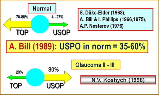
Classical works of S. Duke-Elder (1968) [13], A. P. Nesterov, etc. (1974,1995) [14,15] on determination of the level of consumption of aqueous humor (AH) by USOP in the amount of 4-27% were carried out on the enucleated eyes. In such eyes, apparently, AH could not pass normally through the interwoven spaces of the ciliary muscle due to the lack of any tension in it. I.e. USOP, therefore, were absent or inconspicuous. Therefore, these researchers and were made a natural conclusion that TOP is the main way outflow in the eye (Figure 1).
And this conclusion is absolutely consistent with the conditions of the experiment. Moreover, the results of A. Bill and I. Phillips (1971) showed in vivo the presence of both USOP and TOP in the ratio of 30% and 70%, respectively [16]. And with this not quite correct data, the myth of the superiority of TOP was born. However, much later, A. Bill conducted deeper experiments and in 1987 showed on rhesus macaques that the USOP is 35-60% [17], both in dogs and cats, in the absence of TOP, the consumption of AH along this path is even higher.
Figure 1: Interaction of the trabecular outflow pathway (TOP) with the uveoscleral outflow pathway (USOP) in normal eyes and glaucoma eyes.
The work of O. Stachs., et al. [18,19] on the study of the anatomical size of the ciliary muscle (CM) in different phases of accommodation using high-resolution biomicroscopy showed the following. The scleral spur moves posteriorly (and thus opens the TOP) only at moments of more intense close range work and does not shift at other phases of accommodation when the tone of the CM is not reaching its maximum tone. Thus, the mechanism of TOP is induced in the moments of working at close range, opening this way of the outflow of AH as an additional to ensure the physiological possibility of long-term work at near range.
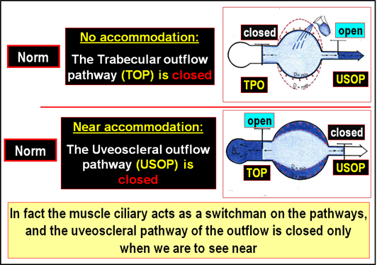
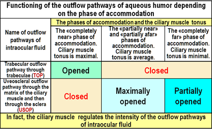
If we systematize all known data today, theoretical ideas and clinical facts on the functioning of the ways of the flow of AH, depending on the tone of the CM, it turns out the following (Figure 2 and Table 1):
- The regulation of accommodation is the determining mechanism for the functioning of TOP and USOP: when looking "completely close", the TOP is closed, and for all other phases of accommodation.
- In the eye, there is an unconditional priority of the accommodation control system over the outflow control system, and the ciliary muscle is actually a regulator on the outflow paths, turning on or off the TOP or USOP.
- Analysis of clinical data revealed functional dominance of the USOP when the eye is set for medium and long distances, while USOP gradually becomes the main outflow pathway during progression of the glaucomatous process.
- Extreme phases of the mechanism of accommodation (far distance or near vision) unfavorable for the USOP.
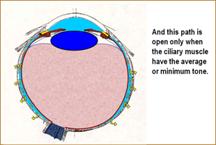
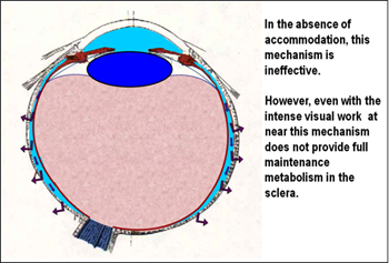
Power supply circuit of the back of the sclera with the help of nutrients coming with water moisture (Figure 3,4) allows identifying the relationship of the intensity of the reproduction of collagen in the posterior portion of the sclera with the tone of the MC. Consider the work of this natural physiological mechanism in the normal.
Figure 3: The delivery of ingredients for collagen in the middle and posterior parts of the sclera in mammals is provided to a large extent, through the USOP.
Figure 4: The second way of delivery of ingredients to the middle and posterior parts of the sclera in mammals is provided by the work of the mechanism of ultrafiltration of intraocular liquid from the vessels of the choroid.
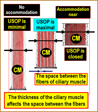
In Figure 5 is shown a scheme of the executive mechanism of USOP regulation by changing the tension of the ciliary muscle, this was created by us using the results of the studies [18,19]. When you look at close objects (right part of the diagram in Figure 5) the ciliary muscle has a minimum length and maximum thickness, but the inter-fiber gaps of the ciliary muscle are overcompressed and the strongly deformed matrix located in them does not allow drainage of aqueous humor. As a result, the necessary ingredients to maintain normal collagen formation are not delivered to the middle and back of the sclera and the metabolic processes in them deteriorate. This is confirmed by numerous morphological sections of the sclera of myopic eyes, when the front part of the sclera has no disturbances in the architectonics of the collagen framework, and its middle and back parts are distorted [20]. Therefore, intensive work in close proximity interrupts normal collagen formation in the posterior part of the sclera.
When looking at objects located at far distance (left part of the diagram in Figure 5) the ciliary muscle is stretched as much as possible and has a minimum thickness, and the inter-fiber gaps are compressed, but not completely, so drainage of aqueous humor can be produced only at a minimum level. Metabolic processes in this case will be significantly lower. And we know well that in conditions of incomplete correction for far and close distance, when the CM is partially or completely relaxed, myopia continues to progress. And a possible physiological reason for this phenomenon is a significant insufficiency of the USOP.
With the average tension of the CM (middle diagram in Figure 5) the inter-fiber gaps are maximally opened, which provides a full outflow of aqueous humor through the matrix of the CM. In this case, collagen formation in the middle and posterior parts of the sclera is maintained in normal state, and there is no reason to weaken its frame. This scheme allows us to understand that it is the average tension of the CM that provides a full nutrition to the sclera, and the extreme phases of accommodation are unfavorable for uveoscleral outflow.
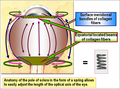
It is also worth noting that the architectonics of the scleral posterior pole has special features that allow the process of adaptation of the optical axis of the eye for long visual loads [21]. In Figure 6 the scheme of fiber arrangement in the scleral frame, in its different segments and in the superficial layers is presented. In the posterior pole of the scleral collagen fibers are located equatorially, like a spring. And it is much easier to stretch the spring than the wire or collagen three-row fiber from which it is made. This structure of the scleral posterior pole frame makes it relatively easy to extend the optical axis of the eye, if necessary, using moments of weakening of the USPO.
This physiological ability of the sclera to adequately stretch the posterior pole is an important part of the executive mechanism of selecting the length of the eye for a required visual load. It should also be noted that normally in the sclera as a whole and in its posterior pole in particular, two opposite but equilibrium processes constantly occur: metabolism and catabolism of collagen [20, 22-24]. Both of these processes are necessary because they allow to constantly update the collagen frame of the sclera, maintaining its elasticity and strength. In cases of temporary slowing of metabolism or the predominance of catabolism processes, the mechanical strength of the posterior pole of the sclera may be weakened, and under the load of the IOP optical axis of the eye (OAE) will be the elongated. The physiological method of controlling the selection of the eyes length in the normal state through a temporary weakening of the intensity of collagen metabolism is hypothetically quite natural and reliable in all animals and humans.
Adaptive Myopia as a Classic Case of Accommodation Predominance over Outflow
The goal of normal adaptation of myopic eyes: All biological systems on Earth are developing by the principle of energy saving - the fundamental principle of adaptation to the environment. The main objective of myopic eye – to reduce power consumption during prolonged and intensive visual work.
The goal of normal adaptation of myopic eyes: All biological systems on Earth are developing by the principle of energy saving - the fundamental principle of adaptation to the environment. The main objective of myopic eye – to reduce power consumption during prolonged and intensive visual work.
The second goal of myopic eye – so to adapt to the visual environment to provide the comfort of performing visual work, i.e., to increase the competitiveness and the survival of the species. Moreover, myopia develops in both children and adults.
We all know that children in the vast majority are born hypermetropic [25,26]. And if the conditions of the surrounding visual environment and the way of life allow, then the person remains for life a hypermetrope. Today, reindeer herders of the Northern territories at the age of about 50 years do not have myopia, and at the age of 18-24 years have, because they have already "tasted the fruits of the display civilization." Myopia among pilots, sailors of the submarine fleet, employees of high-precision industries, bank employees and in other fields of activity has now reached significant levels.
Similarly, we can say about myopia among domestic cats and dogs living in a limited space. A striking example in nature are highly developed monkeys, who are used to looking at close distance and collecting food on the ground. A pack of gorillas are now even forced to put sentries around the perimeter, because of the constant work at near sight so they have become short-sighted and do not see well in the distance! But you'll never see sentries in a pack of wolves that don't work constantly at close range. But the anteater is not in vain of having myopia.
The child by the age of 4-6 has already formed refractive apparatus of the eye and a small general hypermetropic refraction, which, if necessary, can be adapted for more convenient work of the eye at near sight by lengthening the OAE. The executive mechanisms for adequate growth of the OAE due to a controlled directional decrease in the intensity of the process of collagen production in the posterior pole of the sclera can serve as a complete or partial overlap USOP with long-term work at close distance. That is, the" adjustment" of the eyes length to the refraction required by the conditions of the everyday environment that should be associated with the regulation of the level (overlap) of the USOP.
The presence of such an executive mechanism is due to the fact that the anatomical development of animals and humans occurs in a way to perform the necessary work with minimal energy consumption. Therefore, physiologically conditioned adaptive myopisation of the human visual system is aimed at the implementation of the general principle of anatomical development of biological systems - the principle of energy saving. In particular, the work of a hyperopic eye requires a maximum tension of the CM, however in the case of acquired, as a result of refractogenesis, emmetropy or the start of myopia – often requires even below the average tone of the CM.
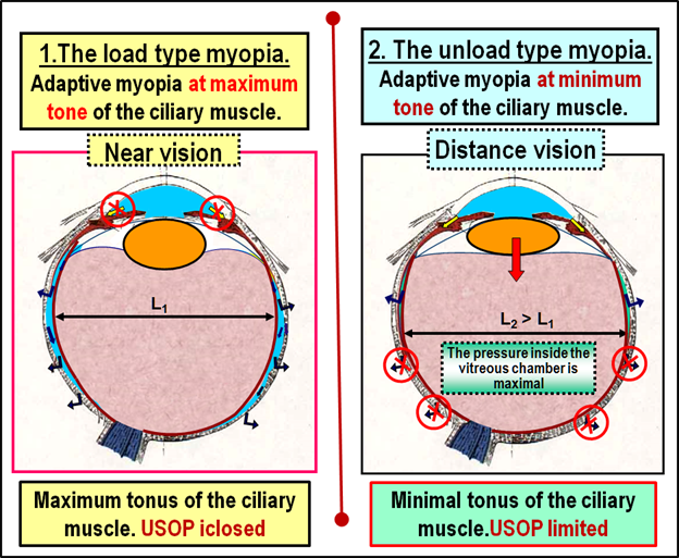
Physiological features of the mechanism of occurrence and progression of acquired myopia by the loading or unloading type: Let us consider the first case of long - term complete closure of the USOP, the result - uncontrolled development of adaptive myopia (AM) by the loading type. Long-term hard work at near sight without preventive correction or in violation of comfortable conditions (regulations) of visual work leads to a spasm of CM and complete shutdown of USOP in the eye with initial hyperopia. Work at close range with the glasses for far sight or with a poor optical correction provokes AM in the eye with an initial spasm of CM and also leads to a complete overlap of the USOP. Both of these cases are largely "man-made" due to inadequate correction or non-compliance with the requirements of the visual regulations and are associated with the lack of mandatory control over the functioning of the natural physiological mechanism of selecting the eye length under the visual load in humans. This case is shown on the left side of the diagram in Figure 7.
It is important to understand that the process of progression of the initial acquired myopia can occur not only by the loading type, but also on the unloading type, since the extreme phases of accommodation are unfavorable for the USOP. Widely used optical under-correction of acquired myopia, which has a good purpose to "reduce tension" of the CM and overlap the negative consequences of spasm of accommodation, can form anatomical and physiological conditions for partial interruption of USOP in the back of the eye and induce the "man-made" mechanism of uncontrolled growth of the OAE.
Let us consider the second case of long - term partial interruption of USOP - the development of myopia by unloading type. The work of the eye with initial myopia in the distance with under-correction glasses leads to relaxation of the MC and a significant deterioration of the USOP in the back of the eye. This is a case of myopia development due to anatomical reasons, when with minimal tone MC the lens is deposited posteriorly, pressed into the surface of the vitreous chamber, causing it to expand in the Equatorial direction.
This significantly reduces the width of suprachoroidal space that leads to difficulties in delivering aqueous humor to the posterior pole of the sclera, and disrupts normal collagen formation. The deterioration in the reproduction of collagen in the posterior pole of the sclera due to the shortage of volume of AH which is necessary for normal metabolism of nutrients required for long-term intensive work in close distance and leads to the weakening of the strength and elongation of the frame of the sclera in the area of the posterior pole.
Summarizing the above, we formulate a physiological hypothesis of anatomical formation of adequate eye length in humans and highly developed mammals: "starting from childhood, the executive mechanism of adequate adaptation of the OAE under visual load is associated with the regulation of the intensity (time interruption) of the uveoscleral outflow path, which allows to optimize the energy consumption of the eye.
Possible pathogenetic links of the physiological mechanism of selection of the value of OAE under visual load, apparently, are the following:
- Originally the eye of a child at the age of 4-5 years has hypermetropic refraction, which leads to stress CM when looking at objects up close;
- In these moments USOP overlaps and nutritional aqueous humor of the middle and posterior parts of the sclera is deteriorating;
- Reproduction of the collagen in the middle and posterior parts of the sclera in these moments is deteriorating, the framework of collagen fibers is weakened, because of the load of IOP a reciprocal increase in OAE appears, where the equatorial orientation of the collagen fibers contributes to adequate lengthening of the axis of the eye in the meridional direction;
- In the process of myopenia gravis of the visual system (adequate to the visual load) due to the lengthening of the OAE, the focus of the eye is shifted to the position in front of the retina, which allows to reduce the tension of the CM and reduce the energy consumption of the eye;
- The eyes of a child – hypermetrope, after the initial myopisation becomes emmetropic or slightly myopic, allowing to perform the necessary visual work at close range comfortable and with less energy conumption.
Thus, the emergence and development of adaptive myopia is primarily associated with the observance of the general principle of energy saving in the development of biological systems, inadequate accommodative system loads, as well as with the unconditional predominance of the control accommodative system over the outflow control system.
Early and Late Refractogenesis and Genetic Theory of Retinal Defocus
The TRDC is based on the idea of growth the optical axis of the eye due to the possible acceleration of processes of genetic refractogenesis even in adulthood:
The TRDC is based on the idea of growth the optical axis of the eye due to the possible acceleration of processes of genetic refractogenesis even in adulthood:
- It is assumed that the speed of genetic mechanisms may increase several times.
- Processes of early and late refractogenesis in TRDC "unified and indivisible": genetic mechanisms refractogenesis in children simply extrapolated to adolescent and adult age.
- According to TRDC executive organs responsible for the growth of the eye is the retina and the sclera. And the retina is the "brain center" of the growth of the eye, even if the optic nerve is cut. Let see how these hypotheses in TRDC are constructive.
The refractogenesis: This issue, unfortunately, in our opinion, in modern ophthalmology is quite complicated. In the arguments put forward by the TRDC hypotheses, was used some experimental data of attaching positive or negative glasses to the developing eye during the early ontogenesis of the young organism, which allegedly leads to the development of hypermetropic or myopic refraction. We will not evaluate the "purity" of these experiments. It is important that, according to the opinion of the authors of the TRDC hypotheses, "there is" the controlling influence of optical effects on "genetic process of refractogenesis" (quote on [8]).
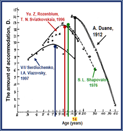
But here we see the mixing or substitution of concepts in the understanding of the physiological mechanisms of early and late refractogenesis. Moreover, the adherents of TRDC, in principle, not distinguish between these stages. And the differences of these stages are little known, but they are significant [27]. In Figure 8 the analysis of the results of studies of various authors on the development of accommodation at a young age. It is clearly seen that after the peak at the age of 12-14 years, the volume of accommodation begins to decline with age.
The early refractogenesis:Note that, unlike in early childhood, a somewhat "slow" youthful genetic code, apparently, cannot provide a quick adjustment of the eyes OAE directly, without the involvement of some other, for example, no longer biochemical, namely physiological executive mechanisms: it is necessary to accelerate the axial growth of the eye in comparison with the already relatively slow growth of the body and brain in early youth.
And this is a key question in the “philosophy” of acquired myopia: if the mechanism of accelerated ocular "axial" genetics does not work in adulthood, what executive mechanismslead to mass and rapid emergence and development of adaptive myopia in their eyes? In other words, most likely, AM is rapidly developing with the help of not genetic, but already physiological executive mechanisms, even in the period of early youth against the background of normal, but already delayed genetic growth of the eye, and even in an adulthood [28].
And here, perhaps, we note the most important thing in TRDC."The authors suggest that at the level of the retina there is a biochemical mechanism of regulation of the axial length of the eye, which, reacting to changes in the value of the defocus of the peripheral retinal image, is able to change the natural, genetically programmed growth of the eye" (quote on [8]).The TRDC hypothesis suggests that the external optical environment is able to regulate (accelerate at times!) the genetic program of growth axis of the eye. From this assumption, in fact, the theory of changes in retinal peripheral defocus, allegedly able to intensively influence the rate of genetic regulation of axial growth of the eye even before the presbyopic period.
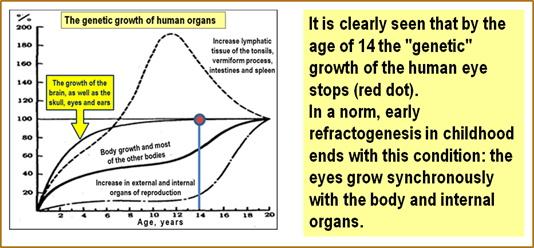
The difference in the growth rate of organs and tissues is shown in Figure 8 [29]. Many studies have shown that"the growth curves of most skeletal and muscular organs repeat the course of the general growth curve. The same applies to changing the size and individual organs: liver, spleen, and kidneys. However, the growth curves of a number of other tissues and organs differ significantly... it is obvious that in the fetal and postnatal periods, the growth rate of the head decreases compared to the growth rate of the legs" (quote on [29]).
Figure 9: Growth curves of individual organs and tissues in comparison with generalized growth curve [29].
In Figure 9 generalized age curves are presented: growth of the lymphatic tissue of the tonsils, appendix, intestine and spleen (I), brain growth, skull, eyes and ears (II), growth of the body and most of the other organs (III), growth of external and internal organs of reproduction (IV). It is clearly seen that by the age of 14, the "genetic" growth of the eye in a person stops (red dot). However, the authors of TRDC suggest that billions of people do not comply by this biological law. Billions of people have eyes (and maybe other organs?!) which should be able to accelerate axial growth at the genetic level from adolescence to the presbyopic period! But clinical evidence of this approach in TRDC is not yet available. There is only hypothesis.
So, we know that in each cub there is a genetic program of gradual growth of the eye "in all directions", according to the corresponding parallel growth of the skull and eye socket. And this program will be executed even if we cut the optic nerve. And normal eye be sure to reach the status of weak hyperopia. This state ends early refractogenesis in normal animals and in human childhood: the eyes grow simultaneously with the body and internal organs.
Those individuals who have a genetic predisposition, a genetic defect in the process of Collagenogenesis in the fibrous shell of the eye can cause a hyperopic "starting line", however it is still crossed in the early stages of refractogenesis, and the axis of such eye during an early "genetic" refractogenesis will be longer due to bad heredity. In such an eye, the axial direction of the fast-growth in childhood will prevail somewhat, since there is nowhere to grow in the other sides of the eye socket. I mean ... the baby in the period of the childhood/adolescence refractogenesis starts not with the level of hyperopia, but with the level of, for example, mild myopia. This, apparently, is hereditary myopia.
The late refractogenesis: The child's hypermetropic eye normally continues to grow in sync with the entire body, according to the genetic code. At the same time, the rate of this growth is gradually slowing down and, finally, it is significantly slowed down in adolescence [29]. However, all animals have different individual-group behavior and different visual environments, in which it is necessary to survive even at a young age. This requires the possibility of additional rapid adjustment to the visual hazard detection mechanism. The same for a wolf or, on the contrary, an anteater. The same goes for almost all the animals in the world.
It should be noted that some of the revolutionary views of the TRDC were, in principle, a step forward, as it was proposed to consider the increase in the axial length of the eye as a controlled process at least at the genetic level. And that is why TRDC adherents began to look for executive mechanisms of the process of direct accelerated impact on the genetic code. And in this way to do without attracting executive biochemical mechanisms was no longer possible. But supporters of TRDC could not imagine AM as an adaptation process: because then we must consider myopia not a disease, but an adaptive reaction to the visual environment.
Still, the adaptation or uncontrolled growth of the eye? Most likely, the authors and adherents of the TRDC hypothesis were simply unaware of possible other adaptive executive mechanisms that regulate the growth of the eyes OAE even without the involvement of genetics. In the USSR in 1965 there was a hypothesis of the origin of myopia V.N. Sorokin and E. S. Avetisov [30]: "myopia should be considered ...as a consequence of the adaptive reaction, and cases of progressive myopia, as a consequence of overregulation, when the appropriate process turns into its opposite" (quote on [20]).
Sorokin-Avetisov's theory, linking visual (read – accommodation) load, weakness of accommodation (unfortunately, the weakness of the accommodation control system as a whole, and not the fatigue of only executor - ciliary muscle!) and the genetic hereditary weakness of the sclera was literally a breakthrough in this area, generalizing a huge range of myopia hypotheses. Today, it has already been revealed in reliable clinical experiments that, for example, "weakness of accommodation is not the cause of development and progression of acquired myopia in children" [31]. But this does not detract from the historical significance of the early submissions of E. S. Avetisov et al. on the mechanisms of development of myopia, but only evolutionarily grinds its other more important facets.
We see today it is not fully justified the views of some researchers that the process of growth of the anterior-posterior axis includes "the entire pathogenic chain of uncontrolled eye growth (highlighted by us)" (quote on [32]), i.e., in fact, is uncontrollable at all stages of acquired myopia and is due to overregulation, the power of which is uncontrolled. But in life there is no uncontrolled growth of healthy human organs in billions of people. At least, we do not yet know such facts.
But if we continue to believe that the adepts of TRDC are right, then why in an amazing way such an uncontrollable re-regulation suddenly turns on in people with perfect vision? After all, AM, as we have already found out, "suddenly" occurs in adult pilots of modern aircraft, crew members of submarines in a long autonomous campaigns, in young and adult sorters in diamond factories and watch factories, microscopes and other workers of intense visual work.
But what should we do today - to look at the above facts as an uncontrollable process of over-regulation?! Is it not better to rise to the level of generalization and remember about ergonomics and energy saving – the biological law of nature, according to which there is an "adjustment" under anatomically-consistent to the size of the living environment?! And on this way, "not genetic", but physiological mechanisms of axial growth of the eye, which have already been listed above, have already been discovered.
The absolute advantages of TRDC include the fact that, finally, there is an understanding of the previously identified by Russian scientists direct connection of the eye axis lengthening with the possible failure of collagen formation in the scleral structures [20,22,24]. Quote on [8]: "the Authors also believe that the flow of chemicals (from the manufacturer-retina - our quote) can cross the vascular membrane to get into the sclera. In the sclera, these substances appear to be able to control the rate of synthesis of proteoglycans and therefore the rate of growth of the sclera".
However, the biochemical mechanisms of the retina proposed by TRDC adepts, which influence the regulation of collagen formation efficiency in the sclera, are more like the "articles of faith" and, in our opinion, do not stand up to criticism. And here we also want to be understood correctly. It is possible that one of the essential ways to remove toxins from the lens, vitreous body and own structures of the retina, directed "in the cut" the pumping function of the outer epithelium of the retina, is the way the toxins go in suprachoroid space, and then to the venous network of the choroid, or outward through the sclera. But so far no answer in this question. And clearly a more precise research at the most modern levels is still required. Fortunately, all the necessary instruments for such studies are already actively used in ophthalmology.
The emergence and adaptive progression of the initial acquired myopia seems to be associated with the manifestation of the usual physiological mechanisms, same in humans and animals [11]. The formation of an adequate length of the eye for the required visual loads to provide the possible low level energy consumption during strenuous and often prolonged visual work at near sight. In the normal state, the adaptive anatomical elongation of the OAE of initially hypermetropic eye with intense visual work at near sight is, apparently, a manifestation of the law of energy saving in the development of biological systems in childhood, adolescence or adulthood.
But if so, it is necessary to make an important "heretical" conclusion: the acquired myopia is not a disease, but a normal adaptation to the visual environment! And then the disease should be considered only a high degree of myopia, including complications. And then the "direction of the main impact" in the prevention and control of AM weak and medium degrees will be as follows: switching off the physiological functional ability of the human or animal eyes to the adaptive elongation of its OAE.
There is another, quite reasonable view. If myopia is an adaptation process, is it necessary to fight it?! After all, clearly all of humanity today is actively adapting to the new conditions of the visual environment. And in the long term, it is likely that we will all be myopes of a weak degree in childhood. And then from childhood we will all be perfectly adapted to work with displays and gadgets. However, we will never be able to see the distant landscapes in their dazzling beauty without additional optics, especially with strong winds and rain. For such a homo myopicus, the second half of the world will be largely lost.
But refractive surgery will also be in demand, not to mention non-surgical methods. And it will not be medicine, but a new type of service - eye cosmetology. Which will fight the discomfort of visual perception in other, non-display areas of human activity related to the agricultural sector, transport, construction, combat capability of the army, etc. An impressive list of prohibited or restricted to the myops professions can be found in any highly developed countrys. Given these circumstances, it is worth considering at least the prevention of AM today, because tomorrow will be too late.
Executive Mechanisms of Axial Eye Growth in the Theory of Defocus
According to TRDC hypotheses, the retina is the "brain center" of eye growth even when the optic nerve is cut. This is a strong but dangerous hypothesis. It turns out that even without connection with the brain, the eyes of animals can rapidly-genetically grow, if the focus "walks" in the right direction. Therefore, the processes of early and late refractogenesis in TRDC "one and not divisible": genetic mechanisms of development in children just extrapolated on the Junior and senior periods.
According to TRDC hypotheses, the retina is the "brain center" of eye growth even when the optic nerve is cut. This is a strong but dangerous hypothesis. It turns out that even without connection with the brain, the eyes of animals can rapidly-genetically grow, if the focus "walks" in the right direction. Therefore, the processes of early and late refractogenesis in TRDC "one and not divisible": genetic mechanisms of development in children just extrapolated on the Junior and senior periods.
And then it is clear why the accommodation control system should not participate at all according to TRDC in the adaptive extension of the eyes axis. We quote from [8]:" ... the corresponding changes in the rate of growth of the axial length occurred even with damage to the optic nerve or nerve nuclei that control accommodation (highlighted by us), which excludes any influence of the feedback mechanism at the Central or cortical level". After all," the output "apologists of TRDC receive the following "explanation" of the process of myopenia: "... since the mechanism of accommodation cannot compensate for a large area of defocus of the image on the retina, induced by convex or concave lenses of significant optical force (used in experiments on animals), the accommodation system in these experiments, in fact, has no effect on the observed results" (highlighted by us, quote on [8]). But these views have discarded TRDC in the XIX century.
It is this vulnerability of the TRDC that has already been noted by various researchers. In particular, "Deserves a lot of attention... ...model of the refractogenesis, developed by S. E. Avetisov (1986), in which accommodation acts as a regulator of this process.... Under the center of eyes growth control E. S. Avetisov meant "not an anatomical, but functional concept-a system of neurohumoral influences that ensure the growth of the eye and the directed formation of its refraction". Thus, myopia can be considered as a consequence of the adaptive reaction of the body ... in the directed extension of the eyeball, carried out on the principle of feedback" (quote on [33]).
Note that humanity has accumulated a mass of knowledge about the connection of accommodation with the current length of the OAE that under this "avalanche" the TRDC in historical perspective will not survive. Separate "overtures" mediated impact of accommodation on the growth of OAE of the authors of the TRDC was seen. To explain the results of various clinical studies on the effect of the degree of correction on the progression of myopia in children, TRDC apologists even used the method of accommodative response, which allows to evaluate the work of the accommodation system at different levels of visual stimulus. Quote on [8]: ".... The nonlinear dependence of the accommodation response on the value of the accommodation stimulus is well known".
Further (quote on [8]): "with full correction, the accommodation system (highlighted by us) is able to compensate for the change in the size of the defocus zone of the image on the retina, and thus everything develops in normal conditions. The value of the retinal defocus is equal to the difference between the value of the stimulus and the value of the accommodative response... with incomplete correction, there is a decrease in the value of the defocus of the peripheral retinal image when translating the sight from a far-away object...to a nearby one...such a decrease in the defocus contributes to the progression of myopia."
This "explanatory" approach further obscures the understanding of the hypotheses proposed by TCRD adherents. On the one hand – a complete denial of the influence of accommodation on the growth of the OAE, and on the other - (quote on [8]) "...since bifocal or multifocal lenses provide accurate focusing of the image of the object, both remote and located near, on the retina, according to the TCRD, wearing these lenses should have a relatively small impact on genetically programmed eye growth". Therefore, "it's right because it's right."
According to TRDC [8]: "the retina has a central-peripheral mechanism for adjusting the axial growth of the eye, which is sensitive to the contrast of the local image on the retina and, consequently, to the defocus of the retinal image ...the increase in the defocus area on the retina (i.e., the transition from a small blur spot to a larger one) increases the excitation of the retinal periphery relative to the center. This excitation causes an increase in the rate of release of neuromodulators by amacrine cells sensitive to changes in contrast in the periphery".
"Neuromodulators such as dopamine translate this increase into increased nerve conduction and the flow of chemicals through the vascular membrane of the eye to the sclera. This, in turn, causes an increase in the synthesis of proteoglycans, which strengthen the integrity of the structure of the sclera (isolated by us). "The strengthening of the structural integrity of the sclera delays the growth of the axial length of the eye, thus causing inhibition of myopia progression. Reducing the area of defocus on the retina has the opposite effect, causing a decrease in the rate of release of neuromodulators, reducing the rate of synthesis of proteoglycans, weakening the structural integrity of the sclera and, as a consequence, the acceleration of the growth of the axial length of the eye. This leads to the progression of myopia". (Quote on [8]).
From a scientific point of view, all of the above is nonsense, because TRDC explanation of the clinical results are both confirmed by contrary views: in some clinical cases, the accommodation plays a "need" for the author’s role in refractogenesis, but not in the others. In addition, "the Authors also believe that the flow of chemicals can cross the vascular membrane to get into the sclera. In sclera these substances are apparently capable of controlling the rate of synthesis of proteoglycans and, consequently, the rate of growth of the sclera" (quote on [8]). That's a really strong assumption. Collagenogenesis in the sclera is directly controlled by the retina, which is able to "push" through the choroid and through the suprachoroidal space through its outer epithelium, its chemical regulators, which control through an unclear physiological mechanism of Collagenogenesis of the sclera.
According to TRDC authors, the flow of " control signals from the retina to the sclera is due to the fact that (quote on [8])" ... amacrin and/or internal plexiform cells with their powerful branches in the outer layer of plexiform cells act depending on the amount of information transmitted to other layers of the retina and, in turn, to other eye membranes, such as the vascular and sclera". Brilliant! It turns out, the retina even without the participation of the brain is able to process and evaluate the amount of visual information transmitted and produce an appropriate amount of chemical elements that will somehow affect the receptors in the sclera responsible for collagen formation in the fibrous membrane of the eye.
But as far as we know, the physiological mechanisms of this kind are not even discovered. But the most important thing is that everything should happen without the participation of the brain with the help of an independent eye growth center located in the retina! But it's only a symbol of faith.
Unfortunately, our knowledge of the mechanisms of nutrition of the retina structures is still very scarce. It is assumed that the retina gets nutrition from two sources: its internal six layers receive it from the central retinal artery and the outer four from choriocapillaris layer choroid. And while we do not know all the ways and mechanisms of delivery to the retina of nutrients in aqueous humor (AH), located in suprachoroidal the cracks.
But it is already well known about the close connection of the degree of AM with the prevalence of dystrophic changes on the retina, as well as the possibility of effective inhibition of these dystrophic changes with the proper choice of optical correction. Today, from these clinical facts, it is clear that the retina require vital nutrients for its normal metabolism, delivered, including, and through uveoscleral pathway. Therefore, it i absolutely clear that not only the capillary network of the retina is receiving power. However, as far as we know, no in-depth studies were performed in the world of ophthalmology. We assume that normally this is the case.
Apparently, the outer epithelium of the retina further "draws" the necessary nutrients from aqueous humor, located in suprachoroidal space. Because we have never observed the detachment of retina outwards to the side of the sclera. On the contrary, the "detachment cavity" always grows inside the vitreous chamber and is filled with the contents that came from the outside of the supraciliary and suprachoroidal space through the retinal epithelium.
Our point of view has already been expressed above, but most likely such a mechanism exists. Half a century ago, a study on rabbits S. A. Nikitin and I. F. Kovalev (1955) showed using the method of radiometry that after the introduction of radioactive phosphorus into the vitreous chamber, within an hour it was found in the uveoscleral slit and in the optic nerve [34].
But this does not mean that these isotopes have got into the suprachoroid gap from the outer layers of the retina. It is possible that this is another important unexplored link in the metabolism of intraocular structures, including the retina. We are talking about the ways of disposing of "waste products of metabolism" in the retina and the adjacent vitreous chamber. One venous network of the retina with this task is clearly not cope. After all, similarly, a huge part of the spent in the eye nutrients are removed by uveoscleral route through the sclera, despite the presence of a developed venous network in the actual vascular membrane.
Sum up. "According to the authors of TRDC, their theory gives a simple and physiologically real mechanism explaining how lenses of great positive force, complete correction or incomplete correction with progressive myopia can contribute to changes in the axial length of the eye by hypermetropic, emmetropic or myopic type, respectively" (quote on [8]). In fact, this is still a hypothesis without clinical evidence of the proposed executive mechanisms and at the same time the opposite of the key approaches. But let's try to maintain objectivity.
1. Pros for TRDC:
1.1. This is another historical attempt to explain the refractogenesis of AM using the mismatch in the mechanisms of collagenopathy in the sclera. However, about an almost half a century ago it was already told and described by means of tens of morphological sections in classical dissertation work of T. E. Nikolaeva, 1974 [20]. And the direct connection of accommodation with outflow was noted by us in 1997 [35].
1.2. The attempt of Hung G. K. and Ciuffreda K. J. A. to explain the mechanism of "focusing on acuity" by means of neuromodulators sensitive to changes in the contrast of the image on the retina should certainly be welcomed. This is a promising way, although it is also not the first, but an important attempt. Until recently, we did not fully understand how the feedback mechanism from the brain to the ciliary muscle is organized, changing its tone and allowing us to direct our eyes to sharpness. It was clear to us and other researchers [36] that this is not a" blurring of the image", but something else related to the comparative sensitivity of the excitation fields on the retina. And the contrast of the incoming optical signal, most likely, may be one of the additional factors that provide greater efficiency of such a mechanism.
1.3. The attempt of Hung G. K. and Ciuffreda K. J. A. to find direct Executive mechanisms of visual load influence on the processes of collagen formation in the back of the sclera deserves respect. However, this attempt is clearly not successful, but its great importance is that it forms the public opinion of the professional community of ophthalmologists and other eye researchers: certainly will be made a lot of parallel research of this scientific direction in different clinics at the most modern level. And, then, we will accelerate in understanding the essence of AM.
2. Cons TRDC:
2.1. The implication of the absence of the brain especially during late refractogenesis contrary to the vast number of clinical facts. And in the long term will bring more harm than good, because a significant part of researchers in the world will be distracted by the test of this physiologically incorrect hypothesis.
2.2. The hypothesis of the existence of a separate center of eye growth in the retina from the brain is the most incorrect. In the future, we are immersed in the field of mythology, remaining in the two-dimensional (retina), and not in the three-dimensional world of a higher control system, called the brain with its periphery in the form of a receiver-retina. And by and large to blame for this authors not worth, for they have made this so "as have been able", and their need to thank for courage.
After all, in the world of ophthalmology for centuries have not been put forward workable physiological hypotheses that allow to fully understand what physiological principles are based on the functioning of the control system of accommodation and what its true actuators are. The first two authors for many years "made their way" to the formation of viable hypotheses in this area, and it is possible that to some extent it happened.
2.3. The very idea that the retina produces, and then delivers on its own ways to the sclera chemicals that provide at the genetic level, the regulation of the speed of refractogenesis, certainly taken from the field of science fiction. Yes, today the biochemical processes occurring in the retina are described in detail, although not completely. But nowhere will we find reports that the retina is a biochemical "machine" that directly controls the growth of the sclera.
And again, we will not blame the authors, because they, apparently, like many other ophthalmologists, are still in captivity of the myths about the second-rate of the uveoscleral way of the outflow of AH - the only one in the eye of a huge number of species of the animal world (and leading at medium and long distances in humans and 4 species of highly developed monkeys), do not assume that the sclera is the main filter for the withdrawal of waste AH out of the eye cavity: for this in the sclera there are prostaglandin receptors that regulate its permeability and mechanoreceptors, regulating the volume of the eye, and do not understand that it is the Main carrier of metabolites, necessary to maintain collagen formation in the sclera, as well as for the noticeable maintenance of normal metabolism of the retina structures. And that changes in the architectonics of the anterior segment of the sclera in AM were not detected by morphologists.
In order for the TRDC to become a working theory, the authors not only need to find sufficiently powerful "mechanisms for the development" of specific inhibitors and growth catalysts of the sclera and their "storage-cistern" in the retina, but most importantly - to reliably detect the ways of their delivery to the sclera through the external epithelium of the retina and suprachoroid space.
2.4. A serious disadvantage of TRDC is the low rate of response to changes in the visual environment (years), as well as the lack of clear ideas about the functioning of one of the alleged links of the General genetic mechanism of eye growth mainly along its optical axis. We do not even talk about the absence of any consideration of the law of energy saving and ergonomics in the development of biological systems by the authors of TRDC.
2.5. Understanding the courage of the hypotheses proposed by the authors of TRDC, in General, it should be noted their obvious one-sidedness and limitation.
References
- Brien A Holden., et al. “American Academy of Ophthalmology”. (2016):
- Kaptsov VA and Deynego VN. “Analytical review: Light-biological safety and risks of eye diseases among school child in classrooms with led light sources". Proceedings of 3rd Global Pediatric Ophthalmology Congress 9 (2018): 58-59.
- Kumar S and Fu Y. “Age Related Macular Degeneration: a Complex Pathology”. Austin Journal of Genetics and Genomic Research1.1 (2014): 1-5.
- Wong WL., et al. “Global prevalence of age-related macular degeneration and disease burden projection for 2020 and 2040: a systematic review and meta-analysis”. The Lancet Global Health 2 (2014): 106-116.
- Hung GK and Ciuffreda KJ. “An incremental retinal defocus theory of the development of myopia”. Comments on Theoretical Biology 8 (2003): 511-538.
- Hung GK and Ciuffreda KJ. “Differential retinal-defocus magnitude during eye growth provides the appropriate direction signal”. Medical Science Monitor 6.4 (2000): 791-795.
- Hung GK and Ciuffreda KJ. “Incremental retinal defocus theory predicts experimental effect of under-correction on myopic progression”. JBO3 (2004): 59-63.
- Lagase JP. “Theory of retinal defocus changes and the progression of myopia”. Optometry bulletin 1 (2011): 48-57.
- Svetova OV and Koshits IN. “Biomechanical aspects of possible common causes of hereditary and acquired myopia”. Helmholtz Moscow research Institute of Eye diseases (2001): 77-78.
- Koshits I., et al. “Physiological and biomechanical features of the interconnected functioning of the systems of accommodation, and aqueous humor production and outflow systems. Hypotheses and executive mechanisms of the growth of the eye’s optical axis in the metabolic theory of adaptive myopia and in the theory of retinal defocus”. Journal of Clinical & Experimental Ophthalmology 9 (2018): 44-51.
- Svetova OV. “Functional features of interaction of sclera, accommodation and drainage systems of the eye in glaucoma and myopic pathology”. Diss. doctor of medical science (2010): 186.
- “Pediatric Ophthalmology”.
- Duke-Elder S. “System of Ophthalmology // Vol. IV: The Physiology of the Eye and of Vision”. (1968):
- >Nesterov AP. “Glaucoma”. Medicine (1995): 19-32.
- Nesterov AP., et al. “Intraocular pressure”. Science (1974): 381.
- Bill A and Phillips I. “Uveoscleral drainage of aqueous humor in human eyes”. Experimental Eye Research21 (1971): 275-281.
- Bill A. “Uveoscleral drainage of aqueous humor: physiology and pharmacology”. Progress in clinical and biological research312 (1989): 417-427.
- Stachs O., et al. “Monitoring accommodative ciliary muscle function using three - dimensional ultrasound”. Graefe's Archive for Clinical and Experimental Ophthalmology240.11 (2002): 906-912.
- Stachs O. “Monitoring the Human Ciliary Muscle Function during Accommodation”. (2003): 105-118.
- Nikolaeva TE. “Histological, histochemical and electron microscopic studies of sclera in myopia”. (1974): 216.
- Svetlova OV., et al. “Morphological and physiological features of the structure of the sclera of the human eye as a key element in shaping the level of intraocular pressure in normal and glaucoma”. Morphology 136.5 (2009): 5-10.
- Andreeva LD. “Structural features of sclera in myopia and emmetropia”. (1981): 171.
- Vit VV. “The Structure of the human visual system.- Odessa, Astroprint”. (2003): 655.
- Volokolakova R Yu. “Structural biomechanical and biochemical properties of sclera and their significance in the pathogenesis of progressive myopia”. (1980): 214.
- Tron E Zh. “Variability of elements of the optical apparatus of the eye and its importance for the clinic”. (1947): 238.
- Falluch SS and Rozenblyum Yu. “Static and dynamic refraction in the area of future visions for various methods of research”. Proceedings of Dynamic refraction of the eye in health and disease (1981): 87-89.
- Rozenblyum Yu Z., et al. “Accommodation at a young age. Norm and pathology”. Bulletin of the Russian Academy of medical Sciences 2 (2003): 17-22.
- Koshits IN and Svetlova OV. “The Mechanism of formation of adequate eye length in the norm and the metabolic theory of pathogenesis of acquired myopia”. Journal of Ophthalmology 5 (2011): 4-23.
- “Proceedings of 3rd Global Pediatric Ophthalmology Congress”. (2018):
- Sorokin VN and Avetisov ES. “On a new hypothesis of the origin of myopia. In the book: "Materials of the scientific conference dedicated to the 90th anniversary of academician V. P. Filatov””. (1965):
- Pospelov VI and Petrushenko OV. “Absolute accommodation at different eye refraction in children. Topical issues of clinic, diagnosis and treatment in ophthalmology: Materials of the scientific and practical”. Conf. - Barnaul-Belokurikha: "Akimirka" (2009): 121-124.
- Tarutta EP and Khojabekyan N. “In. Filinova O. B. Current understanding of the role of accommodation in refractogenesis.- In the book: Accommodation: A guide for doctors”. (2012): 35-39.
- “Accommodation: A guide for doctors”. (2012): 136.
- Nikitin SA and Kovalev IF. “Dynamics of the distribution of phosphorus isotope (32P) in eye tissues and media at different ways of its introduction”. Ophthalmology journal 3 (1955): 152-157.
- Volkov VV., et al. “Biomechanical features of interaction of accommodation and drainage regulatory systems of the eye in normal and contusion subluxation of the lens”. Ophthalmology journal 113.3 (1997): 5-7.
- Zykov LI., et al. “Experimental eye model for testing intraocular lenses and demonstrations”. Helmholtz Moscow research Institute of Eye diseases (2005): 186-190.
Citation:
Ivan N Koshits., et al. “The Physiological Causes for the Eye’s Optical Axis Growth and Executive Mechanisms in the Metabolic
Theory of Adaptive Myopia and the Theory of Retinal Defocus”. Ophthalmology and Vision Science 2.2 (2018): 251-268.
Copyright: © 2018 Ivan N Koshits., et al. This is an open-access article distributed under the terms of the Creative Commons Attribution License, which permits unrestricted use, distribution, and reproduction in any medium, provided the original author and source are credited.


 Scientia Ricerca is licensed and content of this site is available under a Creative Commons Attribution 4.0 International License.