Case Report
Volume 2 Issue 3 - 2018
Goldenhar Syndrome: Case Report
1Professor of Medicine at UNIFESO - Teresópolis – RJ, Ophthalmologist, Ophthalmology Service, Hospital São José – Teresópolis, Chairman of the Medical Ethics Committee of the Hospital São José - Teresópolis
2Resident Doctor of Ophthalmology of the Center for Studies and Associated Optical Research - Rio de Janeiro - Rio de Janeiro; Director of Knowledge of the League of Ophthalmology of Teresópolis - Rio de Janeiro; Trainee at the Ophthalmology Service of the São José Hospital - Teresópolis - Rio de Janeiro. Hospital São José - HSJ - Teresópolis (RJ), Brazil
2Resident Doctor of Ophthalmology of the Center for Studies and Associated Optical Research - Rio de Janeiro - Rio de Janeiro; Director of Knowledge of the League of Ophthalmology of Teresópolis - Rio de Janeiro; Trainee at the Ophthalmology Service of the São José Hospital - Teresópolis - Rio de Janeiro. Hospital São José - HSJ - Teresópolis (RJ), Brazil
*Corresponding Author: João Maria Ferreira, Chairman of the Medical Ethics Committee of the Hospital São José – Teresópolis, Brazil.
Received: September 19, 2018; Published: November 10, 2018
Abstract
The authors present a case of Goldenhar syndrome, with its characteristic features and variations in the town of Teresopolis/RJ, a male child of four years old at the Ophthalmology Service, Hospital São José the diagnosis was completed due to external and internal changes of the patient, after being evaluated by pediatrics, genetics, otolaryngology and ophthalmology. The Oculo-Auriculo-Vertebral spectrum (OAVS) known as Goldenhar Syndrome is a rare, complex and phenotypically variable condition, of still unknown origin is characterized by dermoid cysts epibulbar, auricular appendices and mandibular hypoplasia. We aim to case report, given the rarity of this syndrome and varieties of presentation spectrum, increase knowledge of the medical profession on this subject, to facilitate recognition and help conduct before future cases.
Keywords: Goldenhar syndrome; Oculo-Auriculo-Vertebral dysplasia; Goldenhar/diagnosis syndrome; Goldenhar/dermoid cysts syndrome
Introduction
The Oculo-Auriculo-Vertebral spectrum (EOAV) known as Goldenhar's syndrome was described by Goldenhar in 1952 and completed by Gorlin in 1936. It is a syndrome because of its peculiarities that permeate symptoms ranging from atrial involvement (in particular, microtia/anotia and pre-auricular appendages), face (hemifacial microsomal), eyes (epibulbar dermoid) and (microphthalmia) to the spine (vertebral alterations). It is a rare syndrome with an estimated prevalence of 1: 26,000 live births with a higher prevalence in males (3: 2).
Studies have shown that its etiology is characterized by the involvement of the first gill arches. In the literature we have reported cases that suggest the existence of genetic factors, with an autosomal recessive and autosomal dominant inheritance pattern.
There are descriptions of genetic abnormalities and gestational exposures such as: exposure to thalidomide, retinoic acid and to diabetes mellitus, mimicking its phenotype.
We report here a case of Goldenhar's syndrome, a syndrome with few descriptions in the literature, passing its presentation, its diagnosis and the conduct of the ophthalmological complaint.
Case Report
A.A.S.R, male, 4 years old, brown, native of Teresópolis/RJ. On 01/14/2015, the mother went to the Ophthalmology Department of the São José Hospital, stating that her son, A.A.S.R. presents the diagnosis of Goldenhar's syndrome and developed "cysts in both eyes with discomfort and photophobia", without additional complaints.
The mother reported in a directed anamnesis that the diagnosis was established by pediatrics, genetics and otolaryngology in the first year of her son's life. He also reported that the minor was operated 30 days ago to treat a urethral fistula. According to the mother there is no report of family cases, parents are healthy. The patient attends the nursery, however, does not speak fluently and obeys the commands. The mother reports that she has gone to other ophthalmology services and that they did not indicate surgery, despite the complaints reported by the minor's mother as: Lacrimation, hyperemia, secretion and constant pruritus.
During the examination, it was not possible to perform visual acuity because the patient did not cooperate and did not respond to the drawings presented.
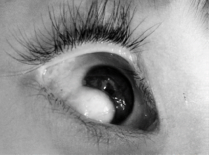
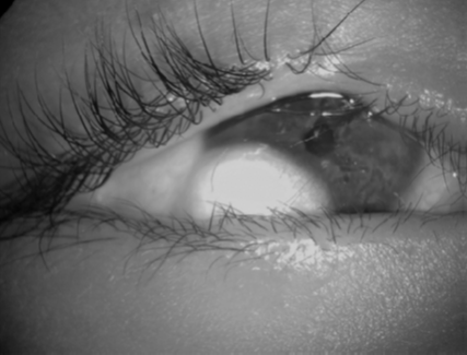
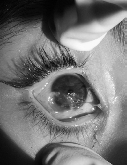
Regarding biomicroscopy, this presence was observed in both eyes with the appearance of a raised, whitish, vascularized tumor lesion with hair follicles on its entire surface (Figures 1, 2 and 3). Through the trauma of the eyelashes of the upper eyelid, there were sharp corneal ulcerations in the right eye. The diagnosis of dermoid cysts in both eyes is suggested.
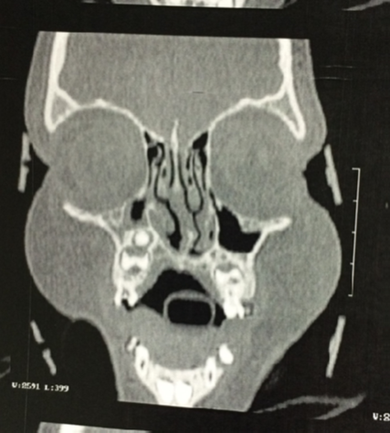
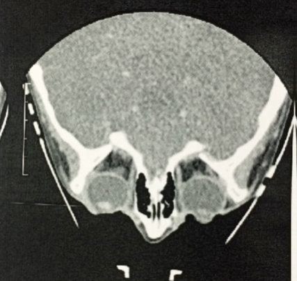
Computed tomography of both orbits showed no continuity solution of the posterior wall of the dermoid cysts with the corneal surface and the sclera. He only showed hypoplasia of mandible (figure 4) and evident dermoid cyst (Figure 5).
Surgery for excision of the dermoid cyst was scheduled for 06/02/2015.
The surgical procedure was performed under general anesthesia, with total removal of the dermoid cysts to the surface of the cornea, where the epithelial layer of the cornea was removed. There was cauterization of the deep vessels of the dermoid lesions and perilimbar region, without need of stitches, with immediate postoperative without intercurrences (Figure 6).
The surgical procedure was performed under general anesthesia, with total removal of the dermoid cysts to the surface of the cornea, where the epithelial layer of the cornea was removed. There was cauterization of the deep vessels of the dermoid lesions and perilimbar region, without need of stitches, with immediate postoperative without intercurrences (Figure 6).
Tobramycin eye drops and dexamethasone 1% x 4x daily eye drops were prescribed for topical use for a period of 15 days.
In the postoperative period scheduled for 02/23/2015, the patient presented no signs of infection, showing inflammatory signs, but without complaints.
In consultation on February 25, 2015, the patient presented without corneal ulcerations, without complaints and regression of the inflammatory signs. The postoperative medications were suspended, prescribed only ocular lubricant (sodium hyaluronate eye drops).
We advised the mother to follow up the child and return once a month for 6 months to evaluate the prognosis of the lesion.
Discussion
Goldenhar's syndrome is part of the oculomotor-vertebral spectrum (OAVA) being recognized as a syndrome by the presence of the same classic triad of ocular, auricular and vertebral alterations. It is characterized mainly by atrial appendages, epibulbar dermoid cysts and hemifacial microsomes, as well as cardiac, genital, renal, pulmonary alterations, including the central nervous system and, in rare cases, corpus callosum lipomas are described 7,8.
Goldenhar's syndrome was detected in 1845 by Von Arlt and recognized as a clinical entity in 1952 by the doctor Maurice Goldenhar who described it as a child. It was also named as "first arch syndrome" and "Gorlin syndrome". It is currently better known as oculo-auriculo-vertebral dysplasia.1,15
Of unknown origin, it is known today that there is involvement of the first branchial arches 1. Of the pathophysiological mechanisms that have been attributed to Goldenhar's syndrome, there is a hypothesis of reduced blood supply or focal hemorrhage in the region of development of the first and second gill arches, occurring around 30-45 days of gestation in the period of blastogenesis. These alterations would explain the abnormalities of the external ear, since the first branchial arch gives rise to the primordium of the anterior part of the auricles and the second branchial arch to the primordium of the posterior part of the ears 3,4,5.
Another hypothesis raises the idea that its etiology may be related to an abnormality of neural crest cell migration. Some studies suggest a pattern of autosomal inheritance recessive and autosomal dominant. In the literature there are descriptions of chromosomal abnormalities and gestational exposures such as thalidomide, retinoic acid and diabetes mellitus, mimicking its phenotype 6,10,11.
The diagnosis is based on clinical data: anamnesis, physical examination and results of complementary tests. The diagnosis of Goldenhar's syndrome can be performed during pregnancy by fetal ultrasound and genetic studies and, after birth, by ultrasound and nuclear magnetic resonance imaging 1,3.
There are diagnostic criteria such as the criteria adopted by Strämland and cols⁴ (2007) that are given when there are two or more characteristics in the oro-craniofacial, ocular, auricular and vertebral areas. We also have the criteria of Digilio and cols⁶ where at least two of the following findings must be present: unilateral microtia, unilateral mandibular hypoplasia, epibulbar dermal cysts (unilateral or bilateral) or vertebral malformations. Nonspecific symptoms have not been reported 7,8.
It is important to highlight that one of its characteristics is the extreme variability of expression of the affected individuals. Some patients have a wide variety of anomalies. In others, only a discrete and simple anomaly, such as a preauricular appendix or a moderately dysplastic ear, is observed 1,8. About 10% of the patients present with minty delay 1,8,14.
Macrostomia, open bite, crossbite, deep bite, high palate, cleft lip and palate, facial paralysis, micrognathia/retrognatia, unilateral hypoplasia of the mandibular and condyle branches, geographic language, hypoplastic tongue, tongue and bifid uvula, agenesis of the salivary gland with appearance of fistulae, atrophy or hypoplasia of masseter, temporal and pterygoid, pharyngeal anomaly, tracheoesophageal fistula, aplasia or malformation of external, middle and inner ear, pre-auricular dermal appendages, blind fistulas in the pretragus region, unilateral microtia and atresia of the external auditory canal 1,7,8,10,11,15.
Eye abnormalities occur in about 50% of the cases and can present as small dermoid spots, epibulbar lipodermoids, coloboma (usually in the upper part of the eyes), palpebral fissure, nasolacrimal obstruction, ophthalmoplegia or Dnam syndrome, atrophied cataract, enophthalmia, dermoid cysts, dry fistulas, microphthalmia and anophthalmia. The most frequent ocular anomalies are epibulbar dermoids and lipodermoids 11,12.
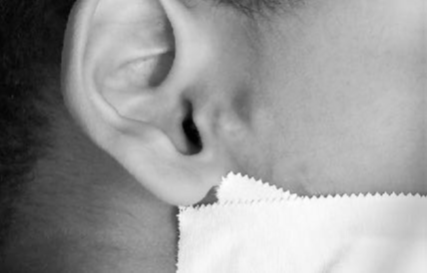
Atrial malformations may range from complete aplasia to deformities in the outer ear, middle and inner, resulting in hearing loss. Often, there are pre-auricular fibromatous appendages and cavities in front of the ear or in the line between the ear and the corner of the mouth. In the auricular malformations, the ear canal may be completely absent, implying deafness (unilateral) in approximately 40% of the cases 4,6,10,11,12. The patient in the case described had dermoid cysts, atrial appendages (figure 7), urethral fistula and delayed development.
One study has shown the prevalence of congenital heart defects (CHD) in patients with Goldenhar syndrome, where we have a prevalence of 5 to 58%. In this study, all patients with EOAV in the sample died before the end of the second year of life due to complications directly related to their DCC. It is known that these defects represent the main cause of death of patients with OAVE, which usually occurs in the first years of life 1,8,5,9,10,13.
The treatment is performed within each area with the objective of providing the best treatment, based on the literature and experience of the professionals involved. In the case reported, dermoid cysts impaired the patient's vision beyond aesthetic impairment, and the approach performed at the Ophthalmology service was extremely important.
Further preventive control in Goldenhar Syndrome should encompass assessments of vision, hearing, cervical mobility, renal, cardiac function, and dentition.
Eating difficulties usually do not persist beyond the first year of life. Sometimes it is necessary to collaborate with speech therapists and dentists when there are palate dysfunctions and dental anomalies. Taking a multidisciplinary approach 2,5,6,8,9,13.
Goldenhar syndrome remains very rare and difficult to follow beyond the complexity of its treatment. Its facial changes, mainly micrognathia, are usually treated surgically. Ocular changes treated with excision of dermoid or lipodermoid cysts. Deficits, especially auditory deficits, can also be treated with surgical procedures and follow-up with the audiologist. The variety of disease presentation spectrum, with varying degrees of impairment, is an aggravating factor in the diagnosis and follow-up of patients 1,2,10,11,15.
Conclusion
Because it is a rare syndrome commonly diagnosed a priori by paediatricians and otolaryngologists, the syndrome often occurs without the specialist in the area being sought initially, which we can observe in this case study specifically.
In this case, we observed a surgical need since the dermoid cysts cause a palpebral retraction leading to a entropion with ulcerated lesions of the cornea and consequent significant impairment of vision. The reported patient presented a postoperative without any type of complication with cicatrization of the ulcerated corneal lesions without relapses until the last follow-up.
In the case described, the approach to injury was a source of great satisfaction and relief for parents who were anxious for incisive medical intervention that could improve their child's visual quality.
We infer from this study that the publications related to rare diseases, such as the one described in this report, are important, given that such descriptions allow greater assistance to professionals who may be present to this type of patient, thus being able to better serve them, facilitating a behavior and diagnosis.
The present study makes possible a more detailed analysis of the syndrome in order to offer more specific elements about its etiology, diagnosis and surgical treatment.
References
- Cohen MM Jr., et al.“Oculoauriculovertebral spectrum: an updated critique". The Cleft Palate-Craniofacial Journal 26.4 (1989): 276-286.
- Morrison PJ., et al.“Cardiovascular abnormalities in the oculo-auriculo-vertebral spectrum (Goldenhar syndrome)”. American Journal of Medical Genetics 44.4 (1992): 425-428.
- Castori M., et al.“Antenatal presentation of the oculo-auriculo-vertebral spectrum (OAVS)”. American Journal of Medical Genetics140.14 (2006): 1573-1579.
- Strömland K., et al. “Oculo-auriculo-vertebral spectrum: associated anomalies, functional deficits and possible developmental risk factors”. American Journal of Medical Genetics 143A.12 (2007): 1317-1325.
- Kumar A., et al. “Pattern of cardiac malformation in oculo-auriculo-vertebral spectrum”. American Journal of Medical Genetics46.4 (1993): 423-426.
- Digilio MC., et al. “Congenital heart defects in patients with oculo-auriculo-vertebral spectrum (Goldenhar syndrome)”. American Journal of Medical Genetics 146A.14 (2008): 1815-1819.
- Rollnick BR., et al.“Oculoauriculovertebral dysplasia and variants: phenotypic characteristic of 294 patients”. American Journal of Medical Genetics 26.2 (1987): 361-375.
- Tasse C., et al. “Oculo- auriculo-vertebral spectrum (OAVS): clinical evaluation and severity scoring of 53 patints and proposal for a new classification”. European Journal of Medical Genetics48.4 (2005): 397-411.
- Friedman S and Saraclar M. “The high frequency of congenital heart disease in oculo-auriculo-vertebral dysplasia (Goldenhar's syndrome)”. The Journal of Pediatrics 85.6 (1974): 873-874.
- Greenwood RD., et al.“Cardiovascular malformations in oculoauriculovertebral dysplasia (Goldenhar syndrome)”. The Journal of Pediatrics 85.6 (1974): 816-818.
- Touliatou V., et al. “Clinical manifestations in 17 Greek patients with Goldenhar syndrome”. Journal of Genetic Counseling17.3 (2006): 359-370.
- Lisbôa RC., et al. “Síndrome de Goldenhar e variantes: relato de sete pacientes”. Revista AMRIGS 31 (1987): 265-269.
- Bustamante LN., et al. “Síndrome de Goldenhar. Relato de cinco casos em associação com malformações cardíacas”. Arquivos Brasileiros de Cardiologia 53 (1989): 287-290.
- Verona LL., et al. “Monozygotic twins discordant for Goldenhar syndrome”. The Journal of Pediatrics 82.1 (2006): 75-78.
- Lima FT., et al. “Alterações fonoaudiológicas presents em um caso de síndrome de Goldenhar”. Revista da Sociedade Brasileira de Fonoaudiologia 12.2 (2007): 141-145.
Citation:
João Maria Ferreira and Jéssica Gonzaga. “Goldenhar Syndrome: Case Report”. Ophthalmology and Vision Science 2.3 (2018):
271-278.
Copyright: © 2018 João Maria Ferreira and Jéssica Gonzaga. This is an open-access article distributed under the terms of the Creative Commons Attribution License, which permits unrestricted use, distribution, and reproduction in any medium, provided the original author and source are credited.



 Scientia Ricerca is licensed and content of this site is available under a Creative Commons Attribution 4.0 International License.