Research Article
Volume 3 Issue 1 - 2019
Effect of Amblyopia on Grades of Binocular Single Vision
Optometry, the University of Faisalabad, Faisalabad, Pakistan
*Corresponding Author: Asma Batool, Optometry, the University of Faisalabad, Faisalabad, Pakistan.
Received: July 02, 2019; Published: July 31, 2019
Abstract
Purpose: To determine the relationship between amblyopia and grades of BSV in diagnosed amblyopic patients and patients on occlusion therapy (Two weeks to 4 months) by synaptophore in patients with different forms of amblyopia.
Design: A comparative cross-sectional study
Main result measures: The in 15 patients with diagnosed amblyopia and 15 patients on amblyopia therapy.
Methodology: Total 30 patients were included in the study selected through non convenience sampling technique. All the patients included in the study have amblyopia with its all type, 15 patients with diagnosed amblyopia and 15 patients on amblyopia therapy. Objective assessment of refractive error was evaluated through auto-refractometer to check to promote assessment of amblyopia. Visual acuity was taken through Snellen chart (HUVITZ HDC-900 N) monocular and binocular. Binocular single vision tests were also performed to assess binocularity through Synoptophore.
Results: A total 30 amblyopic patients age from5-10 years includes ten subjects (N=10) shows percentage of 33.3%from 11-15 years includes ten subjects (N=10)33.3% from age16-20 years includes seven subjects (N=7) and shows percentage of 23.3%. Age from 21-25 years includes three subjects (N=3) shows percentage of 10% patients with patch therapy were 16(53.3)% patients without patch therapy were 14(46.7)% there were no significant difference in grades of patients with and without patch therapy.
Conclusion: Our study concluded that amblyopic patient’s stereo-acuity was affected mostly in strabismus amblyopia as compared to aniso-metropic amblyopia so, the study suggests that grades of amblyopic patients should be monitored regularly. Follow up plan must be given to the patients and they must be advised to visit their eye physicians for assessment of grades of BSV.
Keywords: Amblyopia; BSV; Simultaneous perception; Fusion; Stereopsis
Introduction
The term amblyopia is derived from the Greek language and means decreased vision after best correction. Amblyopia is a the reduction of best corrected Visual Acuity to less than 6/9 in snellen optotypes or at least two lines difference in log Mar optotypes between the eyes(Donnelly et al.,2005). “It refers to the unilateral or rarely bilateral decrease of best corrected visual acuity caused by form vision deprivation or abnormal binocular interaction, for which there is no pathology of eye or visual pathway”.
There are many types of amblyopia. First one is Strabismus amblyopia: In this case one eye move in one position and the other eye move in other direction brain cannot fuse image from two eyes into single image so patient have double vision, the brain rejects image from one eye it leads to unilateral amblyopia. (Khurana, 2015) Strabismus amblyopia: In this case there is misalignment of one or both eyes and the brain rejects the poor image and hence the eye becomes lazy and continuously rejects that image. (Mondal., 2014). Second is Stimulus Deprivation Amblyopia: It is due to traumatic cataract or corneal haziness it can be congenital restriction of light to retina hence distorted or no image formed on the retina of the eye. No visual learning will happen it can cause both unilateral and bilateral amblyopia. (Khurana., 2015).
Deprivation or occlusion amblyopia: The ocular media is opaque and image is restricted to lie on retina such as cataract. (Mondal., 2014).Third is Anisometropic amblyopia: It is caused by different focusing power in both eyes, one eye gives clear view than the other the brain start to ignore the blur image. This type of amblyopia cause unilateral amblyopia (Khurana., 2015).
Refractive or anisometric amblyopia: It is due to difference in refractive error the clearer image is accepted and the other eye image rejects and it becomes amblyopic. (Mondal, 2014). Forthisiso-ametropic amblyopia: It is due to bilateral uncorrected high refractive error it is usually occur in children it cause bilateral amblyopia. Fifth one is meridional amblyopia: It occurs in children with amblyopia due to uncorrected astigmatism. The visual acuity is decreased younger the child better the prognosis. It works best when child under 3 years of age. (Khurana., 2015)
Tobacco Amblyopia: Tobacco amblyopia is caused by excessive use of tobacco by smoking and chewing. There is degeneration of ganglion cells of retina especially macular region. It typically occurs in men pipe smokers, heavy drinkers, diet deficiencies in protein and vitamin B complex deficiencies. Toxic agents’ cyanide found in Tabaco, excessive cyanide in blood leads to degeneration of ganglionic cells, particularly in macular lesions. (Kaiti.,2014).
It can cause decrease in binocular visual acuity. The binocular visual acuity of 20/40 in amblyopic eye or worse with the difference of at least 3 log-Mar lines. The contrast sensitivity will reduce pursuit moments will be abnormal. The accommodation will be defective. Two lines difference between normal and amblyopic eye. The Crowding may improve visual acuity when tested with single letter rather than lines. (Jain, 2014).The Amblyopia and strabismus may occur as a result of complication due to anisometropia. (Hashemi et al., 2013). Milestone of BSV Development 2 to 3 weeks, In this duration the infant to fixate a target by moving his head during 4 to 5 weeks the infant can maintain monocular fixation of large near objects during first 1 to 3 months the infant can superimpose images by the third month binocular fusion is developed, stereopsis is developed during 3 to 6 months and after 6 month stereopsis becomes 60 arc sec. Development of horopter & vergence is also influenced by amazing changes in the size of eyeball& the position of orbit during infancy (Sharma., 2015).
Development of Binocular Vision: During the early life, definite normal structural and functional situation are essential for the progression of binocular vision. The aspects involved in the development of Binocular vision that allows the coordinated use of both eyes. Anatomical factors: which comprise that the both eyes are positioned in the orbit so that the visual axis is directed in the similar direction?
Biological factors include the advancement of binocular vision relies on definite standard physiological binocular reflexes. The responses can also be by birth or attained as consequence of suitable stimulation. The numerous binocular reactions are: Fixation reflexes including Compensatory fixation impulse which is also known as gravitational reflex its role is to maintain the eyes in a one location which is looking in the necessary direction recompensing for the motility of the body, skull, appendages etc.
Orientation fixation reflex -It can be established by the eye tracking a moving thing or scene, therefore showing a relatively sluggish movement of sustained fixation but not a quick jerk fixation. This reflex relates to horizontal axis.
Accommodation convergence is defined that appropriately positioning the eyes and maintain them fixated on the target which includes vergence fixation reflex, accommodation reflex and fusional vergence reflex, Second is the refixation reflex it relates the eye back to the original location point or to the new direction point and the third is he pupillary response (Bhola, 2006).
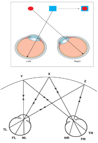
Normal Retinal Correspondence: In normal binocular single vision there is a accurate physiological relationship between two retinal corresponding points which means the fovea’s of both eyes have the same visual direction and act as corresponding points (Khurana, 2007).
Figure 1.1: Shows Normal Retinal Correspondence in which FR and FL are fovea’s of right and left eye showing correspondence and TL and MR are temporal and medium retinas of left and right eye respectively having correspondence and similarly TR and ML showing correspondence.
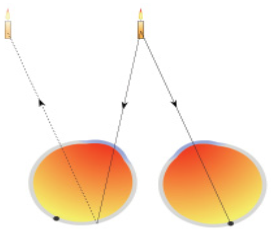
Anomalies of binocular single vision include Confusion which occurs when the corresponding retinal points are stimulated by two different targets and fusion cannot take place. The two images emerge to be on top of one another and projected to the same position in space (Barker et al., 2016).
The person may adapt the anomalies of binocular single vision. Adaptations to anomalies of binocular single vision are stated as Abnormal Retinal Correspondence: Anomalous retinal correspondence (ARC) is a neuronal adaptation due to misaligning of eye in which retinal areas that are different and connected in the visual cortex to give fusion by both eyes (wong et al., 2000).
Suppression is the phenomenon in which the person ignores the image from one eye to overcome confusion of diplopia. It is also a cortical adaptation.
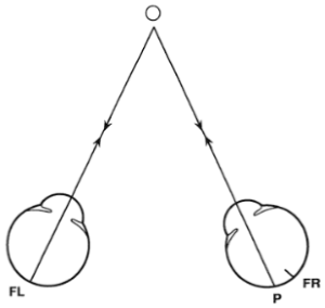
Figure 1.3: Shows abnormal retinal correspondence in right eye which is deviated inwards. O shows fixating target. Fovea’s of both eyes are FL an FR respectively and pseudo fovea of right eye is P. Due to ARC right eye undergoes sensory adaptation therefore FL and P becomes corresponding points.
Abnormal Head Posture is to elevate or depress the chin, take movement of right or left, tilting head to right shoulder or left shoulder, it can happen to facilitate development of binocular single vision, sustain binocularity in cases of attained squint, overcome symptoms like avoid painful eye movements, improve vision in the presence of nystagmus, bilateral ptosis and increase separation of diplopia so that the ignorance of second image occurs (Barker et al., 2016).
Grades of Binocular Vision: There are three grades are classified as Grade I that is simultaneous perception, which is the ability to identify two images simultaneously, one produced on each retina (Rowe, 2012).
Grade II is Fusion is a phenomenon in which two images are perceived as one, each formed on the retina of both eyes.
It has two forms: Sensory and motor fusion.
Sensory fusion: It is process of merging the image formed on each retina in the cortex into a single binocular image (Wright et al., 2006).
Sensory fusion: It is process of merging the image formed on each retina in the cortex into a single binocular image (Wright et al., 2006).
Motor Fusion is the capability to bring into line the eyes in such a way that sensory fusion can be sustained. The target for these fusion eye movements includes retinal discrepancy that is away from thepanum’s area and the eyes showing movement in different direction (vergence) (Bhola, 2013).
Grade III is the stereopsis (Disparity Sensitivity)which is defined as the capability to attain an impression of depth when two pictures are superimposed of the similar which have been taken from somewhat different angles (Bowling and Kanski, 2011).
Rationale of study
Former studies conclude the effect of degrading binocular single vision on fine vasomotor skill task performance. They evaluate the impact of degrading BSV. They used Frisby stereoacuity test on 80 persons. Results showed that degrading motor fusion and stereopsis significantly affects the performance in many fine vasomotor tasks.
Former studies conclude the effect of degrading binocular single vision on fine vasomotor skill task performance. They evaluate the impact of degrading BSV. They used Frisby stereoacuity test on 80 persons. Results showed that degrading motor fusion and stereopsis significantly affects the performance in many fine vasomotor tasks.
This study assessed the Grades of BSV in amblyopic patients having no treatment of amblyopia and with patch therapy from (1 months to onward). The results interpreted that grades were showing no improvement after patch therapy and the grades should be assessed to improve the stereopsis to eliminate reverse or residual amblyopia.
This work showed that we can eliminate the residual amblyopia and recurrent amblyopia if grades were assessed and improved during patching therapy through different exercises.
Objectives of study
- To determine the relationship between grades of BSV in amblyopic patient on therapy (1 month to onward) and diagnosed amblyopia patients without treatment
- To determine the relationship between types of amblyopia and BSV
Material and Methods
Study design
A comparative cross-sectional study was conducted in a time period from November 2017 to june 2018 in the Ophthalmology Department of Madina Teaching Hospital Faisalabad.
A comparative cross-sectional study was conducted in a time period from November 2017 to june 2018 in the Ophthalmology Department of Madina Teaching Hospital Faisalabad.
Sampling technique
Total 30 patients were included in the study selected through non convenience sampling technique. All the patients included in the study have amblyopia with its all type, 15 patients with diagnosed amblyopia and 15 patients on amblyopia therapy.
Total 30 patients were included in the study selected through non convenience sampling technique. All the patients included in the study have amblyopia with its all type, 15 patients with diagnosed amblyopia and 15 patients on amblyopia therapy.
Sample size
30 patients included in the study15 patients with diagnosed amblyopia and 15 patients on amblyopia therapy.
30 patients included in the study15 patients with diagnosed amblyopia and 15 patients on amblyopia therapy.
Inclusion criteria
The patients are included with amblyopia without treatment and amblyopia with treatment and the age ranges from 5-25 years having no other ocular pathology.
The patients are included with amblyopia without treatment and amblyopia with treatment and the age ranges from 5-25 years having no other ocular pathology.
Exclusion criteria
The exclusive criteria of our study included the patients less than the age of 5 years and greater than 25 years who had aphakia, disease or had a history of cataract or refractive surgery, any phorias greater than 4 prism diopter and tropias, and other ocular pathologies (retinal diseases, keratoconus, media opacity, ocular trauma, ocular muscle palsies)
The exclusive criteria of our study included the patients less than the age of 5 years and greater than 25 years who had aphakia, disease or had a history of cataract or refractive surgery, any phorias greater than 4 prism diopter and tropias, and other ocular pathologies (retinal diseases, keratoconus, media opacity, ocular trauma, ocular muscle palsies)
Instruments used
The materials used in this study were:
The materials used in this study were:
- Torch.
- Auto-refractometer
- Ophthalmoscope (HEINE BETA200)
- Trial box
- Snellen chart (HUVITZ HDC-900 N)
- Synopto-phore (Haag-streit Clement Clarke 2003)
Procedure for data collection
Informed consent was taken prior to the tests for their inclusion in the study. The data collected through the Performa which includes chief complain (for assessing any other condition associated), ocular history, medical history and family history in accordance to the inclusion criteria of the study. Objective assessment of refractive error was evaluated through auto-refractometer to check to promote assessment of amblyopia. Visual acuity was taken through Snellen chart (HUVITZ HDC-900 N) monocular and binocular. Torch examination was done on the patients in accordance to the exclusion criteria of the study. Ophthalmoscope were also performed to rule out any pathology and subjects inclusion in our study.
Informed consent was taken prior to the tests for their inclusion in the study. The data collected through the Performa which includes chief complain (for assessing any other condition associated), ocular history, medical history and family history in accordance to the inclusion criteria of the study. Objective assessment of refractive error was evaluated through auto-refractometer to check to promote assessment of amblyopia. Visual acuity was taken through Snellen chart (HUVITZ HDC-900 N) monocular and binocular. Torch examination was done on the patients in accordance to the exclusion criteria of the study. Ophthalmoscope were also performed to rule out any pathology and subjects inclusion in our study.
Binocular single vision tests were also performed to assess binocularity through Synoptophore.
Results
Descriptive statistics:
| Chi-Square Test | |||
| Value | df | Asymp. Sig. (2-sided) | |
| Pearson Chi-Square | 4.447a | 3 | .217 |
| Likelihood Ratio | 5.257 | 3 | .154 |
| Linear-by-Linear Association | 1.445 | 1 | .229 |
| N of Valid Cases | 30 | ||
Chi-square test was applied to analyze the data with p value less than 0.005 was considered significant. After applying test statistically results were found significant given below
| Frequency Distribution Od Grades Of BSV | |||||
| Frequency | Percent | Valid Percent | Cumulative Percent | ||
| valid | grade 1 | 7 | 23.3 | .3 | 23.3 |
| grade 2 | 2 | 6.7 | 6.7 | 30.0 | |
| grade 3 | 11 | 36.7 | 36.7 | 66.7 | |
| all grades absent | 10 | 33.3 | 33.3 | 100.0 | |
| Total | 30 | 100.0 | 100.0 | ||
Table 4.1: Frequency Distribution of Grades of BSV.
Table 4.1 demonstrating frequency and percentage of grades of BSV in 30 amblyopic patients (N=30) in which first grade the simultaneous perception is present in seven patients (N=7) and grade second fusion is present in two patients (N=2).grade third is present in eleven patients (N=11). All grades absent in ten patients (N=10).
| Types of Amblyopia | |||||
| Frequency | Percent | Valid Percent | Cumulative Percent | ||
| Valid | Anisometropic | 13 | 43.3 | 43.3 | 43.3 |
| Strabismic | 10 | 33.3 | 33.3 | 76.7 | |
| stimulus deprivatie | 7 | 23.3 | 23.3 | 100.0 | |
| Total | 30 | 100.0 | 100.0 | ||
Table 4.2: Frequency and Percentage Distribution According to Types of Amblyopia.
Description of table 4.2 demonstrating frequency and percentage of Types of amblyopia out of 30 amblyopic patients (N=30) in which anisometropic amblyopic were 13 from 30 patients including in our study (N=13) and the patient present with strabismic amblyopia were 10 out of 30 patients. Patients with stimulus deprivation amblyopia were found (N=7).
| Grades Of BSV | Grades | Treatment | Total | |
| With Patch | Without Patch | |||
| Grade 1 | 4 | 3 | 7 | |
| Grade 2 | 2 | 0 | 2 | |
| Grade 3 | 7 | 4 | 11 | |
| All Grades Absent | 3 | 7 | 10 | |
| Total | 16 | 14 | 30 | |
Table 4.3: Relationship between grades of BSV and amblyopia patients on treatment with patch therapy and without patch therapy.
Grade 1 was present in 4 with patch treatment and 3 without patch treatment. Grade 2 was present in 2 subjects with patch treatment and 0 in without patch treatment. Grades 3 was present in 7 subjects with patch treatment and 4 in without patch treatment. All grades were present in 3 subjects with patch treatment and 7 in without patch treatment.
| Association Of Grades Of BSV in different types of amblyopia | |||||
| Types Of Amblyopia | Total | ||||
| Grades of BSV | Anisometropic amblyopia | Strabismic amblyopia | Stimulus Deprivatie amblyopia | ||
| Grades Of BSV Present | Grade 1 | 3 | 1 | 3 | 7 |
| Grade 2 | 2 | 0 | 0 | 2 | |
| Grade 3 | 6 | 4 | 1 | 11 | |
| All Grades Absent | 2 | 5 | 3 | 10 | |
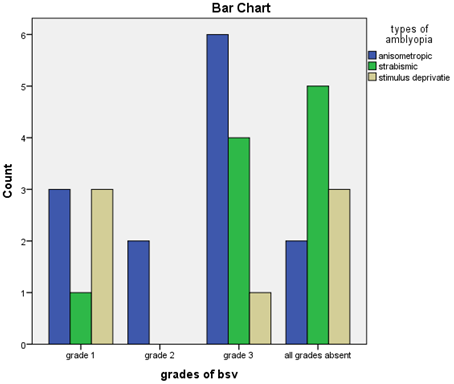
| Total | 13 | 10 | 7 | 30 | |
Table 4.4: “Grades of BSV * Type of Amblyopia.
Table 4.4 demonstrating frequency and percentage of treatment in thirty amblyopic patients (N=30) in which the patients on to patch therapy were sixteen (N=16) shows percentage of 53.3% and the patients without patching i.e early diagnosed amblyopic were fourteen patients (N=14) shows percentage of 46.7%.
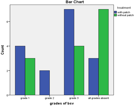
Bar chart 1: Association between the grades of BSV in patient diagnosed with amblyopia on patient with treatment and without treatment.
Bar chart demonstrating the cross tabulation of grades of BSV and treatment it shows that the grade 1 simultaneous perception was present in (N= 4) patients with patch therapy and the patient without patch therapy were (N=3) and second grade was present in (N=2) patients on patch therapy where as in patient without patch therapy it was absent. The grade 3 present in 7 patients in the patient on patch therapy, but the 4 patient without patch grade 3 was present. All grades absent in 3 patients on patch therapy and 7 patient without patch therapy
Bar chart demonstrating the cross tabulation of grades of BSV and Types of amblyopia it shows that Grade 1 (simultaneous perception)was present in 3 anisometropic amblyopic patients, 1 in strabismic amblyopia and 3 in stimulus deprivation amblyopia. Grade 2 (fusion) was present in 2 anisometropic amblyopic patients. Grade 3 (stereopsis) was present in 6 anisometropic amblyopic patients, 4 in strabismic amblyopia and 1 in stimulus deprivation amblyopia. All grades absent in grade 2 anisometropic amblyopia, 5 in strabismic amblyopia and 3 in stimulus deprivation amblyopia
Discussion
All three Grades of binocular single vision are a necessary element in binocularly mature adults for viewing from both eyes and singly with depth perception. Stereopsis is a powerful indication to depth. In the present study the main objectives were to determine relationship between amblyopia and grades of binocular single vision and To determine the grades of BSV in initially diagnosed amblyopic patient (without treatment) and patient on occlusion therapy(with treatment).Results obtained by Webber and Wood, agreed to our results that most commonly seen deficit with amblyopia under binocular viewing conditions is impairment in stereopsis but disagrees when it comes to the point where we find that some of our subjects with amblyopia showed grades of binocular single vision normal while Webber and Wood suggested that all patients with amblyopia had abnormal grade. (Webber and Wood, 2005) In another study Melmoth showed that amblyopes with vision had been corrected but still stereopsis is impaired and there is an risk of recurrent amblyopia which agrees to our study because in case of follow up with amblyopia therapy for a long period we find that there is still defect in grades of binocular single vision is present. (Melmoth et al. 2009)
Similarly according to Levi and Movshon in their observational and cross-sectional study found that in comparison between strabismicalyopes and anisometropic amblyopes. They performed Randot circle test,a test for stereopsis. This test showed that strabismic amblyopia had much more adverse effect on binocular single vision than that of anisometropic amblyopes that contradicts our finding, our study found that most decline in grades of binocular single vision occurs more in anisometropic amblyopes than that of strabismic amblyopes. to (Levi and Movshon,2003).Another study conducted by Webber, Wood, Gole and Brown after finalization of their results suggested that defect in stereopsis adversely affect routine activities of children suffering from amblyopia which agrees to our study.(Webber et al.2008)
One more research conducted in a different age group of 18 to 40 years that’s differing from our study. The purpose was to access the role of reduced binocular single vision on performance of fine visuomotor skill tasks to those that requires speed. Participants with reduced binocular single vision and also having amblyopia were also included in this study. The Frisby stereotest plates were used. Results found that degrading motor fusion and stereopsis significantly affects the area of performance in many fine visuomotor tasks it supports our study in a way that we included both patients with early diagnosed amblyopia and patients on patching therapy. We found that as compared to those patients on patching therapy, grades of binocular single vision were more defective in patients with early diagnosed amblyopia only few patients with initially diagnosed amblyopic had normal grades so our baseline measurements for grades indicated that deficit in binocular single vision grades affects fine visuomotor tasks and also increases the rate of risk of amblyopia reversal so we should initially prescribe some vision therapies for grades of binocular single vision improvement. (Marianne, 2012)
One more research conducted by Cleary in which they used dichoptic videogame play on 12 amblyopic children who did not respond on patching therapy.8 sessions were done. Seven children showed improved visual acuity in high contrast and eight showed improved vision in low contrast. Twelve patients showed significant improvement in stereopsis but there study also concluded that combination of both amblyopia therapy and dichoptic treatment eliminate the risk of persistent amblyopia. On combining all results of dichoptic videogame studies along with amblyopic therapy showed that subjects improved visual acuity and stereopsis better than 160 arcs which agrees to our study that there is some association is present between amblyopia and BSV.so our findings also suggested that while prescribing treatment to amblyopic patient with defective grades as we found this in patients with patching therapy so along with amblyopia therapy also prescribe some vision therapies for improvement in visual acuity and BSV (Cleary et al. 2009)
Conclusion
Our study concluded that amblyopic patient’s stereo-acuity was reduced mostly in anisometropia amblyopia as compared to strabismus amblyopia and shows statistically significant relationship between grades of binocular single vision in different types of amblyopia. There was found significant change in grades of BSV with and without occlusion therapy.
References
- Aggarwal S., et al. “Effect of induced monocular blur on monocular and binocular visual functions”. Indian Journal of Clinical and Experimental Ophthalmology 1.4 (2015): 197-201.
- Andrew KC., et al. “Effect of naturally occurring visual acuity differences between two eyes in stereoacuity”. Ophthalmic and Physiological Optics 16.3 (2002): 189-195.
- Bhola R. “Binocular Vision”. (2013):
- Bi SH., et al. “Stereoacuity and Related Factors: The Shandong Children Eye Study”. PLoS ONE 11.7 (2016): 1-13.
- Birch EE. “Amblyopia and binocular vision”. Progress in Retinal and Eye Research 33.1 (2013): 67-84.
- Bixler OD., et al. “Associations of anisometropia with unilateral amblyopia, interocular acuity difference, and stereoacuity in preschoolers”. Ophthalmology 120.3 (2013): 495-503.
- Candy RT., et al. “The relationship between Anisometropia and Amblyopia”. Progress in Retinal and Eye Research 36.4 (2013): 120-158.
- Chen IS., et al. “The Effect of Monocularly and Binocularly Induced Astigmatic Blur on Depth Discrimination Is Orientation Dependent”. Optometry and Vision Science 82.2 2005): 101-113.
- Cho YA., et al. “Influence of stereopsis and abnormal binocular vision on ocular and systemic discomfort while watching 3D television”. Eye (Lond) 27.11 (2013): 1243-1248.
- Corliss D and Rutstein RP. “Relationship between anisometropia, amblyopia, and binocularity”. Optometry and Vision Science 76.4 (1999): 229-233.
- Dakin CS., et al. “Visual Acuity, Crowding, and Stereo-Vision Are Linked in Children with and without Amblyopia”. Investigative Ophthalmology & Visual Science 53.12 (2012): 7655-7665.
- Dobson V., et al.“Associations between Anisometropia, Amblyopia, and Reduced Stereoacuity in a School-Aged Population with a High Prevalence of Astigmatism”. Investigative Ophthalmology & Visual Science 49.10 (2009): 4427-4436.
- Doi TL., et al. “A push-pull treatment for strengthening the 'lazy eye' in amblyopia”. Current Biology 23.8 (2013): R309-310.
- Donnelly UM., et al. “Prevalence and out-comes of childhood visual disorders”. Ophthalmic Epidemiology 12.1 (2005): 243-250.
- Gagal B., et al.“Correlation between Stereoacuity and Experimentally Induced Graded Monocular and Binocular Astigmatism”. Journal of Clinical and Diagnostic Research 10.5 (2016): 14-17.
- Gargantini A. “Using 3dvision for the diagnosis and treatment of amblyopia in young children”. Proceedings of the Inter-national Conference on Health Informatics (2011): 472-476.
- Gonzalez EG., et al.“Eye position stability in amblyopia and in normal binocular vision”. Investigative Ophthalmology & Visual Science 53.9 (2012): 5386-5394.
- Hess RF and Baker CL. “Assessment of retinal function in severely amblyopic individuals”. Vision Research 24.10 (1984): 1367-1376.
- Hess RF and Pointer JS. “Differences in the neural basis of human amblyopia: the distribution of the anomaly across the visual field”. Vision Research 25.11 (1985): 1577-1594.
- Hess RF., et al. “On the nature of the neural abnormality in human amblyopia; neural aberrations and neural sensitivity loss”. Pflügers Archiv 377.3 (1978): 201-207.
- Hess RF., et al. “The contrast dependence of the cortical fMRI deficit in amblyopia; a selective loss at higher contrasts”. Human Brain Mapping 31.8 (2010a): 1233-1248.
- Hess RF., et al. “A new binocular approach to the treatment of amblyopia in adults well beyond the critical period of visual development”. Restorative Neurology and Neuroscience 28.6 (2010b): 793-802.
- Hess RF., et al. “Binocular vision in amblyopia: structure, suppression and plasticity”. Ophthalmic and Physiological Optics 34.2 (2014): 146-162.
Citation: Asma Batool., et al. “Effect of Amblyopia on Grades of Binocular Single Vision”. Ophthalmology and Vision Science 3.1 (2019): 28-38.
Copyright: © 2019 Asma Batool., et al. This is an open-access article distributed under the terms of the Creative Commons Attribution License, which permits unrestricted use, distribution, and reproduction in any medium, provided the original author and source are credited.


 Scientia Ricerca is licensed and content of this site is available under a Creative Commons Attribution 4.0 International License.