Review Article
Volume 3 Issue 1 - 2020
Open Dissection of the Brachial Artery, Percutaneous Brachial Access, Aortography, Renal Angiography
1Medical Director of Cardiac Catheterization of Collet Foundation, Clinica el Avila, Caracas, Altamira
2Catheterization of Collet Foundation, Clinica el Avila, Caracas, Altamira
3Clinical Pharmacology Unit, Vargas Medical School, Central University of Venezuela, Caracas, Venezuela
4Professor, Director, Clinical Pharmacology Unit, Vargas Medical School, Central University of Venezuela, Caracas, Venezuela
*Corresponding Author: Manuel Velasco MD, Professor, Director, Clinical Pharmacology Unit, Vargas Medical School, Central University of Venezuela, Caracas, Venezuela.
This is an elegant technique for the interventional cardiologist who requires skill and ability. In the 70s and 80s, the brachial approach or Sones technique was the dominant and most popular technique. This technique became popular because ambulatory coronary angiography could be performed and a catheter was used for the right coronary artery, for the left coronary artery, ventriculography, aortography, renal angiography. Currently, this approach is performed in less than 1%, and skills are required for artery dissection and repair. This approach can be performed in patients with severe peripheral vascular disease, Leriche syndrome, thoracic aortic aneurysms, abdominal in patients with chronic anticoagulation and risk of major bleeding, and early ambulation for economic and spatial reasons. It is very important before beginning any procedure in this way to rule out thoracic outflow syndrome such as scalene syndrome, supernumerary ribs, subclavian steal, lusoria retroesophageal artery, previous catheter scars, and takayasu arteritis.
This brachial access has advantages in the congenital patient to access the right cavities, measurement of short circuits. The contra indications of this technique are absence of the brachial pulse, presence of arteriovenous fistula, infection, subclavian obstruction, arthropathy with deformation of the upper limb and dupuytren’s contracture and rejection of the procedure or mental retardation. A complete cardiovascular history and inspection of the brachial plane are required, emphasizing the anatomy of the brachial area, of the antecubital folds, biceps tendon, medial epicondyle, lateral, and humerus.
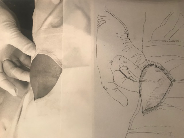
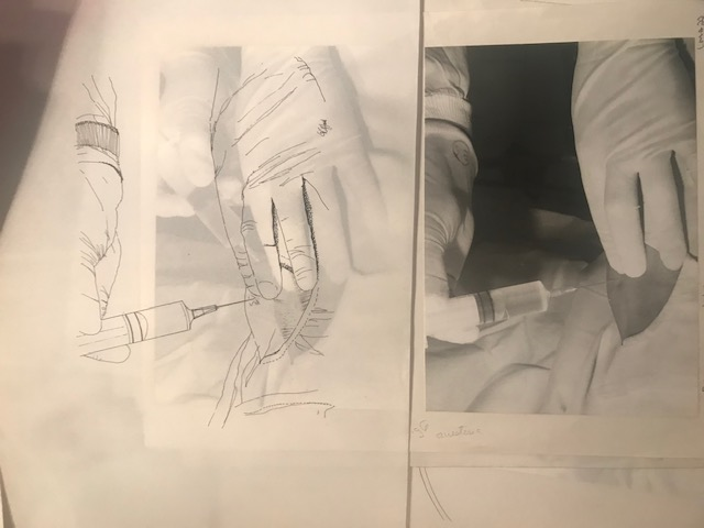
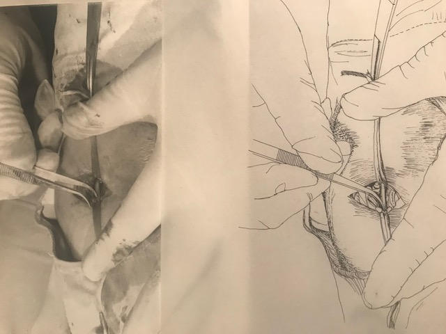
The inspection technique is placed in the supine position and the dissection is performed / laminated 1 approximately two to 3 cm above the antecubital fold. The presence of a radial and ulnar pulse should be evaluated, as well as performing a plethysmography or arterial Doppler to see the patency of the segments. The right upper limb should be abducted, lamina 1 approximately 90° from the body. The brachial artery is palpated, just above the ante-ulnar fossa at the level of the epicondyle of the humerus. After good asepsis and antisepsis, proceed before local anesthesia. Lamina 2 A transverse incision of one to 1.5 cm in length is made and lamina 3 proceeds to dissect the artery and the brachial vein, taking great care with the median nerve (see figure 1). When the isolation of the artery is achieved, umbilical tape is passed under it at the level of its proximal and distal portions (see figure 2) the artery is cleaned with the posterior edge of the scalpel and hemostatic forceps are used under pressure (see figure 3) a small hole is made in the middle of the anterior wall of the artery A scalpel blade number 11 or 15 , then it is widened with an Iris forceps (see figure 4) catheter is inserted, which is advanced under pressure control to prevent a false pathway from being formed.
The patient is told that he will have a burning sensation in the arm, he is instructed to turn his head to the left side and take the pressure and advance the Sones positrol 7.5 catheter and left coronary cannula see photo and later s e right coronary cannula see photo. Other catheters that could be used are the Castillo or Amplatz catheters with curves one 2 and 3. Other catheters have been described for the approach to aortocoronary grafts and guides of 045, 032 can be used for catheter exchange. Multipurpose catheters, aorto-coronary grafts are used. For coronary anomalies, ar2 catheters may be used. At L. 2, after completion of the diagnostic study, repair is performed with the 5 or 6 lamina prolene 11 many Operators place the catheter distally and suction to avoid thrombosis. Plate 12 Arterial repair is performed by controlling proximal bleeding with digital pressure and umbilical tape. Lamina 13 Lamina After artery repair, the distal clamp is removed and the proximal clamp is subsequently removed, maintaining hemostasis with digital pressure while simultaneously palpating the distal pulses. See this lamina complete repair of a brachial arteriotomy. In summary, the brachial artery is isolated and marked proximal and distal with wet umbilical tapes. Incision of the brachial artery with a number 11 scalpel with the cutting edge facing up. Catheter passage through the hole and closure of the arteriotomy. There are other approaches such as percutaneous brachial puncture with a vascular sheath and radial artery puncture the technique preferred by interventionist groups, in Asia 50 percent abd in Europe 40 percent of the cases in USA 7 percent, with this approach you can do PTCA of the coronary, PTCA of renals,15 percent of patients has coronary and simultaneous renal disease and follow the protocol of laroca to detect kidney injury early, The Sones catheter is made of polyurethane with a 5F tip and four lateral holes and it is a catheter for performing coronary angiography and left ventriculography. It is unfortunate that this technique is not performed in postgraduate interventional cardiology and peripheral vascular but Teaching develops surgical skill in an area where procedures are percutaneous. Mason Sones was an American physician whose pioneering work in cardiac catheterization.
References
- Sones. “Circulation” 20 (1959): 773.
- Sones FM Jr and Shirey EK. “Cine coronary arteriography”. Mod Concepts Cardiovasc Dis 31 (1962): 735-738.
- Abrahans. “The complication of coronary arteriography”. Jama (1962).
Citation:
Manuel Velasco MD., et al. “Open Dissection of the Brachial Artery, Percutaneous Brachial Access, Aortography, Renal Angiography”. Therapeutic Advances in Cardiology 3.1 (2020): 01-03.
Copyright: © 2020 Manuel Velasco MD., et al. This is an open-access article distributed under the terms of the Creative Commons Attribution License, which permits unrestricted
use, distribution, and reproduction in any medium, provided the original author and source are credited.


 Scientia Ricerca is licensed and content of this site is available under a Creative Commons Attribution 4.0 International License.