Opinion Article
Volume 3 Issue 1 - 2020
Extraction of Foreign Body with Loop, Basket and Grasping, Via Brachial or Jugular Access is Easy? Or are Images Required to Avoid Radiation?
1Head of the Cardiac Catheterization Unit of the Colllet Foundation
2Catheterization Laboratory Assistant
3Clinical Pharmacology Unit, Vargas Medical School, Central University of Venezuela, Caracas, Venezuela
2Catheterization Laboratory Assistant
3Clinical Pharmacology Unit, Vargas Medical School, Central University of Venezuela, Caracas, Venezuela
*Corresponding Author: Manuel Velasco MD, Professor, Director, Clinical Pharmacology Unit, Vargas Medical School, Central University of Venezuela, Caracas, Venezuela.
The axillary jugular femoral brachial vascular access procedures through catheters, guides and devices are not exempted from their migration due to fractures to different vascular beds. The migration of the guides in the jugular bed terminals to the pulmonary artery is very common when one is trying to take jugular lines in internal medicine rooms for hydratation of patients or when venous pressure is taking at the Intensive Care Room for Jugular Access and subclavian too. In catheterization rooms with femoral access, less catheter fracture due to material fatigue or recycled material is also described. As guide migration is also described, the guide size is 00 35, 00 38, 00 25, 00 32, Amplatz Mitral Valvuloplasty guide, guides in ductus arteriosus occlusion, as well as migration of stent Amplatz devices , ivalom polyvinyl alcohol buffer, lower vena cava filter migration. The technique is different depending on the venous vascular bed, or arterial vascular bed. In the venous vascular bed the main technique is the use of the loop of Goosneck, we can also use a basket or a grasping system, or a biopsy of endomyocardium. These devices are very similar to those of the Gastroenterologist and Urologists. To locate the devices, besides fluoroscopy, film angiography, the time of the fluoroscope and film in different groups has been from half an hour to four hours, to avoid access to open surgery., To locate these devices in 3D images we can use various tools such as intravascular echo, intracavitary echo, fluoroscope, and tomography. It must be remembered that the venous system can use Mullins shirts and through it we can use a guide catheter with the loop or with the Grasping, we have to be very careful in not to dissecting the vein and not to break or induces fracture of the capture device. The arterial route is more complicated when using guide catheters and long shirts such as Mullins, since it presents vasovagal manifestations that could produce hypotension, and cardiac asystole. In capturing electrode fractures in Marcapaso implantation we have been successful without performing traction maneuvers, or using lasers within the heart, we have also been successful in removing fractured guides, stents which mígrate to different vascular beds from the heart, intravascular echo devices, atheroctome olives, coils devices, amplatzer, migration of aortic prostheses, migration of closure devices, such as the biostar Medtronic.
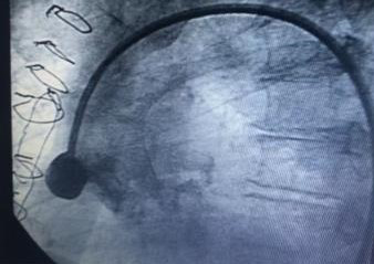
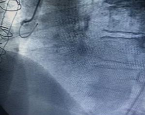
Before accessing the vascular route, it is very important at the present to keep in mind the location of the device when removing it, prior to the study of Doppler echo images, locating the device in the pulmonary arteries if the bed is venous, such as Pigtail catheters, guides, Coils when trying the closure of the ductus arteriosus. In the arterial bed the most frequent is the fracture of catheters as seen in figure 1, and the rescue of guides and devices such as the bio Star, at pleasure which are captured and they were embolized until the right femoral artery and from there it can be removed in the open depending on the device. In our experience of more than 100 extracted cases we have studied in the fluoroscope images by cinema observing the images in posterior position or anterior position, right anterior oblique, left anterior oblique, lateral, especially in coils migration, and anterior position in amplatzer migration, intravascular echo. It is a useful tool to locate if it is in the anterior or posterior wall of the vessel. The intracardiac echo is a useful tool, the transesophageal echo, it guide us in which portion of the ascending aorta or descending aorta the device is located. Experience with multiple images for the transluminal extraction of foreign bodies to decrease the radiation time. And our fuoroscopy and film time, as our clamp is grasping to decrease the radiation time.
Citation:
Manuel Velasco MD., et al. “Extraction of Foreign Body with Loop, Basket and Grasping, Via Brachial or Jugular Access is Easy? Or are Images Required to Avoid Radiation?”. Therapeutic Advances in Cardiology 3.1 (2020): 06-07.
Copyright: © 2020 Manuel Velasco MD., et al. This is an open-access article distributed under the terms of the Creative Commons Attribution License, which permits unrestricted
use, distribution, and reproduction in any medium, provided the original author and source are credited.


 Scientia Ricerca is licensed and content of this site is available under a Creative Commons Attribution 4.0 International License.